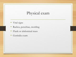
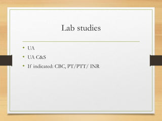
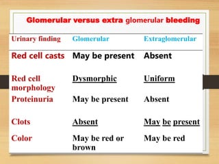
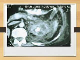
HEMATURIA
Mostafa Magdi Eltawel
Associate professor
Urology
Hematuria
Transient phenomenon
of little significance
Sign of serious
renal disease
Definition
• Macroscopic (gross) Hematuria
• any discolored urine visible to the human eye
• Microscopic Hematuria
• >3-5 RBC/hpf seen under microscope
Classification of
hematuria
Macroscopic - Microscopic
Symptomatic - Symptomless
Transient - Persistent
According to the act of void:
-Initial.
-Terminal.
-Total.
• 25% of patients with gross hematuria have a
life threatening urologic lesion
• 5 to 20% of patients with microscopic
hematuria may have a serious urologic
disorder
• 1% of patients may develop a neoplasm even
though an initial workup is negative within 3-4
years
• Up to 18% of normal individuals may have
some degree of microscopic hematuria
DETECTION OF HEMATURIA
• Dipstick testing 91% sensitivity, 65% specificity for
the detection of Hgb
• False positives caused by:
• Myoglobinuria
• Menstrual bleeding
• Dehydration (increased SG level)
• Outdated or dried dipsticks
• Substances or medications
Substances and medications
• Artificial food coloring
• Beets
• Berries
• Lead or mercury poisoning
• Meds: adriamycin, chloroquine, hydroxychloroquine,
metronidazole, nitrofurantoin, phenazapyridine,
rifampin
Most common causes
• S stones
• I infection
• T tumor
• T trauma
Etiology of Hematuria
• V vascular
• I infectious
• T Tumor/ trauma
• A anatomic anomaly
• M metabolic
• I inherited
• N nonspecific
Vascular
• Aneursym
• Renal vein thrombosis/ Infarct
• Malignant hypertension
• nephritis
Infectious
• Bacterial (remember levaquin)
• Viral
• Fungal
• Parasitic
• TB
Tumor/ trauma
• Kidney, ureteral, bladder, prostate, urethral
malignancy
• Instrumentation
• Radiation
• Exercise
• Foreign body
• Trauma
Anatomic anomaly
• BPH benign prostatic hyperplasia
• Pelvic muscle relaxation
• Renal duplication
• UPJ obstruction
• Polycystic kidney
• Medullary sponge kidney
• Strictures
• Ureterocele or urethrocele
Metabolic
• Kidney stones
• Bladder stones
• Drug induced
Inherited
• IgA nephropathy (Berger’s disease)
• Sickle cell anemia
• Benign familial hematuria
Nonspecific
• Physiologic
• Exercise
• Fever
False Positive
- Endometriosis
- Vaginal bleeding
- factitious
History
• Gross vs. microscopic
• Initial, terminal, or throughout stream
• Painful, irritation, or flank pain
• Recent strep or skin infection
• Medications
• Smoking, occupational exposure
• Trauma/ instrumentation, radiation/ chemo
Physical exam
• Vital signs
• Rashes, petechiae, mottling
• Flank or abdominal mass
• Genitalia exam
Lab studies
• UA
• UA C&S
• If indicated: CBC, PT/PTT/ INR
Radiological
• KUB
• U/S
• C.T
• MRU
Glomerular versus extra glomerular bleeding
Urinary finding Glomerular Extraglomerular
Red cell casts May be present Absent
Red cell
morphology
Dysmorphic Uniform
Proteinuria May be present Absent
Clots Absent May be present
Color May be red or
brown
May be red
Urology referral
If not previously evaluated for hematuria, ALL
patients need a cystoscopic examination of the
bladder mucosa. NO radiologic study or urinary test
(cytology, NMP-22, or BTA-stat) fully evaluates the
bladder.
Nephrology referral
• Significant proteinuria without hematuria
• RBC casts
• Elevated creatinine above 2.5
Follow up
• Somewhat controversial
• Repeat UA, cytology every 6 months for 2 years
followed by yearly exams.
• Re-evaluation for a new episode of gross hematuria
if over 6 months and no previous cause was found.
• 18% of patients may always have hematuria without
a definitive cause
Case 1
• A 65-year-old woman is referred with total painless
haematuria. A midstream specimen of urine (MSU)
has been sent for microscopy, culture and sensitivity.
Microscopy shows a normal white cell count (WCC),
over count red blood cells (RBCs) per high-powered
field and no growth has been found on culture. She
has moderate irritative voiding lower urinary tract
symptoms (LUTS).The patient also gave a history of
anorexia, weight loss, and night sweating . No other
investigations have been performed. How would you
assess this woman?
CASE 2
• A 35-year-old man presents to the emergency
department with intermittent right loin to groin pain
and visible hematuria. He had history of passing
stones . What is the likely cause?
CASE 3
• Male patient 55 years old , diabetic , hypertensive ,
and heavy smoker , presented to you with red
discoloration of urine . The patient gave a history of
taking anti ischemic drugs due to recent myocardial
infarction one month ago, what is the most probably
cause of hematuria , and how to exclude a urological
cause ?!
CASE 4
• A 64-year-old man presents with a 2-month history
of painless visible haematuria, hypertension, weight
loss, loin swelling and anaemia. What is the most
likely diagnosis?
HEMATURIA.ppt

HEMATURIA.ppt

  • 1.
  • 2.
    Hematuria Transient phenomenon of littlesignificance Sign of serious renal disease
  • 3.
    Definition • Macroscopic (gross)Hematuria • any discolored urine visible to the human eye • Microscopic Hematuria • >3-5 RBC/hpf seen under microscope
  • 4.
    Classification of hematuria Macroscopic -Microscopic Symptomatic - Symptomless Transient - Persistent According to the act of void: -Initial. -Terminal. -Total.
  • 5.
    • 25% ofpatients with gross hematuria have a life threatening urologic lesion • 5 to 20% of patients with microscopic hematuria may have a serious urologic disorder • 1% of patients may develop a neoplasm even though an initial workup is negative within 3-4 years • Up to 18% of normal individuals may have some degree of microscopic hematuria
  • 6.
    DETECTION OF HEMATURIA •Dipstick testing 91% sensitivity, 65% specificity for the detection of Hgb • False positives caused by: • Myoglobinuria • Menstrual bleeding • Dehydration (increased SG level) • Outdated or dried dipsticks • Substances or medications
  • 7.
    Substances and medications •Artificial food coloring • Beets • Berries • Lead or mercury poisoning • Meds: adriamycin, chloroquine, hydroxychloroquine, metronidazole, nitrofurantoin, phenazapyridine, rifampin
  • 8.
    Most common causes •S stones • I infection • T tumor • T trauma
  • 9.
    Etiology of Hematuria •V vascular • I infectious • T Tumor/ trauma • A anatomic anomaly • M metabolic • I inherited • N nonspecific
  • 10.
    Vascular • Aneursym • Renalvein thrombosis/ Infarct • Malignant hypertension • nephritis
  • 11.
    Infectious • Bacterial (rememberlevaquin) • Viral • Fungal • Parasitic • TB
  • 12.
    Tumor/ trauma • Kidney,ureteral, bladder, prostate, urethral malignancy • Instrumentation • Radiation • Exercise • Foreign body • Trauma
  • 13.
    Anatomic anomaly • BPHbenign prostatic hyperplasia • Pelvic muscle relaxation • Renal duplication • UPJ obstruction • Polycystic kidney • Medullary sponge kidney • Strictures • Ureterocele or urethrocele
  • 14.
    Metabolic • Kidney stones •Bladder stones • Drug induced
  • 15.
    Inherited • IgA nephropathy(Berger’s disease) • Sickle cell anemia • Benign familial hematuria
  • 16.
    Nonspecific • Physiologic • Exercise •Fever False Positive - Endometriosis - Vaginal bleeding - factitious
  • 17.
    History • Gross vs.microscopic • Initial, terminal, or throughout stream • Painful, irritation, or flank pain • Recent strep or skin infection • Medications • Smoking, occupational exposure • Trauma/ instrumentation, radiation/ chemo
  • 18.
    Physical exam • Vitalsigns • Rashes, petechiae, mottling • Flank or abdominal mass • Genitalia exam
  • 19.
    Lab studies • UA •UA C&S • If indicated: CBC, PT/PTT/ INR
  • 20.
  • 21.
    Glomerular versus extraglomerular bleeding Urinary finding Glomerular Extraglomerular Red cell casts May be present Absent Red cell morphology Dysmorphic Uniform Proteinuria May be present Absent Clots Absent May be present Color May be red or brown May be red
  • 39.
    Urology referral If notpreviously evaluated for hematuria, ALL patients need a cystoscopic examination of the bladder mucosa. NO radiologic study or urinary test (cytology, NMP-22, or BTA-stat) fully evaluates the bladder.
  • 40.
    Nephrology referral • Significantproteinuria without hematuria • RBC casts • Elevated creatinine above 2.5
  • 41.
    Follow up • Somewhatcontroversial • Repeat UA, cytology every 6 months for 2 years followed by yearly exams. • Re-evaluation for a new episode of gross hematuria if over 6 months and no previous cause was found. • 18% of patients may always have hematuria without a definitive cause
  • 42.
    Case 1 • A65-year-old woman is referred with total painless haematuria. A midstream specimen of urine (MSU) has been sent for microscopy, culture and sensitivity. Microscopy shows a normal white cell count (WCC), over count red blood cells (RBCs) per high-powered field and no growth has been found on culture. She has moderate irritative voiding lower urinary tract symptoms (LUTS).The patient also gave a history of anorexia, weight loss, and night sweating . No other investigations have been performed. How would you assess this woman?
  • 43.
    CASE 2 • A35-year-old man presents to the emergency department with intermittent right loin to groin pain and visible hematuria. He had history of passing stones . What is the likely cause?
  • 44.
    CASE 3 • Malepatient 55 years old , diabetic , hypertensive , and heavy smoker , presented to you with red discoloration of urine . The patient gave a history of taking anti ischemic drugs due to recent myocardial infarction one month ago, what is the most probably cause of hematuria , and how to exclude a urological cause ?!
  • 45.
    CASE 4 • A64-year-old man presents with a 2-month history of painless visible haematuria, hypertension, weight loss, loin swelling and anaemia. What is the most likely diagnosis?