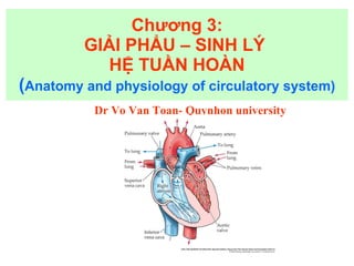
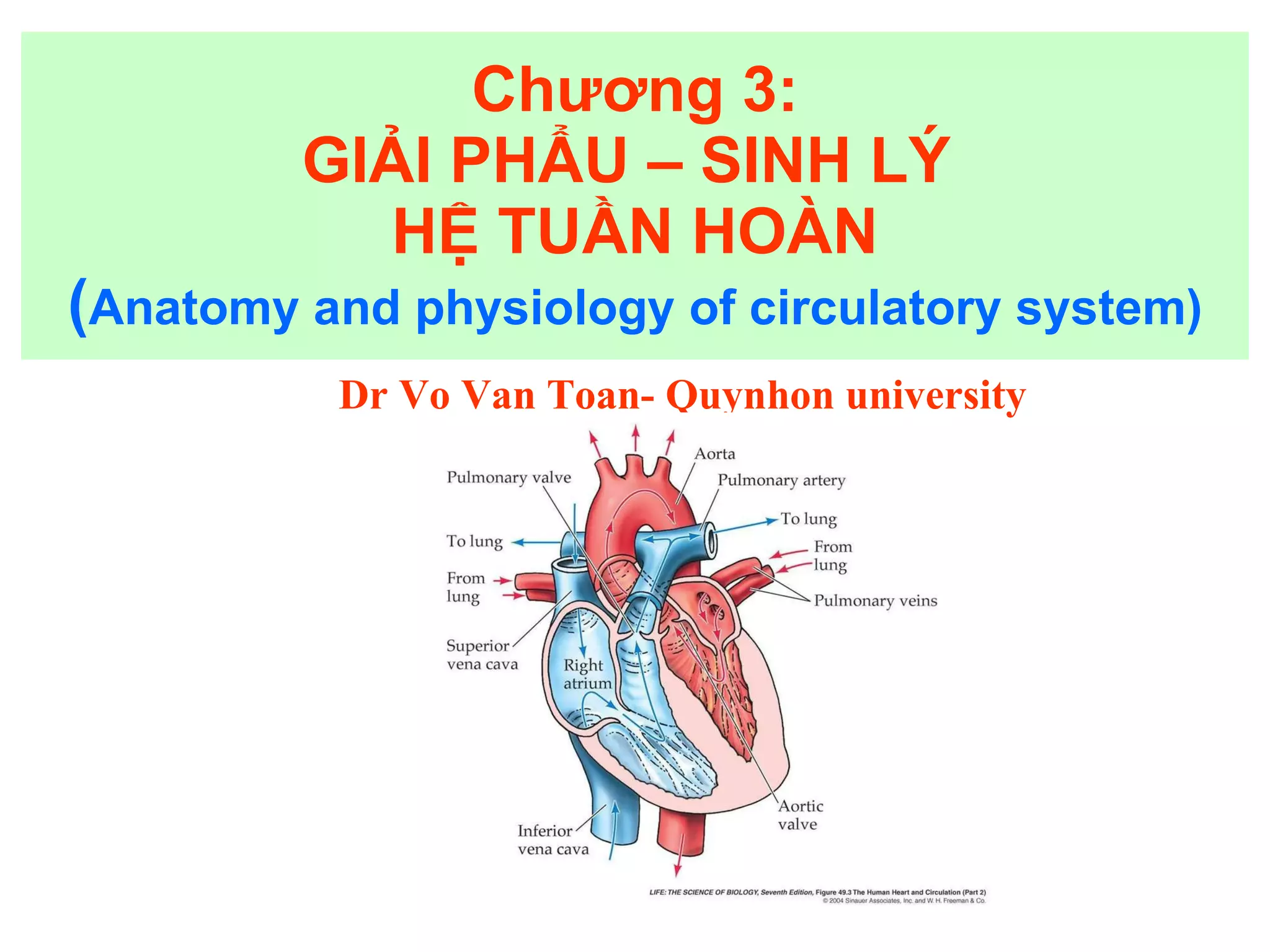
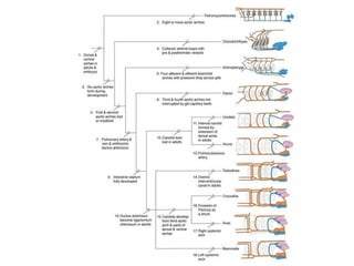
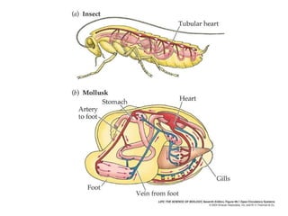
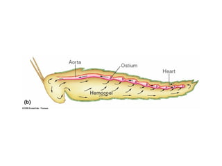
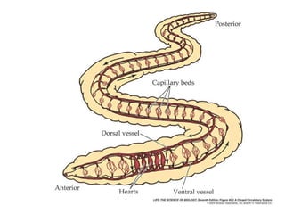
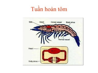
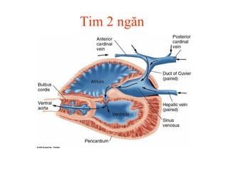
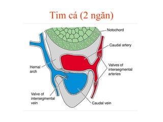
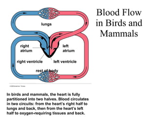
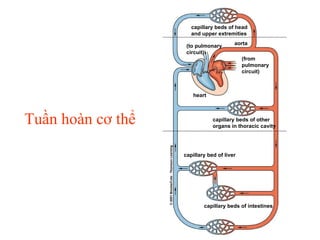
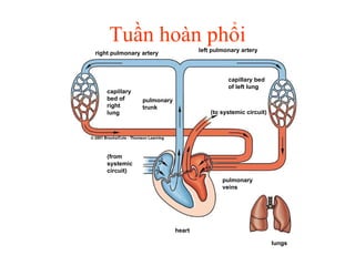
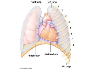
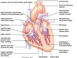
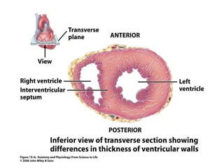
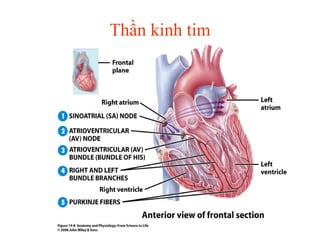
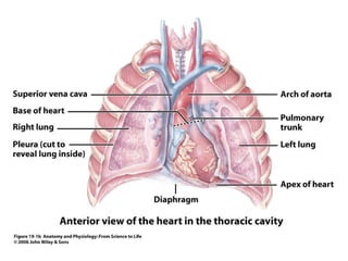
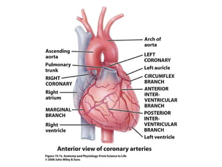
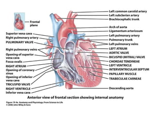
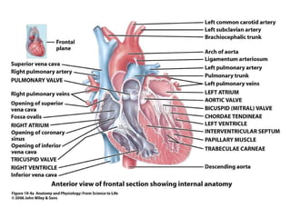
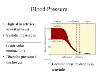
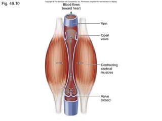
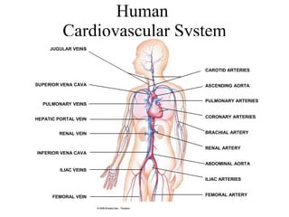
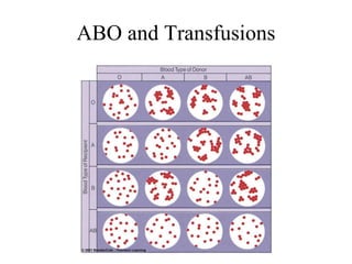
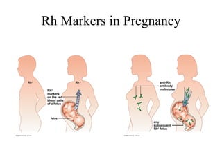
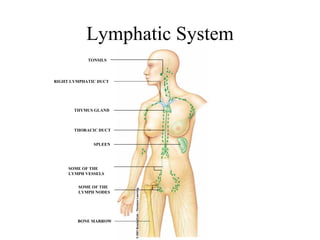
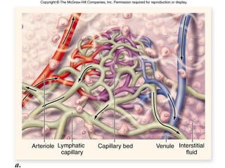
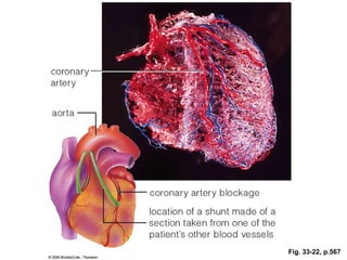
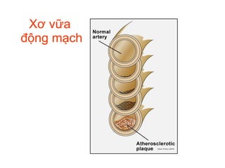
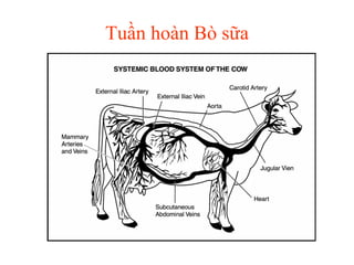
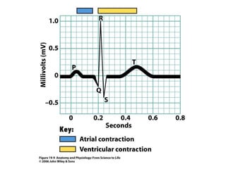
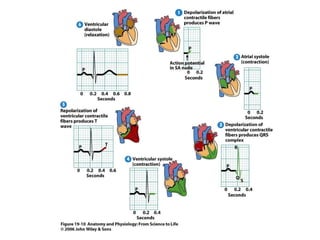
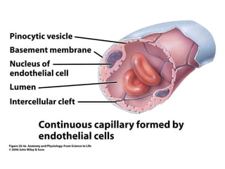
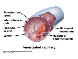
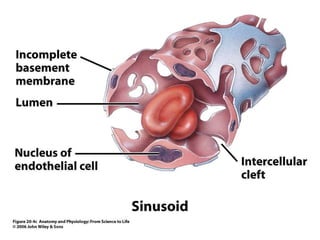
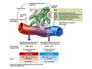
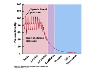
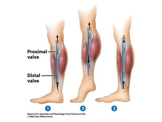
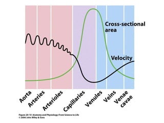
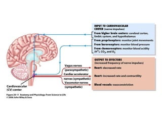
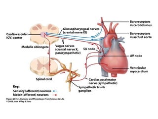
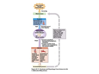
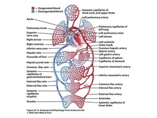
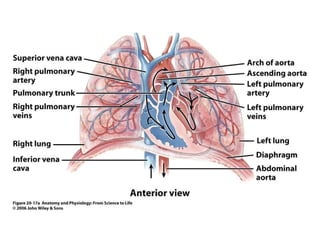
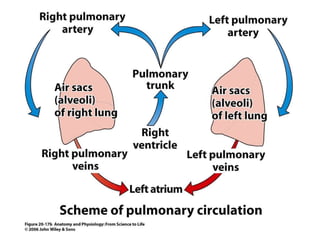
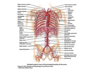
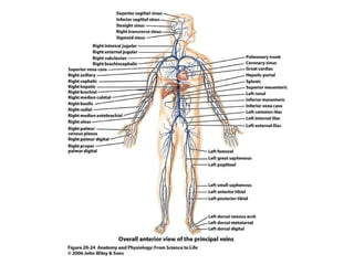
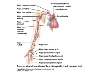
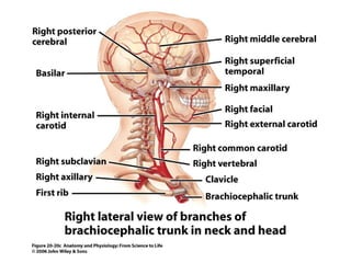
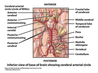
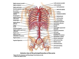
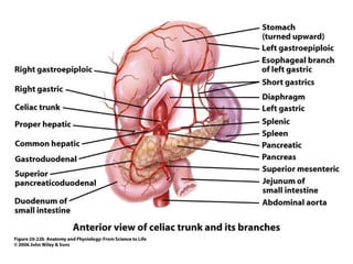
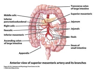
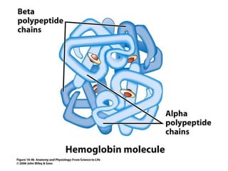
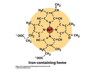
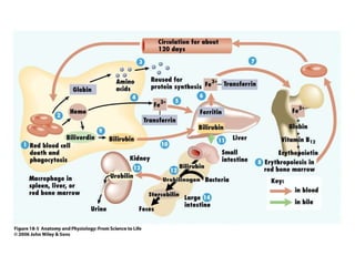
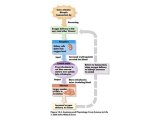
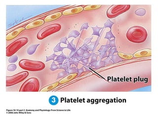
Chương 3: GIẢI PHẨU – SINH LÝ  HỆ TUẦN HOÀN ( Anatomy and physiology of circulatory system) Dr Vo Van Toan- Quynhon university


Tuần hoàn tôm
Tim 2 ngăn
Tim cá (2 ngăn)
Blood Flow in Birds and Mammals rest of body lungs right atrium left atrium right ventricle left ventricle In birds and mammals, the heart is fully partitioned into two halves. Blood circulates in two circuits: from the heart’s right half to lungs and back, then from the heart’s left half to oxygen-requiring tissues and back.
Tuần hoàn cơ thể capillary beds of head and upper extremities (to pulmonary circuit) aorta (from pulmonary circuit) heart capillary beds of other organs in thoracic cavity capillary bed of liver capillary beds of intestines
Tuần hoàn phổi right pulmonary artery left pulmonary artery capillary bed of right lung pulmonary trunk capillary bed of left lung (to systemic circuit) pulmonary  veins lungs (from systemic circuit) heart
 
right lung left lung diaphragm pericardium rib cage 1 2 3 4 5 6 7 8
superior vena cava (from head, upper limbs) right semilunar valve (shown closed); to the pulmonary trunk right pulmonary veins (from lungs) right atrium right AV valve (shown open) right ventricle (muscles that prevent valve from  everting)  inferior vena cava (from trunk, legs) septum (partition between heart's  two halves) myocardium heart’s apex arch of aorta trunk of pulmonary arteries left semilunar valve (shown closed); to aorta left pulmonary veins (from lungs) left atrium left AV valve (shown open) left ventricle endothelium and underlying connective tissue inner layer of pericardium
 
Thần kinh tim
Figure 19.4a
Figure 19.3c
Figure 19.1b
Figure 19.4a
Figure 19.7a
Figure 19.4a
Figure 19.3c
Figure 19.1b
Figure 19.4a
Figure 19.7a
Blood Pressure Highest in arteries, lowest in veins Systolic pressure is __________________(ventricular contraction) Diastolic pressure is the lowest   Greatest pressure drop is in arterioles
 
Fig. 49.10
Human  Cardiovascular System
p.550b
 
 
ABO and Transfusions
Rh Markers in Pregnancy
Lymphatic System
 
Lymph Nodes
 
Fig. 33-22, p.567
Xơ vữa  động mạch
Tuần hoàn Bò sữa
Figure 19.9
Figure 19.10
Figure 20.4a
Figure 20.4b
Figure 20.4c
Figure 20.7
Figure 20.7
Figure 20.8
Figure 20.9
Figure 20.10
Figure 20.11
Figure 20.12
Figure 20.13
Figure 20.16
Figure 20.17a
Figure 20.17b
Figure 20.18a
Figure 20.18b
Figure 20.24
Figure 20.20b
Figure 20.20c
Figure 20.20d
Figure 20.21b
Figure 20.22b
Figure 20.22c
Figure 20.22d
Figure 18.1a
Figure 18.1b
Figure 18.3
Figure 18.4a
Figure 18.4b
Figure 18.4c
Figure 18.5
Figure 18.6
Figure 18.7
Figure 18.3
Fig. 18.10 pt 1
Fig. 18.10 pt 2
Fig. 18.10 pt 3
Fig. 18.11a
Fig. 18.11b
Fig. 18.11c
Fig. 18.11d

H tu-n hoàn p1(1)