graft vein thrombosis after renal transplantation
•Download as PPT, PDF•
0 likes•95 views
in adult renal transplant recipient, the graft vein was anastomosed to the recipient external iliac vein. many factors contribute thrombosis: recombanc postoperative ? ABMR & VASCULITIS SURGICAL ANASTOMOSIS
Report
Share
Report
Share

Recommended
Lytics for Normotensive Submassive PE
- This document summarizes the case of a 45-year-old woman who presented with shortness of breath and chest pressure. Exams found elevated jugular venous pressure and signs of right ventricular strain on EKG.
- Differential diagnosis includes pulmonary embolism. Imaging with CTA and invasive hemodynamics confirmed massive pulmonary embolism with right heart strain. Treatment options discussed include anticoagulation with heparin or thrombolysis. Risks and benefits of thrombolysis versus anticoagulation alone were debated based on the degree of right ventricular dysfunction seen.
Interdepartment Wafarin
This document summarizes an interdepartmental conference discussing the management of a 51-year-old female patient with a history of antiphospholipid syndrome who presented with left-sided weakness and was found to have a large brain infarction and intracranial hemorrhage. The conference discusses treatment to reverse her anticoagulation including vitamin K, fresh frozen plasma, prothrombin complex concentrates and recombinant factor VIIa. It also notes the development of left leg swelling and deep vein thrombosis during her admission and options for anticoagulation or inferior vena cava filter placement.
Presentation1
ACUTE MYOCARDIAL INFARCTION FOLLOWING INTRAVENOUS TISSUE PLASMINOGEN ACTIVATOR FOR
ACUTE ISCHEMIC STROKE : AN UNKNOWN DANGER
arrhythmogenic right ventricular dysplasia/Cardiomyopathy
This case discusses arrhythmogenic right ventricular cardiomyopathy (ARVC) in a 19-year-old female whose sister recently passed away from the condition. ARVC is characterized by replacement of the RV myocardium with fibrofatty tissue and electrical instability. The patient's sister's autopsy confirmed ARVC. The doctor discusses the pathology, diagnosis, treatment including ICDs and screening of relatives of ARVC to help inform the patient of her risk.
ARVD
ARVC is a heritable heart muscle disorder that predominantly affects the right ventricle. It is caused by genetic defects in cardiac desmosomes, which are important for cell-to-cell adhesion. This leads to progressive loss of right ventricular myocardium and replacement by fibrofatty tissue. ARVC can cause dangerous ventricular arrhythmias and is a leading cause of sudden cardiac death in young people. Diagnosis involves imaging tests and electrocardiography to detect right ventricular structural abnormalities and arrhythmias.
Acquired hemophilia a
Acquired hemophilia A is a rare bleeding disorder caused by autoantibodies against factor VIII. It most commonly presents in older patients as severe bleeding and has a high mortality rate if not properly treated. Evaluation involves testing for prolonged aPTT and ruling out an inhibitor through mixing studies. Treatment focuses on controlling bleeding with bypassing agents or factor VIII while also using immunosuppressants to eliminate the autoantibody inhibitor. Proper management can reduce bleeding and inhibitor levels, but monitoring is needed due to the slow response to therapy.
Bruguda syndrome
Brugada Syndrome is a inherited sodium channel disorder leading to life threatening ventricular fibrillation in young population. diagnosis and ICD therapy could be life saving.
Trauma induced coagulopathy
Trauma induced coagulopathy (TIC) starts early due to shock and hypoperfusion which activates the thrombomodulin-protein C pathway. TIC is associated with higher mortality, transfusion requirements, and organ dysfunction. Assessment of TIC requires thromboelastometry rather than standard coagulation tests due to confounding factors like hypothermia, acidosis, and dilution of clotting factors from fluid resuscitation. Treatment focuses on replacing depleted clotting factors, with fibrinogen and prothrombin concentrates highly recommended, as well as consideration of antifibrinolytics and recombinant factor VIIa in specific cases.
Brugada syndrome
Brugada Syndrome is a genetic cardiac condition characterized by an abnormal ECG pattern and increased risk of sudden cardiac death. It is caused by mutations that result in loss of function of cardiac ion channels, most commonly sodium channels encoded by SCN5A. The ECG typically shows ST segment elevation in leads V1-V3. Brugada Syndrome presents variably from asymptomatic to sudden cardiac death, usually during sleep. Drug challenges may help diagnose when the ECG is unclear. An ICD is recommended for those with symptoms or inducible arrhythmias on electrophysiological study to prevent sudden death.
Early repolrisation syndrome(ERS)
This document discusses early repolarization syndrome (ERS). [1] ERS is characterized by J point elevation in the inferior and/or lateral leads of an ECG. [2] It was first described in the 1930s and takes its name from increased repolarization seen in some heart conditions. [3] ERS has three subtypes based on location of J point elevation and risk of arrhythmias. The mechanism involves increased heterogeneity of ventricular repolarization. ERS has been associated with increased risk of fatal arrhythmias, especially when the pattern involves the inferior leads.
EKG Patterns of SCD - Can't Miss EKG Patterns for Generalist & Psychiatrist
This document discusses several electrocardiogram patterns that can indicate risk of sudden cardiac death. It presents six case studies and the corresponding ECG patterns:
1) Wolf-Parkinson-White syndrome seen in a 34-year-old with palpitations, shown by a short PR interval and delta wave.
2) Brugada syndrome in a 36-year-old with chest pain, shown by ST elevation in right precordial leads.
3) Arrhythmogenic right ventricular dysplasia in a 28-year-old with dizziness, shown by epsilon waves and T wave inversion.
4) Long QT syndrome in a 44-year-old with dizziness and
Ectopic Paroxysmal Atrial Tachycardia
A 45-year-old female presented with progressive palpitations over the past year. Holter monitoring showed frequent atrial premature complexes and runs of atrial tachycardia. An electrophysiology study identified a focal source of ectopic beats in the right atrium above the appendage. Radiofrequency catheter ablation was performed at this site, which eliminated the arrhythmia based on follow up Holter monitoring two months later.
Omar babker
Omar Babiker Mohammed Khalifa, a 54-year-old Sudanese male, was admitted to the hospital with microscopic polyangitis and pulmonary vasculitis. He received immunosuppressive treatments including pulse steroids, cyclophosphamide, and plasma exchange. His condition deteriorated, requiring intubation and mechanical ventilation in the ICU for severe dyspnea and hypoxia. Tests showed a massive left pleural effusion which was drained. The patient's kidney function and blood counts fluctuated during his hospital stay while receiving dialysis and treatments for his lung hemorrhage and vasculitis.
Arrhythmogenic right ventricular dysplasia
Arrhythmogenic right ventricular dysplasia (ARVD) is a genetic heart condition characterized by replacement of right ventricular myocardium with fat and fibrous tissue. This causes ventricular arrhythmias which can lead to sudden cardiac death. ARVD is diagnosed based on criteria involving cardiac imaging, biopsy, electrocardiogram findings and genetic testing. A combination of major and minor criteria must be met, including ventricular dysfunction, aneurysms, conduction abnormalities and evidence of fibrofatty tissue replacement on biopsy. ARVD has an autosomal dominant inheritance pattern and is a leading cause of sudden cardiac death in young people.
Brugada Syndrome - Case Study
Brugada Syndrome - Case Study
Precise data regarding Brugada Syndrome with a Case Study for DGNM, B.Sc Nursing & M.Sc Nursing Students.
Arrhythmogenic right ventricular 2003
The document summarizes arrhythmogenic right ventricular dysplasia (ARVD), a condition where the right ventricle of the heart is replaced by fat and fibrous tissue. It affects mostly young males and can cause sudden cardiac death. Genetic factors are involved in many cases. The condition starts with fatty infiltration of the right ventricle and progresses to include fibrosis, thinning of the ventricular wall, and later involvement of the left ventricle. Diagnosis involves criteria related to structural changes, electrocardiogram abnormalities, arrhythmias, and family history.
Repolarization syndromes
Early repolarization (ER), consisting of a J-point elevation, notching or slurring of the terminal portion of the R wave (J wave), and tall/symmetric T wave, is a common finding on the 12-lead electrocardiogram. For decades, it has been considered as benign, barring sporadic case reports and basic electrophysiology research that suggested a critical role of the J wave in the pathogenesis of idiopathic ventricular fibrillation (VF). In 2007-2008, a high prevalence of ER in patients with idiopathic VF was reported and subsequent studies reinforced the results. This PPT describes the current state of knowledge concerning ER syndrome associated with sudden cardiac death.
Tranexamic acid
Tranexamic acid is an antifibrinolytic drug that inhibits fibrinolysis by reversibly binding to plasminogen. It is effective at reducing surgical bleeding across many procedures like cardiac, orthopedic, and cranial surgery. It also reduces bleeding in gynecological conditions like heavy menstrual bleeding and postpartum hemorrhage. Tranexamic acid is generally well tolerated with side effects including headache, nausea, and diarrhea. It is given orally, intravenously, or topically depending on the condition being treated.
Icm aki 194
There are multiple definitions of acute kidney injury (AKI) but no universal consensus. The RIFLE and AKIN classifications provide criteria for staging AKI based on changes in serum creatinine and urine output. AKI occurs in 7-36% of hospitalized or critically ill patients depending on the definition, with 5-6% requiring renal replacement therapy. Even minor increases in creatinine are associated with higher mortality, and requiring RRT carries a mortality risk of 50-70%. Ischemic acute tubular necrosis is a common cause of AKI and results from renal hypoperfusion that overwhelms autoregulatory mechanisms.
Inhibitors in Congenital Hemophilia
This document discusses inhibitors in congenital hemophilia and their treatment. It begins with an overview of hemophilia A and B, risk factors for inhibitor development like family history and treatment intensity, and mechanisms of inhibitor action. Treatment options discussed include high-dose factor replacement, bypassing agents like activated prothrombin complex concentrate and recombinant factor VIIa, and immune tolerance induction regimens to eradicate inhibitors. Two studies directly comparing aPCC and rFVIIa found they achieved similar rates of hemostasis, though one study found rFVIIa in a single 270 μg/kg dose was more effective than aPCC or multiple 90 μg/kg rFVIIa doses. Prophylaxis with
Cardiac chanellopathies
This document discusses several cardiac channelopathies including Brugada syndrome, long QT syndrome, short QT syndrome, catecholaminergic polymorphic ventricular tachycardia, idiopathic ventricular fibrillation, and early repolarization syndromes. It provides details on the electrocardiographic patterns, genetic causes, clinical manifestations, diagnosis and management of these conditions. Specifically, it focuses on the electrocardiographic patterns that define Brugada syndrome, the genetic and ion channel basis for the condition, and its association with sudden cardiac death.
Brugada Syndrome
Brugada Syndrome is a genetic disorder characterized by abnormal ECG patterns and increased risk of ventricular arrhythmias. It is caused by mutations in genes encoding sodium channels. Typical ECG findings include ST elevation in leads V1-V3. Risk factors include spontaneous type 1 ECG pattern, family history of sudden cardiac death, and inducible arrhythmias on electrophysiology study. Diagnosis requires type 1 ECG pattern plus symptoms or family history of events.
Approach to Channelopathies ppt
This document discusses different types of channelopathies, which are inherited arrhythmogenic diseases caused by mutations in ion channel genes. It summarizes the definitions, epidemiology, clinical manifestations, risk stratification approaches, and management recommendations for several major channelopathies:
- Long QT syndrome is associated with prolonged cardiac repolarization and can cause syncope or cardiac arrest. Risk stratification considers clinical factors and genetic testing, and management includes beta-blockers and ICDs.
- Brugada syndrome causes ST segment elevation and is a common cause of sudden cardiac death. Risk is assessed based on symptoms and ECG patterns, and ICDs are often recommended for high-risk patients.
- Cate
Cardiology board mc qs.ppt2
The document discusses several different cardiac conditions and test results including atrial fibrillation with complete heart block, second degree 2:1 heart block, complete heart block, atrial flutter with 2:1 block, amyloidosis indicated by an echocardiogram showing severe LVH, speckled appearance and low voltage limb leads on ECG, inferolateral reversible ischemia of moderate degree found on a graft study for a post-CABG patient which also showed an occluded superior OM and diseased OM2 and OM3 that were stented.
Brugada syndrome
Brugada syndrome is a genetic heart condition characterized by abnormal ECG patterns and risk of sudden cardiac death. It is caused by mutations in genes encoding sodium channels. The condition is diagnosed through ECG showing ST segment elevation in leads V1-V3. Treatment involves implanting an ICD to detect and treat lethal arrhythmias with shocks. Prognosis depends on risk stratification and treatment.
Congenital long qt syndrome
The long QT syndrome (LQTS) is a rare inherited heart condition in which delayed repolarization of the heart following a heartbeat increases the risk of episodes of torsades de pointes (TDP, a form of irregular heartbeat that originates from the ventricles). These episodes may lead to palpitations, fainting and sudden death due to ventricular fibrillation. Episodes may be provoked by various stimuli, depending on the subtype of the condition.The condition is so named because of the appearances of the electrocardiogram (ECG/EKG), on which there is prolongation of the QT interval. In some individuals the QT prolongation occurs only after the administration of certain medications.
The Long QT Syndrome: Overview and Management The Long QT Syndrome: Overvie...
Long QT Syndrome is a genetic disorder characterized by a prolonged QT interval on electrocardiogram that can cause dangerous arrhythmias and sudden cardiac death. Symptoms include unexplained fainting, seizures, or sudden death, especially with exercise or emotions. Treatment involves beta blockers, implantable cardioverter defibrillators, or left stellate ganglionectomy depending on risk level and genotype. Ongoing research seeks to better understand genotype-phenotype relationships and develop mutation-specific therapies.
4. anticoagulation during ecmo #beach2019 (peperstraete)
This workshop will outline the basic principles of extracorporeal life support made easy by key-experts in the field. During the course delegates will gain a good understanding of ECMO in the following areas: Theoretical concepts, basic physiology and pathophysiology, cardiac and respiratory support and monitoring, alarm settings and monitoring, role of cardiac ultrasound during ECMO, newest technologies, circuits and devices, practical hands-on sessions and simulations.
Transplant Nephrectomy Improves Survival following a Failed Renal Allograft (...
This document summarizes a study examining the impact of transplant nephrectomy on mortality rates following kidney allograft failure. The study used data from the United States Renal Data System on over 19,000 patients who returned to dialysis between 1994-2004 after allograft failure. It found that patients who underwent nephrectomy after late graft failure (>1 year) had a 12% lower risk of death compared to those who did not undergo nephrectomy. However, nephrectomy after early graft failure (<1 year) was associated with a 13% higher risk of death.
03 Perioperative Renal Failure In Cardiac Surgery
1) Perioperative acute renal failure occurs in up to 30% of cardiac surgery patients and 1-7% of aortic surgery patients, with mortality over 40%. Acute tubular necrosis is the most common cause.
2) Risk factors include preexisting renal insufficiency, older age over 60, and left ventricular dysfunction. General preventative measures include optimizing hemodynamics, avoiding nephrotoxins, and using isosmolar contrast.
3) Pharmacologic interventions studied to prevent renal failure include fenoldopam, atrial natriuretic peptide, mannitol, and N-acetylcysteine, though evidence is limited. Continuous renal replacement therapy may benefit critically ill
More Related Content
What's hot
Brugada syndrome
Brugada Syndrome is a genetic cardiac condition characterized by an abnormal ECG pattern and increased risk of sudden cardiac death. It is caused by mutations that result in loss of function of cardiac ion channels, most commonly sodium channels encoded by SCN5A. The ECG typically shows ST segment elevation in leads V1-V3. Brugada Syndrome presents variably from asymptomatic to sudden cardiac death, usually during sleep. Drug challenges may help diagnose when the ECG is unclear. An ICD is recommended for those with symptoms or inducible arrhythmias on electrophysiological study to prevent sudden death.
Early repolrisation syndrome(ERS)
This document discusses early repolarization syndrome (ERS). [1] ERS is characterized by J point elevation in the inferior and/or lateral leads of an ECG. [2] It was first described in the 1930s and takes its name from increased repolarization seen in some heart conditions. [3] ERS has three subtypes based on location of J point elevation and risk of arrhythmias. The mechanism involves increased heterogeneity of ventricular repolarization. ERS has been associated with increased risk of fatal arrhythmias, especially when the pattern involves the inferior leads.
EKG Patterns of SCD - Can't Miss EKG Patterns for Generalist & Psychiatrist
This document discusses several electrocardiogram patterns that can indicate risk of sudden cardiac death. It presents six case studies and the corresponding ECG patterns:
1) Wolf-Parkinson-White syndrome seen in a 34-year-old with palpitations, shown by a short PR interval and delta wave.
2) Brugada syndrome in a 36-year-old with chest pain, shown by ST elevation in right precordial leads.
3) Arrhythmogenic right ventricular dysplasia in a 28-year-old with dizziness, shown by epsilon waves and T wave inversion.
4) Long QT syndrome in a 44-year-old with dizziness and
Ectopic Paroxysmal Atrial Tachycardia
A 45-year-old female presented with progressive palpitations over the past year. Holter monitoring showed frequent atrial premature complexes and runs of atrial tachycardia. An electrophysiology study identified a focal source of ectopic beats in the right atrium above the appendage. Radiofrequency catheter ablation was performed at this site, which eliminated the arrhythmia based on follow up Holter monitoring two months later.
Omar babker
Omar Babiker Mohammed Khalifa, a 54-year-old Sudanese male, was admitted to the hospital with microscopic polyangitis and pulmonary vasculitis. He received immunosuppressive treatments including pulse steroids, cyclophosphamide, and plasma exchange. His condition deteriorated, requiring intubation and mechanical ventilation in the ICU for severe dyspnea and hypoxia. Tests showed a massive left pleural effusion which was drained. The patient's kidney function and blood counts fluctuated during his hospital stay while receiving dialysis and treatments for his lung hemorrhage and vasculitis.
Arrhythmogenic right ventricular dysplasia
Arrhythmogenic right ventricular dysplasia (ARVD) is a genetic heart condition characterized by replacement of right ventricular myocardium with fat and fibrous tissue. This causes ventricular arrhythmias which can lead to sudden cardiac death. ARVD is diagnosed based on criteria involving cardiac imaging, biopsy, electrocardiogram findings and genetic testing. A combination of major and minor criteria must be met, including ventricular dysfunction, aneurysms, conduction abnormalities and evidence of fibrofatty tissue replacement on biopsy. ARVD has an autosomal dominant inheritance pattern and is a leading cause of sudden cardiac death in young people.
Brugada Syndrome - Case Study
Brugada Syndrome - Case Study
Precise data regarding Brugada Syndrome with a Case Study for DGNM, B.Sc Nursing & M.Sc Nursing Students.
Arrhythmogenic right ventricular 2003
The document summarizes arrhythmogenic right ventricular dysplasia (ARVD), a condition where the right ventricle of the heart is replaced by fat and fibrous tissue. It affects mostly young males and can cause sudden cardiac death. Genetic factors are involved in many cases. The condition starts with fatty infiltration of the right ventricle and progresses to include fibrosis, thinning of the ventricular wall, and later involvement of the left ventricle. Diagnosis involves criteria related to structural changes, electrocardiogram abnormalities, arrhythmias, and family history.
Repolarization syndromes
Early repolarization (ER), consisting of a J-point elevation, notching or slurring of the terminal portion of the R wave (J wave), and tall/symmetric T wave, is a common finding on the 12-lead electrocardiogram. For decades, it has been considered as benign, barring sporadic case reports and basic electrophysiology research that suggested a critical role of the J wave in the pathogenesis of idiopathic ventricular fibrillation (VF). In 2007-2008, a high prevalence of ER in patients with idiopathic VF was reported and subsequent studies reinforced the results. This PPT describes the current state of knowledge concerning ER syndrome associated with sudden cardiac death.
Tranexamic acid
Tranexamic acid is an antifibrinolytic drug that inhibits fibrinolysis by reversibly binding to plasminogen. It is effective at reducing surgical bleeding across many procedures like cardiac, orthopedic, and cranial surgery. It also reduces bleeding in gynecological conditions like heavy menstrual bleeding and postpartum hemorrhage. Tranexamic acid is generally well tolerated with side effects including headache, nausea, and diarrhea. It is given orally, intravenously, or topically depending on the condition being treated.
Icm aki 194
There are multiple definitions of acute kidney injury (AKI) but no universal consensus. The RIFLE and AKIN classifications provide criteria for staging AKI based on changes in serum creatinine and urine output. AKI occurs in 7-36% of hospitalized or critically ill patients depending on the definition, with 5-6% requiring renal replacement therapy. Even minor increases in creatinine are associated with higher mortality, and requiring RRT carries a mortality risk of 50-70%. Ischemic acute tubular necrosis is a common cause of AKI and results from renal hypoperfusion that overwhelms autoregulatory mechanisms.
Inhibitors in Congenital Hemophilia
This document discusses inhibitors in congenital hemophilia and their treatment. It begins with an overview of hemophilia A and B, risk factors for inhibitor development like family history and treatment intensity, and mechanisms of inhibitor action. Treatment options discussed include high-dose factor replacement, bypassing agents like activated prothrombin complex concentrate and recombinant factor VIIa, and immune tolerance induction regimens to eradicate inhibitors. Two studies directly comparing aPCC and rFVIIa found they achieved similar rates of hemostasis, though one study found rFVIIa in a single 270 μg/kg dose was more effective than aPCC or multiple 90 μg/kg rFVIIa doses. Prophylaxis with
Cardiac chanellopathies
This document discusses several cardiac channelopathies including Brugada syndrome, long QT syndrome, short QT syndrome, catecholaminergic polymorphic ventricular tachycardia, idiopathic ventricular fibrillation, and early repolarization syndromes. It provides details on the electrocardiographic patterns, genetic causes, clinical manifestations, diagnosis and management of these conditions. Specifically, it focuses on the electrocardiographic patterns that define Brugada syndrome, the genetic and ion channel basis for the condition, and its association with sudden cardiac death.
Brugada Syndrome
Brugada Syndrome is a genetic disorder characterized by abnormal ECG patterns and increased risk of ventricular arrhythmias. It is caused by mutations in genes encoding sodium channels. Typical ECG findings include ST elevation in leads V1-V3. Risk factors include spontaneous type 1 ECG pattern, family history of sudden cardiac death, and inducible arrhythmias on electrophysiology study. Diagnosis requires type 1 ECG pattern plus symptoms or family history of events.
Approach to Channelopathies ppt
This document discusses different types of channelopathies, which are inherited arrhythmogenic diseases caused by mutations in ion channel genes. It summarizes the definitions, epidemiology, clinical manifestations, risk stratification approaches, and management recommendations for several major channelopathies:
- Long QT syndrome is associated with prolonged cardiac repolarization and can cause syncope or cardiac arrest. Risk stratification considers clinical factors and genetic testing, and management includes beta-blockers and ICDs.
- Brugada syndrome causes ST segment elevation and is a common cause of sudden cardiac death. Risk is assessed based on symptoms and ECG patterns, and ICDs are often recommended for high-risk patients.
- Cate
Cardiology board mc qs.ppt2
The document discusses several different cardiac conditions and test results including atrial fibrillation with complete heart block, second degree 2:1 heart block, complete heart block, atrial flutter with 2:1 block, amyloidosis indicated by an echocardiogram showing severe LVH, speckled appearance and low voltage limb leads on ECG, inferolateral reversible ischemia of moderate degree found on a graft study for a post-CABG patient which also showed an occluded superior OM and diseased OM2 and OM3 that were stented.
Brugada syndrome
Brugada syndrome is a genetic heart condition characterized by abnormal ECG patterns and risk of sudden cardiac death. It is caused by mutations in genes encoding sodium channels. The condition is diagnosed through ECG showing ST segment elevation in leads V1-V3. Treatment involves implanting an ICD to detect and treat lethal arrhythmias with shocks. Prognosis depends on risk stratification and treatment.
Congenital long qt syndrome
The long QT syndrome (LQTS) is a rare inherited heart condition in which delayed repolarization of the heart following a heartbeat increases the risk of episodes of torsades de pointes (TDP, a form of irregular heartbeat that originates from the ventricles). These episodes may lead to palpitations, fainting and sudden death due to ventricular fibrillation. Episodes may be provoked by various stimuli, depending on the subtype of the condition.The condition is so named because of the appearances of the electrocardiogram (ECG/EKG), on which there is prolongation of the QT interval. In some individuals the QT prolongation occurs only after the administration of certain medications.
The Long QT Syndrome: Overview and Management The Long QT Syndrome: Overvie...
Long QT Syndrome is a genetic disorder characterized by a prolonged QT interval on electrocardiogram that can cause dangerous arrhythmias and sudden cardiac death. Symptoms include unexplained fainting, seizures, or sudden death, especially with exercise or emotions. Treatment involves beta blockers, implantable cardioverter defibrillators, or left stellate ganglionectomy depending on risk level and genotype. Ongoing research seeks to better understand genotype-phenotype relationships and develop mutation-specific therapies.
4. anticoagulation during ecmo #beach2019 (peperstraete)
This workshop will outline the basic principles of extracorporeal life support made easy by key-experts in the field. During the course delegates will gain a good understanding of ECMO in the following areas: Theoretical concepts, basic physiology and pathophysiology, cardiac and respiratory support and monitoring, alarm settings and monitoring, role of cardiac ultrasound during ECMO, newest technologies, circuits and devices, practical hands-on sessions and simulations.
What's hot (20)
EKG Patterns of SCD - Can't Miss EKG Patterns for Generalist & Psychiatrist
EKG Patterns of SCD - Can't Miss EKG Patterns for Generalist & Psychiatrist
The Long QT Syndrome: Overview and Management The Long QT Syndrome: Overvie...
The Long QT Syndrome: Overview and Management The Long QT Syndrome: Overvie...
4. anticoagulation during ecmo #beach2019 (peperstraete)
4. anticoagulation during ecmo #beach2019 (peperstraete)
Similar to graft vein thrombosis after renal transplantation
Transplant Nephrectomy Improves Survival following a Failed Renal Allograft (...
This document summarizes a study examining the impact of transplant nephrectomy on mortality rates following kidney allograft failure. The study used data from the United States Renal Data System on over 19,000 patients who returned to dialysis between 1994-2004 after allograft failure. It found that patients who underwent nephrectomy after late graft failure (>1 year) had a 12% lower risk of death compared to those who did not undergo nephrectomy. However, nephrectomy after early graft failure (<1 year) was associated with a 13% higher risk of death.
03 Perioperative Renal Failure In Cardiac Surgery
1) Perioperative acute renal failure occurs in up to 30% of cardiac surgery patients and 1-7% of aortic surgery patients, with mortality over 40%. Acute tubular necrosis is the most common cause.
2) Risk factors include preexisting renal insufficiency, older age over 60, and left ventricular dysfunction. General preventative measures include optimizing hemodynamics, avoiding nephrotoxins, and using isosmolar contrast.
3) Pharmacologic interventions studied to prevent renal failure include fenoldopam, atrial natriuretic peptide, mannitol, and N-acetylcysteine, though evidence is limited. Continuous renal replacement therapy may benefit critically ill
03 Perioperative Renal Failure In Cardiac Surgery
1) Perioperative acute renal failure occurs in up to 30% of cardiac surgery patients and 1-7% of aortic surgery patients, with mortality over 40%. Acute tubular necrosis is the most common cause.
2) Risk factors include preexisting renal insufficiency, older age over 60, and left ventricular dysfunction. General preventative measures include optimizing hemodynamics, avoiding nephrotoxins, and using isosmolar contrast.
3) Pharmacological interventions studied to prevent renal failure include fenoldopam, atrial natriuretic peptide, mannitol, and N-acetylcysteine, though evidence is limited. Continuous renal replacement therapy may benefit critically ill
Early graft dysfunction
The document discusses early graft dysfunction following kidney transplantation, which can occur within the first six months post-transplant. There are several potential causes of early graft dysfunction, including delayed graft function (DGF) from ischemic acute tubular necrosis, acute rejection, vascular complications, drug toxicity from calcineurin inhibitors, and recurrence of the original kidney disease. DGF is the most common cause and presents as slow or no improvement in kidney function within the first week, often requiring dialysis. Long-term outcomes are negatively impacted by early graft dysfunction. Ongoing monitoring for acute rejection and other potential causes is important to identify issues and optimize graft survival in the early post-transplant period.
Case Report On Dic & Dvt
A 82-year-old woman underwent a total hip arthroplasty and developed acute disseminated intravascular coagulation postoperatively. She experienced profuse bleeding through her surgical drain and had a precipitous drop in blood pressure. Despite aggressive treatment including blood transfusions, coagulation factor replacements, and hemodynamic support, her condition deteriorated over the next two weeks with multi-organ dysfunction. She ultimately died from her disseminated intravascular coagulation 14 days after her surgery. Disseminated intravascular coagulation is a life-threatening condition with high mortality that can rarely complicate total hip arthroplasty as seen in this case.
Stroke recent
1. Thrombolysis using intravenous rt-PA within 3-4.5 hours of stroke onset is the primary reperfusion therapy approved by the FDA.
2. Other reperfusion therapies include intra-arterial thrombolysis, mechanical thrombectomy devices, sonothrombolysis, laser thrombolysis, and percutaneous angioplasty.
3. Neuroprotective therapies aim to reduce excitatory neurotransmitters, reperfusion injury, and neuronal cell death through mechanisms such as decreasing calcium influx and free radical production.
Minha S
This case report describes a late pseudoaneurysm that developed at the puncture site in a 30-year old male with systemic lupus erythematosus (SLE) and antiphospholipid antibody syndrome (APLA) who underwent coronary angiography. Two weeks after the procedure, an enlarging pulsatile bulge was detected at the puncture site. Ultrasound-guided thrombin injection resolved the pseudoaneurysm. The authors speculate that patients with chronic inflammatory diseases treated with anticoagulation and steroids may be predisposed to late pseudoaneurysm formation due to vessel wall weakness. Close follow-up is recommended for high-risk patients.
An Interesting Case of Stuck Mitral Valve by Clot, Post Thrombolysis Develope...
Prosthetic mechanical valve thrombosis is seen in patients with inadequate anticoagulation, irregular medications, and lack of proper follow up. Thrombolysis is a good alternative to surgery in selected cases. Acute limb ischemia may one of the embolic complications that may occur post thrombolysis. Early PAG and catheter directed re- thrombolysis despite having high risk can further save patients from such serious complications and its sequalae.
Endovascular treatment in acute cerebral ischemia
This document discusses ischemic stroke and endovascular management. It notes that ischemic stroke accounts for 87% of strokes and results in significant disability. It reviews cerebral circulation and the limitations of intravenous tissue plasminogen activator (tPA) therapy. The document then discusses endovascular management techniques in more detail, including intra-arterial thrombolysis, mechanical thrombectomy devices, patient selection, imaging guidance, anesthesia options, reperfusion strategies, and clinical trials of endovascular therapy for middle cerebral artery occlusions.
Transcatheter intraarterial infusion of rt pa for
This study evaluated the success and complication rates of using intraarterial recombinant tissue-type plasminogen activator (rt-PA) infusion to treat acute lower extremity artery and bypass graft occlusions in 74 limbs of 70 patients. Thrombolytic success, defined as 95% thrombolysis and return of blood flow, was achieved in 86% of cases. Major bleeding complications occurred in 47% of patients, including bleeding at vascular access sites in 22 patients and remote bleeding in 7 patients. Despite the significant bleeding complications, 30-day mortality was 1% and amputation rate was 6%, resulting in a favorable 30-day amputation-free survival rate of 93%.
Cerebral Venous Sinus Thrombosis 2010 - Dr. Rajiv Jha (Neurosurgeon Nepal)
Cerebral Venous Sinus Thrombosis
Dr Rajiv Jha, MS
Senior Resident M Ch Neurosurgery
National Neurosurgical Referral Center
National Academy Of Medical Sciences
Changes in blood coagulation markers associated with uterine artery embolizat...
Changes in Blood Coagulation Markers Associated with Uterine Artery Embolization for Leiomyomata
Dr. Hilario Martinez of http://www.palmvascular.com
Urban P
The document discusses bleeding as a complication of percutaneous coronary intervention (PCI) and antiplatelet therapy, noting that bleeding is associated with significantly increased mortality and morbidity, and that assessing bleeding risk, optimizing vascular access through the radial approach, and tailoring antiplatelet treatment can help address this problem. Major causes of bleeding include access site complications and prolonged dual antiplatelet therapy beyond 6 months without clear benefits.
San jose 2011
This document discusses endovascular treatment for acute ischemic stroke. It describes the goals of endovascular treatment as expanding the treatment window, including patients resistant to IV treatment or who have exclusion criteria, and increasing recanalization rates. Various endovascular treatments are discussed such as thrombolytic drugs, mechanical thrombectomy devices, and angioplasty. Complications of treatment like hemorrhagic transformation are also reviewed. Clinical trials demonstrating the safety and efficacy of endovascular therapies like the MERCI Retriever and Solitaire device are summarized.
Acute Renal Failure Lecture
The document discusses the history and development of acute renal failure treatment from the first dialysis machines in the 1920s to modern biomarkers and therapies. It covers key events and discoveries like Dr. Kolff's first patient surviving hemodialysis in 1943. Classification systems for acute renal failure like RIFLE are presented along with the limitations of creatinine as a marker. Biomarkers like KIM-1 and NGAL are proposed as more accurate indicators of kidney injury. Differential diagnosis and treatment options such as renal replacement therapy are also summarized.
Dissolution trial
Thrombus aspiration during percutaneous coronary intervention (PCI) for ST-segment elevation myocardial infarction (STEMI) is said to reduce PCI-induced distal occlusion.
In an attempt to enhance its effectiveness, thrombus aspiration is often coupled with glycoprotein IIb/IIIa (GP IIb/IIIa) inhibitors, although conflicting results with this strategy have been reported.
GP IIb/IIIa antagonists inhibit the final common pathway that leads to platelet aggregation and leukocyte plugging, which are the main components of fresh thrombi.
Normal Pressure Hydrocephalus
A 73-year-old man presented with a 3-year history of progressive memory loss, poor balance, and recent urinary incontinence. Examination showed impaired memory and difficulties with calculations and gait. Brain imaging showed enlarged ventricles and white matter changes. Laboratory tests did not reveal a treatable cause of dementia. The patient's gait improved transiently after lumbar puncture. This document discusses normal pressure hydrocephalus (NPH) and reviews tests to diagnose NPH such as CSF pressure measurement, CSF removal tests, and CSF resistance measurement. It also discusses predictors of shunt response and treatment options for NPH.
Early recurrence and cerebral bleeding in patients with acute ischemic stroke...
(1) This study evaluated the risk of recurrent stroke and bleeding in patients with acute ischemic stroke and atrial fibrillation treated with different types and timings of anticoagulation.
(2) It found that initiating anticoagulation between 4-14 days had the lowest risks, with a 7.6% risk of recurrent stroke and 3.6% risk of symptomatic bleeding.
(3) Oral anticoagulants alone were associated with the lowest bleeding risk, while low molecular weight heparin followed by oral anticoagulants or low molecular weight heparin alone had higher bleeding risks.
AKI Lecture 2010
The document discusses the history and development of dialysis for treating acute renal failure. It describes how Dr. Haas invented the first dialysis machine for humans in 1928 but all 6 of his initial patients died. Dr. Kolff then created the second human dialysis machine in 1943 and was able to successfully treat his first patient. The document also examines biomarkers like KIM-1 and NGAL that can help diagnose acute kidney injury earlier than creatinine. It analyzes the RIFLE criteria for classifying the severity of acute renal failure.
Pulmonary Thromboembolism | Jindal Chest Clinic
Pulmonary Thromboembolism is a blood clot that originates in a deep vein in the leg and travels to the lung, blocking blood flow to an artery in the lung. A clot in a different vein is an uncommon condition known as deep vein thrombosis (DVT). For more information, please contact us: 9779030507.
Similar to graft vein thrombosis after renal transplantation (20)
Transplant Nephrectomy Improves Survival following a Failed Renal Allograft (...
Transplant Nephrectomy Improves Survival following a Failed Renal Allograft (...
An Interesting Case of Stuck Mitral Valve by Clot, Post Thrombolysis Develope...
An Interesting Case of Stuck Mitral Valve by Clot, Post Thrombolysis Develope...
Cerebral Venous Sinus Thrombosis 2010 - Dr. Rajiv Jha (Neurosurgeon Nepal)
Cerebral Venous Sinus Thrombosis 2010 - Dr. Rajiv Jha (Neurosurgeon Nepal)
Changes in blood coagulation markers associated with uterine artery embolizat...
Changes in blood coagulation markers associated with uterine artery embolizat...
Early recurrence and cerebral bleeding in patients with acute ischemic stroke...
Early recurrence and cerebral bleeding in patients with acute ischemic stroke...
More from drsalwa22000
Irm 3
Plasmapheresis is a medical procedure that separates blood components to remove plasma. There are three main types: autologous, therapeutic exchange, and donation. Autologous plasmapheresis removes a patient's own plasma, treats it, and returns it to remove antibodies, immune complexes, or toxins. It is used to treat various neurological, hematological, metabolic, dermatological, and renal diseases. Complications can include hypotension, allergic reactions, hemorrhage, hypocalcemia, and infections from replacement fluids. Plasmapheresis removes drugs and proteins from plasma like IVIG and monoclonal antibodies but not drugs like steroids that are widely distributed in tissues. It is used pre- and
immunomodulation part 1
immunomodulation either stimulation or suppression has a crucial role in clinical practice dealing with either malignancy or infection
organ transplantation also need
atypical presentationS of COVID 19
THIS is a PowerPoint presentation denoting different clinical picture and case report of atypical prese tation
the question is: is it coindenance or autiological relation
New microsoft power point presentation
This document provides recommendations for fasting during Ramadan for those with diabetes. It categorizes risk levels as very high, high, moderate, and low based on factors like blood sugar control and medical history. For each risk level, it recommends whether fasting is advised against or can be done with/without caution. It also reviews common diabetes medications and provides guidance on any dosage adjustments or precautions needed when taking them during Ramadan fasting. Finally, it outlines the religious opinion of the Egyptian Fatwa Authority, which is that fasting should avoid hardship and harm for all patients with diabetes.
Immunosupp
immunosuppressive drugs after renal transplantation need more adjustment, need immunometery test that help in assessment of immunity state
Irm
This document discusses various topics related to immunosuppressive drugs used in renal transplantation. It begins by reviewing mTOR inhibitors used in combination with calcineurin inhibitors, noting their synergistic effect. It then discusses a study finding lower rates of discontinuation and better graft function with mTORi-CNI compared to MMF/MPA-CNI. Finally, it briefly touches on induction immunosuppression methods, tolerance without immunosuppression, and withdrawal of steroids and calcineurin inhibitors.
immunosuppressive drugs
This document discusses immunosuppressive therapy for renal transplantation. It covers various types of immunosuppressive drugs used for induction and maintenance, including calcineurin inhibitors (CNIs), mTOR inhibitors, steroids, and antiproliferatives. It provides information on monitoring drug levels, drug toxicities, and strategies to improve graft survival like avoiding high intrapatient drug level variability. It also addresses the impact of immunosuppressive drugs on male reproduction and pregnancy.
Diabetes
This document discusses post-transplant diabetes mellitus (PTDM), covering its definition, risk factors, diagnostic criteria, management, and the effects of various immunosuppressive and antidiabetic drugs. It provides questions and answers on topics like the most common cause of end-stage kidney disease in the US, risk factors for PTDM, criteria for diagnosing diabetes and prediabetes, preferred tests for diagnosing diabetes after transplantation, and factors that affect immunosuppressant and antidiabetic drug levels.
New microsoft office power point 97 2003 presentation
Membranoproliferative glomerulonephritis (MPGN) is characterized by three histopathologic findings: proliferation of mesangial and endothelial cells, thickening of peripheral capillary walls, and mesangial interposition into capillary walls. It can present with asymptomatic proteinuria and hematuria, nephrotic syndrome, acute nephritic syndrome, or recurrent gross hematuria. Treatment involves immunosuppression, antiplatelet agents, anticoagulants, and anti-inflammatory drugs. Recurrent disease is common after kidney transplantation.
New microsoft office power point 97 2003 presentation
Membranoproliferative Glomerulonephritis (MPGN) is a type of glomerular disease characterized by three histological findings: proliferation of mesangial and endothelial cells, thickening of capillary walls, and mesangial interposition into capillary walls. It accounts for 6-12% of renal biopsies performed to evaluate glomerular disease. Presentations include asymptomatic proteinuria and hematuria, nephrotic syndrome, acute nephritic syndrome, and recurrent gross hematuria. Recurrence is common after kidney transplantation.
Crrt livertx (1)
This document discusses the role of continuous renal replacement therapy (CRRT) in liver transplantation. It provides details on how to prescribe CRRT, including blood flow, dose, mode, substitution fluid, dialysate flow, fluid removal, fluid addition, and anticoagulation. It compares CRRT to intermittent hemodialysis. It then presents a case scenario of a patient undergoing liver transplantation who develops hepatorenal syndrome and requires CRRT intraoperatively and postoperatively based on their condition of massive blood loss, hypotension, and anuria. The CRRT prescription and monitoring of the patient over one week is described, showing recovering kidney function.
Crrt livertx
This document discusses the role of continuous renal replacement therapy (CRRT) in liver transplantation. It provides details on how to prescribe CRRT, including blood and dialysate flow rates, substitution fluid used, and anticoagulation. It notes CRRT is useful for hemodynamically unstable patients undergoing liver transplant due to complications like blood loss and hypotension. The case scenario describes using CRRT intraoperatively and postoperatively for a patient with liver and kidney failure undergoing liver transplant who experienced blood loss and hypotension.
Sexual tx
This document discusses sexual and reproductive health issues after kidney transplantation. It notes that more than half of male patients with renal failure have biochemical hypogonadism, which is a risk factor for sexual dysfunction. Several medications commonly used after transplantation can negatively impact erectile function and fertility in men. Treatment with phosphodiesterase 5 inhibitors can help erectile dysfunction. Female patients may experience ovulatory and menstrual irregularities due to hypothalamic-pituitary changes from chronic kidney disease. Pregnancy is possible after transplantation if kidney function is good with no proteinuria or hypertension and at least one year post-transplant. Fertility preservation techniques before transplantation like ovum pickup and preservation are discussed.
Donor esnt
This document discusses living kidney donors for transplantation. It notes that living donation is beneficial as it provides superior graft survival and reduces waiting lists compared to deceased donation. Potential living donors are screened based on health criteria like being over 21, having normal kidney function, and no uncontrolled medical issues. While criteria have been expanded, donors must be carefully monitored long term to ensure no excess mortality or health issues result from donation.
Journal
During a 6-year period, 499 patients with morbid obesity and either end-stage renal disease or chronic kidney disease were evaluated for possible sleeve gastrectomy surgery. A total of 243 patients, 198 with end-stage renal disease and 45 with chronic kidney disease, underwent sleeve gastrectomy from December 2011 through February 2018. Immunosuppressive drugs, graft biopsy, and median follow-up period of 5.8 years were examined.
Presentation
The document discusses hepatitis C virus (HCV) infection in patients with kidney disease. It covers several topics:
1) HCV is highly prevalent among patients undergoing dialysis, with rates ranging from 1.4-28.3% in developed countries and 4.7-41.9% in developing countries.
2) HCV can accelerate progression of chronic kidney disease and increase risk of end-stage renal disease. Successful treatment of HCV with antiviral therapy can improve kidney function and reduce dialysis risk.
3) Several direct-acting antiviral regimens, including paritaprevir/ritonavir/ombitasvir/dasabuvir, paritaprevir/
I.vc
1) The document discusses using ultrasound measurement of the inferior vena cava (IVC) diameter to assess volume status in patients.
2) Measuring the IVC is a fast, reliable, and non-invasive method to detect volume status changes compared to other methods like central venous pressure measurement.
3) The document reviews the technique for measuring IVC diameter using ultrasound and interpreting the results to help guide clinical decisions like fluid administration in patients with abnormal volume status.
More from drsalwa22000 (20)
New microsoft office power point 97 2003 presentation
New microsoft office power point 97 2003 presentation
New microsoft office power point 97 2003 presentation
New microsoft office power point 97 2003 presentation
Recently uploaded
Phone Us ❤8107221448❤ #ℂall #gIRLS In Dehradun By Dehradun @ℂall @Girls Hotel...
Phone Us ❤8107221448❤ #ℂall #gIRLS In Dehradun By Dehradun @ℂall @Girls Hotel...chandankumarsmartiso
Phone Us ❤8107221448❤ #ℂall #gIRLS In Dehradun By Dehradun @ℂall @Girls Hotel With 100% Satisfaction
Light House Retreats: Plant Medicine Retreat Europe
Our aim is to organise conscious gatherings and retreats for open and inquisitive minds and souls, with and without the assistance of sacred plants.
The Electrocardiogram - Physiologic Principles
These lecture slides, by Dr Sidra Arshad, offer a quick overview of the physiological basis of a normal electrocardiogram.
Learning objectives:
1. Define an electrocardiogram (ECG) and electrocardiography
2. Describe how dipoles generated by the heart produce the waveforms of the ECG
3. Describe the components of a normal electrocardiogram of a typical bipolar lead (limb II)
4. Differentiate between intervals and segments
5. Enlist some common indications for obtaining an ECG
6. Describe the flow of current around the heart during the cardiac cycle
7. Discuss the placement and polarity of the leads of electrocardiograph
8. Describe the normal electrocardiograms recorded from the limb leads and explain the physiological basis of the different records that are obtained
9. Define mean electrical vector (axis) of the heart and give the normal range
10. Define the mean QRS vector
11. Describe the axes of leads (hexagonal reference system)
12. Comprehend the vectorial analysis of the normal ECG
13. Determine the mean electrical axis of the ventricular QRS and appreciate the mean axis deviation
14. Explain the concepts of current of injury, J point, and their significance
Study Resources:
1. Chapter 11, Guyton and Hall Textbook of Medical Physiology, 14th edition
2. Chapter 9, Human Physiology - From Cells to Systems, Lauralee Sherwood, 9th edition
3. Chapter 29, Ganong’s Review of Medical Physiology, 26th edition
4. Electrocardiogram, StatPearls - https://www.ncbi.nlm.nih.gov/books/NBK549803/
5. ECG in Medical Practice by ABM Abdullah, 4th edition
6. Chapter 3, Cardiology Explained, https://www.ncbi.nlm.nih.gov/books/NBK2214/
7. ECG Basics, http://www.nataliescasebook.com/tag/e-c-g-basics
Top-Vitamin-Supplement-Brands-in-India List
Swisschem Dermacare provides the Top 10 Vitamin Supplement Brands in India. To know more about us give us call at our official number
Local Advanced Lung Cancer: Artificial Intelligence, Synergetics, Complex Sys...
Overall life span (LS) was 1671.7±1721.6 days and cumulative 5YS reached 62.4%, 10 years – 50.4%, 20 years – 44.6%. 94 LCP lived more than 5 years without cancer (LS=2958.6±1723.6 days), 22 – more than 10 years (LS=5571±1841.8 days). 67 LCP died because of LC (LS=471.9±344 days). AT significantly improved 5YS (68% vs. 53.7%) (P=0.028 by log-rank test). Cox modeling displayed that 5YS of LCP significantly depended on: N0-N12, T3-4, blood cell circuit, cell ratio factors (ratio between cancer cells-CC and blood cells subpopulations), LC cell dynamics, recalcification time, heparin tolerance, prothrombin index, protein, AT, procedure type (P=0.000-0.031). Neural networks, genetic algorithm selection and bootstrap simulation revealed relationships between 5YS and N0-12 (rank=1), thrombocytes/CC (rank=2), segmented neutrophils/CC (3), eosinophils/CC (4), erythrocytes/CC (5), healthy cells/CC (6), lymphocytes/CC (7), stick neutrophils/CC (8), leucocytes/CC (9), monocytes/CC (10). Correct prediction of 5YS was 100% by neural networks computing (error=0.000; area under ROC curve=1.0).
#cALL# #gIRLS# In Dehradun ꧁❤8107221448❤꧂#cALL# #gIRLS# Service In Dehradun W...
#cALL# #gIRLS# In Dehradun ꧁❤8107221448❤꧂#cALL# #gIRLS# Service In Dehradun W...chandankumarsmartiso
#cALL# #gIRLS# In Dehradun ꧁❤8107221448❤꧂#cALL# #gIRLS# Service In Dehradun Women Seeking Service
Best Ayurvedic medicine for Gas and Indigestion
Here is the updated list of Top Best Ayurvedic medicine for Gas and Indigestion and those are Gas-O-Go Syp for Dyspepsia | Lavizyme Syrup for Acidity | Yumzyme Hepatoprotective Capsules etc
Dehradun #ℂall #gIRLS Oyo Hotel 8107221448 #ℂall #gIRL in Dehradun
Dehradun #ℂall #gIRLS Oyo Hotel 8107221448 #ℂall #gIRL in Dehradun
The Best Ayurvedic Antacid Tablets in India
Treat the symptoms of indigestion, heartburn and stomach reflux with the 10 Best Ayurvedic Antacid Tablets in India.
NVBDCP.pptx Nation vector borne disease control program
NVBDCP was launched in 2003-2004 . Vector-Borne Disease: Disease that results from an infection transmitted to humans and other animals by blood-feeding arthropods, such as mosquitoes, ticks, and fleas. Examples of vector-borne diseases include Dengue fever, West Nile Virus, Lyme disease, and malaria.
Top 10 Best Ayurvedic Kidney Stone Syrups in India
we have mentioned the best 10 ayurvedic Kidney Stone Syrups. You can check all these products and choose the best one for yourself.
Role of Mukta Pishti in the Management of Hyperthyroidism
Muktapishti is a traditional Ayurvedic preparation made from Shoditha Mukta (Purified Pearl), is believed to help regulate thyroid function and reduce symptoms of hyperthyroidism due to its cooling and balancing properties. Clinical evidence on its efficacy remains limited, necessitating further research to validate its therapeutic benefits.
8 Surprising Reasons To Meditate 40 Minutes A Day That Can Change Your Life.pptx
8 Surprising Reasons To Meditate 40 Minutes A Day That Can Change Your Life.pptxHolistified Wellness
We’re talking about Vedic Meditation, a form of meditation that has been around for at least 5,000 years. Back then, the people who lived in the Indus Valley, now known as India and Pakistan, practised meditation as a fundamental part of daily life. This knowledge that has given us yoga and Ayurveda, was known as Veda, hence the name Vedic. And though there are some written records, the practice has been passed down verbally from generation to generation.Recently uploaded (20)
Phone Us ❤8107221448❤ #ℂall #gIRLS In Dehradun By Dehradun @ℂall @Girls Hotel...
Phone Us ❤8107221448❤ #ℂall #gIRLS In Dehradun By Dehradun @ℂall @Girls Hotel...
Light House Retreats: Plant Medicine Retreat Europe
Light House Retreats: Plant Medicine Retreat Europe
Thyroid Gland- Gross Anatomy by Dr. Rabia Inam Gandapore.pptx
Thyroid Gland- Gross Anatomy by Dr. Rabia Inam Gandapore.pptx
Local Advanced Lung Cancer: Artificial Intelligence, Synergetics, Complex Sys...
Local Advanced Lung Cancer: Artificial Intelligence, Synergetics, Complex Sys...
#cALL# #gIRLS# In Dehradun ꧁❤8107221448❤꧂#cALL# #gIRLS# Service In Dehradun W...
#cALL# #gIRLS# In Dehradun ꧁❤8107221448❤꧂#cALL# #gIRLS# Service In Dehradun W...
Dehradun #ℂall #gIRLS Oyo Hotel 8107221448 #ℂall #gIRL in Dehradun
Dehradun #ℂall #gIRLS Oyo Hotel 8107221448 #ℂall #gIRL in Dehradun
NVBDCP.pptx Nation vector borne disease control program
NVBDCP.pptx Nation vector borne disease control program
Top 10 Best Ayurvedic Kidney Stone Syrups in India
Top 10 Best Ayurvedic Kidney Stone Syrups in India
Role of Mukta Pishti in the Management of Hyperthyroidism
Role of Mukta Pishti in the Management of Hyperthyroidism
Vestibulocochlear Nerve by Dr. Rabia Inam Gandapore.pptx
Vestibulocochlear Nerve by Dr. Rabia Inam Gandapore.pptx
8 Surprising Reasons To Meditate 40 Minutes A Day That Can Change Your Life.pptx
8 Surprising Reasons To Meditate 40 Minutes A Day That Can Change Your Life.pptx
graft vein thrombosis after renal transplantation
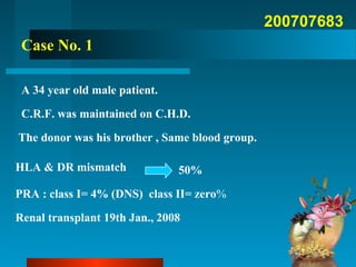
- 1. 200707683 Case No. 1 A 34 year old male patient. C.R.F. was maintained on C.H.D. The donor was his brother , Same blood group. HLA & DR mismatch PRA : class I= 4% (DNS) class II= zero% Renal transplant 19th Jan., 2008 50%
- 2. Operative details Artery internal iliac artery Vein external iliac vein Ischemia time : 45 minutes Immediate duiresis &
- 3. Smooth immediate postoperative course Maintained on steroid , CSA, AZA S.Cr. On discharge : 1.1mg/dl
- 4. One month Post transplantation • oliguria • ↑ S.cr.1.1 →3.2 mg/dl Clinical picture: Differential diagnosis: ?rejection ?Obstruction ?Vascular occlusion
- 5. Graft Doppler & U.S. Patchy perfusion Absent diastolic flow Basal On day of event
- 6. BiopsyBiopsy Picture of Acute Humoral Rejection Focal +ve C4d StainingATN like picture
- 7. Marked right Lower limb oedema. Doppler US Sluggish flow in the deep venous system Next Day:
- 8. MRA Thrombosed graft vein. Thrombosed RT external iliac vein. Patent IVC Patent graft artery Thrombosed RT common femoral vein.
- 9. OPTIONS FOR TREATMENT: Local injection of streptokinase Exploration & thrombectomy Graft nephrectomy GRAFT VEIN THROMBOSIS Systemic injection of streptokinase Combined local & systemic injection of streptokinase
- 16. Follow up MRA
- 17. Angiography was done Local injection of streptokinase 750 000 IU Systemic injection of Streptokinase 100 000IU/hr LMWH Plus oral anti-coagulant LMWH stopped when target INR 2-3 achieved GRAFT VEIN THROMBOSIS
- 18. CurrentlyCurrently Recovered graft functionRecovered graft function:: S. cr = 0.8 mg/dlS. cr = 0.8 mg/dl UU.O.P = 5 L/day.O.P = 5 L/day Patient under treatment of CMV disease
- 20. Graft vein thrombosis RVT has incidence of 0.55:3.4% One third of early allograft losses. One case reported just compression of the vein due to abnormal position of the graft (Nephron 1996;73:480-481)
- 21. diagnosis time management reference Renal vein compression 3rd day Exploration& nephropexy Nephron 1996 Partial thrombosis of graft vein 11months Selective intra- arterial administration of streptokinase Nephrol Dial Transplant 1998 Renal vein thrombosis 8th day Exploration& thrombectomy Nephrol Dial Transplant 2006
- 22. •Number of transplanted cases : 1969 •2003: one case suffered from both graft artery & vein thrombosis managed by nephrectomy •2008 One case of graft vein thrombosis managed by local & systemic injection of streptokinase
- 23. Conclusion: Close follow up Early detection & diagnosis Perfect intervention & cooperation Recent advances in treatment of vascular rejection =Good outcome