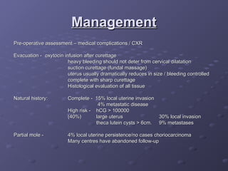
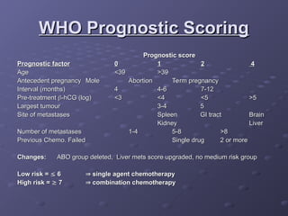
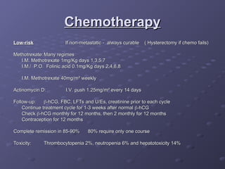
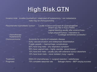
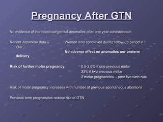
Gestational
Gestational
Trophoblastic Disease
Current management
Current management
Background
Background
Incidence –
Incidence – U.S. and Europe 1/1500
U.S. and Europe 1/1500
South East Asia 1/150
South East Asia 1/150 (
(↓
↓ Carotene, animal fat and
Carotene, animal fat and
Vit. A)
Vit. A)
Can follow any gestational event – abortion, miscarriage, ectopic, normal
pregnancy
pregnancy
Curable in vast majority – chemotherapy 1956
Complete & partial mole
Invasive mole
Invasive mole
Placental-site trophoblastic tumour
Choriocarcinoma ( always if follows term pregnancy 1/50,000 )
Latter 3
Latter 3 usually
usually derive from molar pregnancy
derive from molar pregnancy
Complete Mole
Complete Mole
Pathology:
Pathology: Generalized hydatidiform swelling and trophoblastic hyperplasia
Generalized hydatidiform swelling and trophoblastic hyperplasia
fetal/embryonic tissue absent
Karyotype:
Karyotype: 90% 46XX haploid sperm fertilizes ovum and duplicates
90% 46XX haploid sperm fertilizes ovum and duplicates
ovum nucleus either absent or inactivated
10% 46XY
10% 46XY
Clinical:
Clinical: Vaginal bleeding
Vaginal bleeding 95%
95% (prune juice, anaemia)
(prune juice, anaemia)
Enlarged uterus
Enlarged uterus 50%
50%
Theca lutein cysts
Theca lutein cysts 50%
50% (often >6cm.- may take 3 months)
(often >6cm.- may take 3 months)
Hyperemesis
Hyperemesis 25%
25%
PET
PET 25%
25% (no reported case of eclampsia)
(no reported case of eclampsia)
Hyperthyroidism
Hyperthyroidism 5%
5% (if free T4
(if free T4↑ - B-blockade)
↑ - B-blockade)
Trophoblastic emboli 2%
Diagnosis:
Diagnosis: U/S usually very sensitive – generalized swelling (snow-storm )
U/S usually very sensitive – generalized swelling (snow-storm )
Complete Mole
Complete Mole
Complete Mole Histology
Partial Mole
Partial Mole
Triploid karyotype – extra haploid set paternal
Sometimes fetus present – usually triploid
Sometimes fetus present – usually triploid growth restricted / multiple anomalies
growth restricted / multiple anomalies
Pathology differs from complete:
Pathology differs from complete: focal hydatidiform swelling
focal hydatidiform swelling
varying size of chorionic villi
marked villous scalloping
focal trophoblastic hyperplasia
identifiable embryonic or fetal tissues
Clinical:
Clinical: Usually as a regular incomplete or missed abortion
Usually as a regular incomplete or missed abortion
Excessive uterine enlargement / PET very rare
No hyperemesis / hyperthyroidism / theca-lutein cysts
Diagnosis:
Diagnosis: U/S may detect focal cystic spaces of varying diameter
U/S may detect focal cystic spaces of varying diameter
Diagnosis on histology of curettings
Partial Mole Histology
Management
Management
Pre-operative assessment – medical complications / CXR
Evacuation -
Evacuation - oxytocin infusion after curettage
oxytocin infusion after curettage
heavy bleeding should not deter from cervical dilatation
suction curettage (fundal massage)
uterus usually dramatically reduces in size / bleeding controlled
complete with sharp curettage
Histological evaluation of all tissue
Natural history:
Natural history: Complete - 15% local uterine invasion
Complete - 15% local uterine invasion
4% metastatic disease
High risk - hCG > 100000
(40%)
(40%) large uterus
large uterus 30% local invasion
30% local invasion
theca lutein cysts > 6cm.
theca lutein cysts > 6cm. 9% metastases
9% metastases
Partial mole -
Partial mole - 4% local uterine persistence/no cases choriocarcinoma
4% local uterine persistence/no cases choriocarcinoma
Many centres have abandoned follow-up
Follow up
Follow up
Weekly
Weekly 
-hCG (syncytiotrophoblast)
-hCG (syncytiotrophoblast)
levels
levels until
until
normal for 3 consecutive weeks
Can take 12-14 weeks
Then monthly until normal
for 6 months
for 6 months
Contraception:
Contraception: Immediate
Immediate
Oral / barrier / permanent
No IUCD until hCG normal (perforation)
No
No 
 persistent disease on OCP and
persistent disease on OCP and
regression time not influenced
GTN Follow-up
GTN Follow-up
WHO Prognostic Scoring
Original assessment and scoring system 1984 changed in 2000
Metastatic disease occurs in 4% patients with molar pregnancy
Plateau:
Plateau: 4 values over 3 weeks
4 values over 3 weeks
Rise:
Rise: 
 10% for 3 values over 2 weeks
10% for 3 values over 2 weeks
Clinical examination – especially pelvis, vagina and vulva
U/S to exclude pregnancy
Brain – MRI superior to CT scan
Chest – CXR adequate for counting metastases / CT scan also acceptable
Abdomen – CT scan
Abdomen – CT scan
WHO Prognostic Scoring
Prognostic score
Prognostic score
Prognostic factor
Prognostic factor 0
0 1
1 2
2 4
4
Age
Age <39
<39 >39
>39
Antecedent pregnancy
Antecedent pregnancy Mole
Mole Abortion
Abortion Term pregnancy
Term pregnancy
Interval (months)
Interval (months) 4
4 4-6
4-6 7-12
7-12
Pre-treatment
Pre-treatment 
-hCG (log)
-hCG (log) <3
<3 <4
<4 <5
<5 >5
>5
Largest tumour
Largest tumour 3-4
3-4 5
5
Site of metastases
Site of metastases Spleen GI tract
Spleen GI tract Brain
Brain
Kidney
Kidney Liver
Liver
Number of metastases
Number of metastases 1-4
1-4 5-8
5-8 >8
>8
Previous Chemo. Failed
Previous Chemo. Failed Single drug 2 or more
Single drug 2 or more
Changes:
Changes: ABO group deleted, Liver mets score upgraded, no medium risk group
ABO group deleted, Liver mets score upgraded, no medium risk group
Low risk =
Low risk = 
 6
6 
 single agent chemotherapy
single agent chemotherapy
High risk =
High risk = 
 7
7 
 combination chemotherapy
combination chemotherapy
FIGO Staging
FIGO Staging
Stage 1
Stage 1 Tumour confined to uterus
Tumour confined to uterus
Stage 2
Stage 2 Tumour confined to pelvis
Tumour confined to pelvis
Stage 3
Stage 3 Metastases to lung ( with/without pelvic metastases )
Metastases to lung ( with/without pelvic metastases )
Stage 4
Stage 4 Distant organ metastases ( with/without lung metastases )
Distant organ metastases ( with/without lung metastases )
Chemotherapy
Chemotherapy
Low-risk
Low-risk If non-metastatic - always curable ( Hysterectomy if chemo fails)
If non-metastatic - always curable ( Hysterectomy if chemo fails)
Methotrexate:
Methotrexate: Many regimes
Many regimes
I.M. Methotrexate 1mg/Kg days 1,3,5,7
I.M./ P.O. Folinic acid 0.1mg/Kg days 2,4,6,8
I.M. Methotrexate 40mg/m
I.M. Methotrexate 40mg/m²
² weekly
weekly
Actinomycin D:
Actinomycin D: I.V. push 1.25mg/m
I.V. push 1.25mg/m² every 14 days
² every 14 days
Follow-up:
Follow-up: 
-hCG, FBC, LFTs and U/Es, creatinine prior to each cycle
-hCG, FBC, LFTs and U/Es, creatinine prior to each cycle
Continue treatment cycle for 1-3 weeks after normal
Continue treatment cycle for 1-3 weeks after normal 
-hCG
-hCG
Check
Check 
-hCG monthly for 12 months, then 2 monthly for 12 months
-hCG monthly for 12 months, then 2 monthly for 12 months
Contraception for 12 months
Complete remission in 85-90%
Complete remission in 85-90% 80% require only one course
80% require only one course
Toxicity:
Toxicity: Thrombocytopenia 2%, neutropenia 6% and hepatotoxicity 14%
Thrombocytopenia 2%, neutropenia 6% and hepatotoxicity 14%
High Risk GTN
High Risk GTN
Invasive mole:
Invasive mole: invades myometrium / diagnosed at hysterectomy / can metastasize
invades myometrium / diagnosed at hysterectomy / can metastasize
mets may be choriocarcinoma
Placental site trophoblastic tumour:
Placental site trophoblastic tumour: Locally invasive composed of cytotrophopblast
Locally invasive composed of cytotrophopblast
small if any rise in hCG (<3000)
vaginal bleeding usually after amenorrhoea
Large polypoid tumour / insensitive to
chemotherapy
chemotherapy Curettage sometimes successful /
Curettage sometimes successful /
Hysterectomy
Hysterectomy
Choriocarcinoma:
Choriocarcinoma: Accounts for majority of metastatic disease
Accounts for majority of metastatic disease
Early vascular invasion and widespread dissemination
Fragile vessels
Fragile vessels 
 haemorrhagic complications
haemorrhagic complications
80% have lung mets – any respiratory symptom
30% have vaginal mets – highly vascular (avoid biopsy)
10% have liver mets – usually only with extensive tumour elsewhere
10% have brain mets – never isolated ( lung / vagina)
Treatment:
Treatment: EMA-CO chemotherapy +/- surgical resection / radiotherapy
EMA-CO chemotherapy +/- surgical resection / radiotherapy
Prognosis:
Prognosis: 75% complete response rate
75% complete response rate Salvage chemo – BEP varying success
Salvage chemo – BEP varying success
Pregnancy After GTN
Pregnancy After GTN
No evidence of increased congenital anomalies after one year contraception
Recent Japanese data –
Recent Japanese data – Women who concieved during follow-up period < 1
Women who concieved during follow-up period < 1
year
year
No adverse effect on anomalies nor preterm
delivery
delivery
Risk of further molar pregnancy:
Risk of further molar pregnancy: 0.5-2.5% if one previous molar
0.5-2.5% if one previous molar
33% if two previous molar
3 molar pregnancies – poor live birth rate
Risk of molar pregnancy increases with number of previous spontaneous abortions
Previous term pregnancies reduce risk of GTN
Conclusions
Conclusions
GTN is rare
GTN is rare
Ultrasound diagnosis becoming more common
Senior staff should perform ERPC ( suction and sharp curettage)
Follow-up – clinical and serum
Follow-up – clinical and serum 
-hCG measurements in specialized clinics
-hCG measurements in specialized clinics
Chemotherapy curative in vast majority low risk patients

Gestational Trophoblastic Disease....ppt

  • 1.
  • 2.
    Background Background Incidence – Incidence –U.S. and Europe 1/1500 U.S. and Europe 1/1500 South East Asia 1/150 South East Asia 1/150 ( (↓ ↓ Carotene, animal fat and Carotene, animal fat and Vit. A) Vit. A) Can follow any gestational event – abortion, miscarriage, ectopic, normal Can follow any gestational event – abortion, miscarriage, ectopic, normal pregnancy pregnancy Curable in vast majority – chemotherapy 1956 Curable in vast majority – chemotherapy 1956 Complete & partial mole Complete & partial mole Invasive mole Invasive mole Placental-site trophoblastic tumour Placental-site trophoblastic tumour Choriocarcinoma ( always if follows term pregnancy 1/50,000 ) Choriocarcinoma ( always if follows term pregnancy 1/50,000 ) Latter 3 Latter 3 usually usually derive from molar pregnancy derive from molar pregnancy
  • 3.
    Complete Mole Complete Mole Pathology: Pathology:Generalized hydatidiform swelling and trophoblastic hyperplasia Generalized hydatidiform swelling and trophoblastic hyperplasia fetal/embryonic tissue absent fetal/embryonic tissue absent Karyotype: Karyotype: 90% 46XX haploid sperm fertilizes ovum and duplicates 90% 46XX haploid sperm fertilizes ovum and duplicates ovum nucleus either absent or inactivated ovum nucleus either absent or inactivated 10% 46XY 10% 46XY Clinical: Clinical: Vaginal bleeding Vaginal bleeding 95% 95% (prune juice, anaemia) (prune juice, anaemia) Enlarged uterus Enlarged uterus 50% 50% Theca lutein cysts Theca lutein cysts 50% 50% (often >6cm.- may take 3 months) (often >6cm.- may take 3 months) Hyperemesis Hyperemesis 25% 25% PET PET 25% 25% (no reported case of eclampsia) (no reported case of eclampsia) Hyperthyroidism Hyperthyroidism 5% 5% (if free T4 (if free T4↑ - B-blockade) ↑ - B-blockade) Trophoblastic emboli 2% Trophoblastic emboli 2% Diagnosis: Diagnosis: U/S usually very sensitive – generalized swelling (snow-storm ) U/S usually very sensitive – generalized swelling (snow-storm )
  • 5.
  • 6.
  • 7.
    Partial Mole Partial Mole Triploidkaryotype – extra haploid set paternal Triploid karyotype – extra haploid set paternal Sometimes fetus present – usually triploid Sometimes fetus present – usually triploid growth restricted / multiple anomalies growth restricted / multiple anomalies Pathology differs from complete: Pathology differs from complete: focal hydatidiform swelling focal hydatidiform swelling varying size of chorionic villi varying size of chorionic villi marked villous scalloping marked villous scalloping focal trophoblastic hyperplasia focal trophoblastic hyperplasia identifiable embryonic or fetal tissues identifiable embryonic or fetal tissues Clinical: Clinical: Usually as a regular incomplete or missed abortion Usually as a regular incomplete or missed abortion Excessive uterine enlargement / PET very rare Excessive uterine enlargement / PET very rare No hyperemesis / hyperthyroidism / theca-lutein cysts No hyperemesis / hyperthyroidism / theca-lutein cysts Diagnosis: Diagnosis: U/S may detect focal cystic spaces of varying diameter U/S may detect focal cystic spaces of varying diameter Diagnosis on histology of curettings Diagnosis on histology of curettings
  • 8.
  • 9.
    Management Management Pre-operative assessment –medical complications / CXR Pre-operative assessment – medical complications / CXR Evacuation - Evacuation - oxytocin infusion after curettage oxytocin infusion after curettage heavy bleeding should not deter from cervical dilatation heavy bleeding should not deter from cervical dilatation suction curettage (fundal massage) suction curettage (fundal massage) uterus usually dramatically reduces in size / bleeding controlled uterus usually dramatically reduces in size / bleeding controlled complete with sharp curettage complete with sharp curettage Histological evaluation of all tissue Histological evaluation of all tissue Natural history: Natural history: Complete - 15% local uterine invasion Complete - 15% local uterine invasion 4% metastatic disease 4% metastatic disease High risk - hCG > 100000 High risk - hCG > 100000 (40%) (40%) large uterus large uterus 30% local invasion 30% local invasion theca lutein cysts > 6cm. theca lutein cysts > 6cm. 9% metastases 9% metastases Partial mole - Partial mole - 4% local uterine persistence/no cases choriocarcinoma 4% local uterine persistence/no cases choriocarcinoma Many centres have abandoned follow-up Many centres have abandoned follow-up
  • 10.
    Follow up Follow up Weekly Weekly -hCG (syncytiotrophoblast) -hCG (syncytiotrophoblast) levels levels until until normal for 3 consecutive weeks normal for 3 consecutive weeks Can take 12-14 weeks Can take 12-14 weeks Then monthly until normal Then monthly until normal for 6 months for 6 months Contraception: Contraception: Immediate Immediate Oral / barrier / permanent Oral / barrier / permanent No IUCD until hCG normal (perforation) No IUCD until hCG normal (perforation) No No   persistent disease on OCP and persistent disease on OCP and regression time not influenced regression time not influenced
  • 11.
  • 12.
    WHO Prognostic Scoring WHOPrognostic Scoring Original assessment and scoring system 1984 changed in 2000 Original assessment and scoring system 1984 changed in 2000 Metastatic disease occurs in 4% patients with molar pregnancy Metastatic disease occurs in 4% patients with molar pregnancy Plateau: Plateau: 4 values over 3 weeks 4 values over 3 weeks Rise: Rise:   10% for 3 values over 2 weeks 10% for 3 values over 2 weeks Clinical examination – especially pelvis, vagina and vulva Clinical examination – especially pelvis, vagina and vulva U/S to exclude pregnancy U/S to exclude pregnancy Brain – MRI superior to CT scan Brain – MRI superior to CT scan Chest – CXR adequate for counting metastases / CT scan also acceptable Chest – CXR adequate for counting metastases / CT scan also acceptable Abdomen – CT scan Abdomen – CT scan
  • 13.
    WHO Prognostic Scoring WHOPrognostic Scoring Prognostic score Prognostic score Prognostic factor Prognostic factor 0 0 1 1 2 2 4 4 Age Age <39 <39 >39 >39 Antecedent pregnancy Antecedent pregnancy Mole Mole Abortion Abortion Term pregnancy Term pregnancy Interval (months) Interval (months) 4 4 4-6 4-6 7-12 7-12 Pre-treatment Pre-treatment  -hCG (log) -hCG (log) <3 <3 <4 <4 <5 <5 >5 >5 Largest tumour Largest tumour 3-4 3-4 5 5 Site of metastases Site of metastases Spleen GI tract Spleen GI tract Brain Brain Kidney Kidney Liver Liver Number of metastases Number of metastases 1-4 1-4 5-8 5-8 >8 >8 Previous Chemo. Failed Previous Chemo. Failed Single drug 2 or more Single drug 2 or more Changes: Changes: ABO group deleted, Liver mets score upgraded, no medium risk group ABO group deleted, Liver mets score upgraded, no medium risk group Low risk = Low risk =   6 6   single agent chemotherapy single agent chemotherapy High risk = High risk =   7 7   combination chemotherapy combination chemotherapy
  • 14.
    FIGO Staging FIGO Staging Stage1 Stage 1 Tumour confined to uterus Tumour confined to uterus Stage 2 Stage 2 Tumour confined to pelvis Tumour confined to pelvis Stage 3 Stage 3 Metastases to lung ( with/without pelvic metastases ) Metastases to lung ( with/without pelvic metastases ) Stage 4 Stage 4 Distant organ metastases ( with/without lung metastases ) Distant organ metastases ( with/without lung metastases )
  • 15.
    Chemotherapy Chemotherapy Low-risk Low-risk If non-metastatic- always curable ( Hysterectomy if chemo fails) If non-metastatic - always curable ( Hysterectomy if chemo fails) Methotrexate: Methotrexate: Many regimes Many regimes I.M. Methotrexate 1mg/Kg days 1,3,5,7 I.M. Methotrexate 1mg/Kg days 1,3,5,7 I.M./ P.O. Folinic acid 0.1mg/Kg days 2,4,6,8 I.M./ P.O. Folinic acid 0.1mg/Kg days 2,4,6,8 I.M. Methotrexate 40mg/m I.M. Methotrexate 40mg/m² ² weekly weekly Actinomycin D: Actinomycin D: I.V. push 1.25mg/m I.V. push 1.25mg/m² every 14 days ² every 14 days Follow-up: Follow-up:  -hCG, FBC, LFTs and U/Es, creatinine prior to each cycle -hCG, FBC, LFTs and U/Es, creatinine prior to each cycle Continue treatment cycle for 1-3 weeks after normal Continue treatment cycle for 1-3 weeks after normal  -hCG -hCG Check Check  -hCG monthly for 12 months, then 2 monthly for 12 months -hCG monthly for 12 months, then 2 monthly for 12 months Contraception for 12 months Contraception for 12 months Complete remission in 85-90% Complete remission in 85-90% 80% require only one course 80% require only one course Toxicity: Toxicity: Thrombocytopenia 2%, neutropenia 6% and hepatotoxicity 14% Thrombocytopenia 2%, neutropenia 6% and hepatotoxicity 14%
  • 16.
    High Risk GTN HighRisk GTN Invasive mole: Invasive mole: invades myometrium / diagnosed at hysterectomy / can metastasize invades myometrium / diagnosed at hysterectomy / can metastasize mets may be choriocarcinoma mets may be choriocarcinoma Placental site trophoblastic tumour: Placental site trophoblastic tumour: Locally invasive composed of cytotrophopblast Locally invasive composed of cytotrophopblast small if any rise in hCG (<3000) small if any rise in hCG (<3000) vaginal bleeding usually after amenorrhoea vaginal bleeding usually after amenorrhoea Large polypoid tumour / insensitive to Large polypoid tumour / insensitive to chemotherapy chemotherapy Curettage sometimes successful / Curettage sometimes successful / Hysterectomy Hysterectomy Choriocarcinoma: Choriocarcinoma: Accounts for majority of metastatic disease Accounts for majority of metastatic disease Early vascular invasion and widespread dissemination Early vascular invasion and widespread dissemination Fragile vessels Fragile vessels   haemorrhagic complications haemorrhagic complications 80% have lung mets – any respiratory symptom 80% have lung mets – any respiratory symptom 30% have vaginal mets – highly vascular (avoid biopsy) 30% have vaginal mets – highly vascular (avoid biopsy) 10% have liver mets – usually only with extensive tumour elsewhere 10% have liver mets – usually only with extensive tumour elsewhere 10% have brain mets – never isolated ( lung / vagina) 10% have brain mets – never isolated ( lung / vagina) Treatment: Treatment: EMA-CO chemotherapy +/- surgical resection / radiotherapy EMA-CO chemotherapy +/- surgical resection / radiotherapy Prognosis: Prognosis: 75% complete response rate 75% complete response rate Salvage chemo – BEP varying success Salvage chemo – BEP varying success
  • 17.
    Pregnancy After GTN PregnancyAfter GTN No evidence of increased congenital anomalies after one year contraception No evidence of increased congenital anomalies after one year contraception Recent Japanese data – Recent Japanese data – Women who concieved during follow-up period < 1 Women who concieved during follow-up period < 1 year year No adverse effect on anomalies nor preterm No adverse effect on anomalies nor preterm delivery delivery Risk of further molar pregnancy: Risk of further molar pregnancy: 0.5-2.5% if one previous molar 0.5-2.5% if one previous molar 33% if two previous molar 33% if two previous molar 3 molar pregnancies – poor live birth rate 3 molar pregnancies – poor live birth rate Risk of molar pregnancy increases with number of previous spontaneous abortions Risk of molar pregnancy increases with number of previous spontaneous abortions Previous term pregnancies reduce risk of GTN Previous term pregnancies reduce risk of GTN
  • 18.
    Conclusions Conclusions GTN is rare GTNis rare Ultrasound diagnosis becoming more common Ultrasound diagnosis becoming more common Senior staff should perform ERPC ( suction and sharp curettage) Senior staff should perform ERPC ( suction and sharp curettage) Follow-up – clinical and serum Follow-up – clinical and serum  -hCG measurements in specialized clinics -hCG measurements in specialized clinics Chemotherapy curative in vast majority low risk patients Chemotherapy curative in vast majority low risk patients