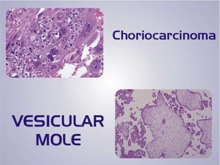
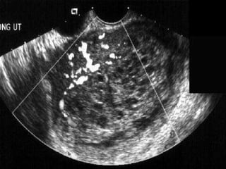
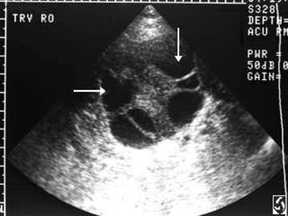
Gestational trophoblastic diseases
They arise from the trophoblast of the balstocyst and are classified as: *  hydatidifrom mole  (molar pregnancy) (80%) Complete (classic) Incomplete (piratical) * gestational trophoblatic neoplasia  Non-metastatic  = invasive mole = chorio –adenoma destruens  (16%) Metastatic   =Choriocarcinoma = chorion epithelioma (4%) Low – risk High – risk
Choriocarcinoma
Pathology : Incidence : 1: 250-5000 pregnancies in Asia and 1:4000 in the west. Origin:   *  Choriocarcinoma is a malignant tumour of the trophoblast.  1- About 50% of cases follow molar pregnancy. 2-  25% follow abortion 3- 23% follow normal pregnancy  4- 2% follow ectopic pregnancy. *  In rare cases, the tumour arises as a teratoma in the ovary or testicle.
 
Macroscopic appearnce: The tumor arises in the endometrium as  a soft friable dark red hemorrhagic mass projecting into the uterine cavity and may from a polyp.  Malignant tissue may be buried within the myometrium , inaccessible to the curette, or hidden in a distant metastasis.   However, any of these tumor patterns secretes (hCG) which causes cystic changes of the ovaries in about 30% of cases..
Microscopic appearance: The tumour consists of cyto-and synecytiotrophoblasts showing malignant characters, invading the myometrium and blood vessels.  Chorionic villi are absent  this differentiates Choriocarcinoma from invasive mole.
 
Mode of spread: direct spread: to the parametrium, tubes and ovaries. Blood spread: occurs early to distant organs. The commonest sites are lunges (80%), vegina (30%), brain (10%) and liver (10%)
FIGO classification :   Stage I  confined to uterine corpus. Stage II  metastases to pelvis and vagina  Stage III  metastases to lung Stage IV  metastases to other organs .
multiple single chemotheraby 9 >8 4-8 1-4 n.Of metastis 8 Brain Liver-GIT Spleen -kidney site of metastis 7 >5 3-5 Tuomer d 6 B-AB O-A Abo group 5 >100000 10000-100000 1000- 10000 1000 HCG iu/l 4 >12 7-12 4-6 4 Interval month 3 Full term abo V.M Associated pregnancy 2 <39 ≥ 39 Age (years) 1 S4 S2 S1 S 0 Prognostic factor
Bagshwe score Total score  ≤ 4  low risk 5-7 moderate risk >8 high risk
Diagnosis: A- symptoms: 1-  Persistent or irregular vaginal bleeding: it is the commonest symptom occurring after labor, abortion or evacuation of a vesicular mole. Bleeding can occur within days or months but rarely after 2 years. 2- Vaginal discharge: which is blood stained and offensive due to ulceration and infection of the growth . 3- amenorrhea: may be present due to continuous hCG production.
4- Acute abdominal pain: due to intraperitoneal haemorrhage as a result of perforation of the uterus by the growth. 5- Abdominal or vaginal swelling: may develop. 6- Symptoms of metastases: as dysponea, haemoptesis, jaundice and neurological symptoms as headache may be the first manifestation of the tumor.
B- signs: (1) cachexia and severe anaemia. (2) fever may be present due to infection and necrosis  (3) the uterus may be normal size or enlarged and soft. (4) the ovaries: may be enlarged and eystic. (5) metaststic nodules: in the vulva or vagina
C- investigations : (1) uterine curettage: should be done in every case of persistent or irregular uterine bleeding after labour, abortion or molar pregnancy. However, intramural tumour cannot be detected by curettage. (2)  serum β- subunite of hCG: persistent or rising titres in absence of pregnancy are indicative of trophoblastic neoplasia . (3) biopsy: from metastatic valvar or vaginal lesions.
(4)  imaging : a- plain X-ray chest: may show secondaries in the form of &quot; cannon balls&quot; or &quot;snowstorm&quot; appearance. b- ultrasonography: to detect tumour, cystic ovaries and exclude remnants of conception. c- CT scan: for lungs, liver, brain and bone. (5) lumbar puncture : plasma hCG/ CSF hCG ratio less than 60 strongly CNS involvement my metastases  (6) blood studies : a- complete blood picture including platelet count  b- Renal, liver and thyroid function tests  c- Blood group.
 
 
Treatment:  The treatment of choice  chemotheraphy
Hysterectomy  may be indicated in the following conditions:  severe uterine bleeding perforation of the uterus with intraperitoneal haemorrhage.  Massive haemorrhage from the bowel  Torsion of a theca lutein cyst. Durg resistance or toxicity. Persistant localised metastases in the vagina, lung or brain after chemotherapy
Chemotherapy: (I)  low- risk group score ≤ 4 : Single cytotoxic drug either methotrexate or actinomycin D  (II)  High-risk group score >8  : Multiple cytotoxic drugs
Thank you

Choriocarcinoma

  • 1.
  • 2.
    They arise fromthe trophoblast of the balstocyst and are classified as: * hydatidifrom mole (molar pregnancy) (80%) Complete (classic) Incomplete (piratical) * gestational trophoblatic neoplasia Non-metastatic = invasive mole = chorio –adenoma destruens (16%) Metastatic =Choriocarcinoma = chorion epithelioma (4%) Low – risk High – risk
  • 3.
  • 4.
    Pathology : Incidence: 1: 250-5000 pregnancies in Asia and 1:4000 in the west. Origin: * Choriocarcinoma is a malignant tumour of the trophoblast. 1- About 50% of cases follow molar pregnancy. 2- 25% follow abortion 3- 23% follow normal pregnancy 4- 2% follow ectopic pregnancy. * In rare cases, the tumour arises as a teratoma in the ovary or testicle.
  • 5.
  • 6.
    Macroscopic appearnce: Thetumor arises in the endometrium as a soft friable dark red hemorrhagic mass projecting into the uterine cavity and may from a polyp. Malignant tissue may be buried within the myometrium , inaccessible to the curette, or hidden in a distant metastasis. However, any of these tumor patterns secretes (hCG) which causes cystic changes of the ovaries in about 30% of cases..
  • 7.
    Microscopic appearance: Thetumour consists of cyto-and synecytiotrophoblasts showing malignant characters, invading the myometrium and blood vessels. Chorionic villi are absent this differentiates Choriocarcinoma from invasive mole.
  • 8.
  • 9.
    Mode of spread:direct spread: to the parametrium, tubes and ovaries. Blood spread: occurs early to distant organs. The commonest sites are lunges (80%), vegina (30%), brain (10%) and liver (10%)
  • 10.
    FIGO classification : Stage I confined to uterine corpus. Stage II metastases to pelvis and vagina Stage III metastases to lung Stage IV metastases to other organs .
  • 11.
    multiple single chemotheraby9 >8 4-8 1-4 n.Of metastis 8 Brain Liver-GIT Spleen -kidney site of metastis 7 >5 3-5 Tuomer d 6 B-AB O-A Abo group 5 >100000 10000-100000 1000- 10000 1000 HCG iu/l 4 >12 7-12 4-6 4 Interval month 3 Full term abo V.M Associated pregnancy 2 <39 ≥ 39 Age (years) 1 S4 S2 S1 S 0 Prognostic factor
  • 12.
    Bagshwe score Totalscore ≤ 4 low risk 5-7 moderate risk >8 high risk
  • 13.
    Diagnosis: A- symptoms:1- Persistent or irregular vaginal bleeding: it is the commonest symptom occurring after labor, abortion or evacuation of a vesicular mole. Bleeding can occur within days or months but rarely after 2 years. 2- Vaginal discharge: which is blood stained and offensive due to ulceration and infection of the growth . 3- amenorrhea: may be present due to continuous hCG production.
  • 14.
    4- Acute abdominalpain: due to intraperitoneal haemorrhage as a result of perforation of the uterus by the growth. 5- Abdominal or vaginal swelling: may develop. 6- Symptoms of metastases: as dysponea, haemoptesis, jaundice and neurological symptoms as headache may be the first manifestation of the tumor.
  • 15.
    B- signs: (1)cachexia and severe anaemia. (2) fever may be present due to infection and necrosis (3) the uterus may be normal size or enlarged and soft. (4) the ovaries: may be enlarged and eystic. (5) metaststic nodules: in the vulva or vagina
  • 16.
    C- investigations :(1) uterine curettage: should be done in every case of persistent or irregular uterine bleeding after labour, abortion or molar pregnancy. However, intramural tumour cannot be detected by curettage. (2) serum β- subunite of hCG: persistent or rising titres in absence of pregnancy are indicative of trophoblastic neoplasia . (3) biopsy: from metastatic valvar or vaginal lesions.
  • 17.
    (4) imaging: a- plain X-ray chest: may show secondaries in the form of &quot; cannon balls&quot; or &quot;snowstorm&quot; appearance. b- ultrasonography: to detect tumour, cystic ovaries and exclude remnants of conception. c- CT scan: for lungs, liver, brain and bone. (5) lumbar puncture : plasma hCG/ CSF hCG ratio less than 60 strongly CNS involvement my metastases (6) blood studies : a- complete blood picture including platelet count b- Renal, liver and thyroid function tests c- Blood group.
  • 18.
  • 19.
  • 20.
    Treatment: Thetreatment of choice chemotheraphy
  • 21.
    Hysterectomy maybe indicated in the following conditions: severe uterine bleeding perforation of the uterus with intraperitoneal haemorrhage. Massive haemorrhage from the bowel Torsion of a theca lutein cyst. Durg resistance or toxicity. Persistant localised metastases in the vagina, lung or brain after chemotherapy
  • 22.
    Chemotherapy: (I) low- risk group score ≤ 4 : Single cytotoxic drug either methotrexate or actinomycin D (II) High-risk group score >8 : Multiple cytotoxic drugs
  • 23.