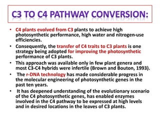
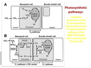
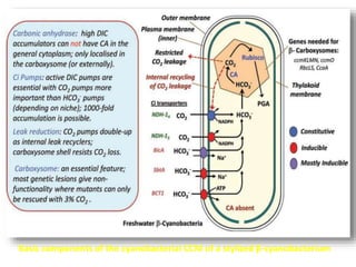
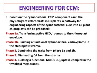
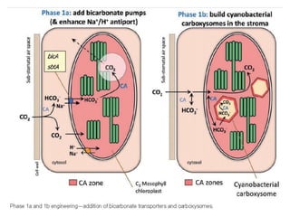
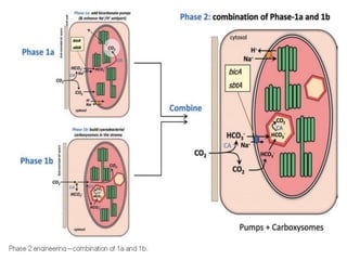
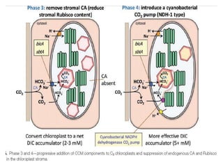
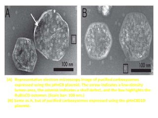
• Crop yields need to nearly double over the next 35
years to keep the pace with projected population
growth.
• The improvement of crop yields during the last 50
years was attributed to better crop architecture, an
increased harvest index and increased application of
fertilizers & pesticides.
• Recently, Genetic Engineering Strategies are
considered as a promising target for crop
improvement with regards to increasing yield .
• Improving Photosynthesis, Nitrogen Assimilation And
Nutrient Uptake Efficiency of plant species has been
recognized as an additional option to achieve desired
targets.
• Engineering higher photosynthetic efficiency for
greater crop yields has gained significant attention
among plant biologists and breeders.
• A number of metabolic targets and canopy architectural
features have been identified that can be modified to
achieve enhanced rates of photosynthesis, e.g.
 altering Rubisco kinetics,
 manipulation of photoprotection,
 rebalancing the carbon metabolic processes,
 introducing photorespiratory bypass pathways,
 introducing inorganic carbon transporters in mesophyll cells,
 transfer of C4 photosynthetic pathways characters &
 selecting for crops with more erect leaves.
Schematic overview of the major relations between leaf anatomy and
photosynthetic efficiency. Anatomical traits are indicated in yellow, Continuous
arrows-direct biophysical relations & dotted arrows-regulatory relationships.
Fig:Factors influencing the delivery of CO2 to Rubisco. The flux of atmospheric CO2 (Ca)
into the leaf is limited by the stomatal conductance (gs). Inside the leaf, the flux B/W CO2
inside the intercellular airspace (Ci) and at the site of carboxylation in the chloroplast (Cc)
is limited by the mesophyll conductance (gm). A large amount of chloroplast surface that
is directly exposed to intercellular airspace (Sc) would increase gm by increasing the
surface area for diffusion. In addition, the CO2 flux can be enhanced by lowering the
resistance of individual components in diffusion pathway. Potential targets can be
engineered to gain increased delivery of CO2 to Rubisco are indicated in red.
Crop rank is based on 2008 production values in US dollars (given in parentheses),
according to the United Nations Food and Agriculture Organization (FAOstat 2011).
• C4 plants evolved from C3 plants to achieve high
photosynthetic performance, high water and nitrogen-use
efficiencies.
• Consequently, the transfer of C4 traits to C3 plants is one
strategy being adopted for improving the photosynthetic
performance of C3 plants.
• This approach was available only in few plant genera and
most C3-C4 hybrids were infertile (Brown and Bouton, 1993).
• The r-DNA technology has made considerable progress in
the molecular engineering of photosynthetic genes in the
past ten years.
• It has deepened understanding of the evolutionary scenario
of the C4 photosynthetic genes, has enabled enzymes
involved in the C4 pathway to be expressed at high levels
and in desired locations in the leaves of C3 plants.
Simplified
illustrations of the
C3 photosynthetic
pathway (A) and the
C4 photosynthetic
pathway of the
NADP-ME (NADP-
malic enzyme) type
C4 plants (B).
Photosynthetic
pathways:
• Overproduction of a single C4 enzyme can alter the carbon
metabolism of C3 plants, BUT does not show any positive effects on
photosynthesis.
• Transgenic C3 plants overproducing multiple enzymes are now being
produced for improving the photosynthetic performance of C3
plants.
• Possible routes and progress to suppressing photorespiration by
introducing C4 photosynthesis in C3 crop plants is feasible, as the
evolution of C3–C4 intermediates can be used as a blueprint for
engineering C4 photosynthesis, which pathway for the C4 cycle
might be introduced and the extent to which processes and
structures in C3 plant might require optimization.
• The physiological impacts of the overproduction in potato, tobacco
(Nicotiana tabacum) and Arabidopsis thaliana as well as rice have
previously been reviewed in detail by HaÈusler et al. (2002).
A hierarchical framework for C3 plant to form C4 plant via either bioengineering
or natural evolution. The top level identifies the engineering goal, second level-
some key traits that will need to be modified to introduce a C4 system into a C3
plant. Each of the listed traits shows the mechanisms underlying each trait. Ex.,
the transport function module can be expanded as changes in triose phosphate
(TP), oxaloacetate (OAA) or amino acid (AA) transporters.
Mechanisms show changes to key enzyme systems in the C4 metabolic cycle, ex.
Pyruvate orthophosphate dikinase (PPDK), PEP carboxylase (PEPC), malate
dehydrogenase (MDH) & the NADP-malic enzyme (NADP-ME), as would occur in an
NADP-ME species such as maize. Genetic level changes that alter itskinetics and location
of expression, Peterhansel (2011) in this issue and Gowik and Westhoff (2011).
• To improve inefficiencies in plants : by improving Rubisco,
modifying photorespiration or by introducing CO2-concentrating
mechanisms.
• An alternative strategy, used by cyanobacteria, involved the
development of active CCMs to turbo-charge the CO2 supply to
Rubisco, although at a minor metabolic cost. In order for a CCM
to function in a plant cell, seven criteria need to be met (Badger
and Spalding, 2000; Leegood, 2002). These are:
(i) An active, photosynthetically driven: CO2 capture system,
(ii) A supply of photosynthetic energy,
(iii) An intermediate pool of captured CO2,
(iv) A mechanism to release CO2 from the intermediate pool,
(v) A compartment in which to concentrate CO2 around Rubisco,
(vi)A means to reduce leakage of CO2 from the site of CO2 elevation &
(vii) Modification of the kinetic properties of Rubisco
Basic components of the cyanobacterial CCM of a stylized β-cyanobacterium
• Based on the cyanobacterial CCM components and the
physiology of chloroplasts in C3 plants, a pathway for
engineering aspects of the cyanobacterial CCM into C3 plant
chloroplasts can be proposed:
Phase 1a. Transferring active HCO3
– pumps to the chloroplast
envelope.
Phase 1b. Building a functional cyanobacterial carboxysome in
the chloroplast stroma.
Phase 2. Combining the traits from phase 1a and 1b.
Phase 3. Eliminating CA from the stroma.
Phase 4. Building a functional NDH-1 CO2 uptake complex in the
thylakoid membranes.
• TITLE:
Modularity of a carbon-fixing protein organelle
• AUTHOR:
Walter Bonaccia, Poh K. Tengb, Bruno Afonsoa,
Henrike Niederholtmeyera, Patricia Grobc,
Pamela A. Silvera,d, and David F. Savageb
Bacterial microcompartments are proteinaceous
complexes that catalyze metabolic pathways in a
manner reminiscent of organelles.
Microcompartment structure is well understood, but
less known about their assembly and function in vivo.
Here, carboxysomes CO2-fixing microcompartments
encoded by 10 genes, was heterologously produced in
Escherichia coli.
Expression of carboxysomes in E. coli resulted in the
production of icosahedral complexes similar to those
from the native host. In vivo, the complexes were
capable of both assembling with carboxysomal
proteins and fixing CO2.
Characterization of purified synthetic carboxysomes indicated
that they were well formed in structure, contained the expected
molecular components and were capable of fixing CO2 in vitro.
 A genetic system capable of producing modular carbon-fixing
microcompartments in a heterologous host was developed.
So, they lay the groundwork for understanding these elaborate
protein complexes and for the synthetic biological engineering
of self-assembling molecular structures.
• One biological assembly that could form the basis of such a
synthetic organelle is the bacterial microcompartment (BMC).
• The CO2-fixing carboxysome (CB) was the first BMC to be
discovered and remains a model system for elucidating how
BMCs assemble and function in the cell.
• Inside the lumen are the enzymes RuBisCO and carbonic
anhydrase (CA), which are directed to this location by protein–
protein interactions with the shell.
• Biochemically, the CB plays a central role in the CCM .
• In the context of the CCM, it is postulated that the CB lumen
contains elevated levels of CO2 and that RuBisCO is able to fix
CO2 near its Vmax with high specificity.
• After fixation, the product 3-phosphoglycerate diffuses out of
the carboxysome and enters the central carbon metabolism
Physical and genetic organization of the carboxysome: (A) Molecular surface
representation of a partial model of the carboxysome showing RuBisCO (green),
carbon anhydrase (orange), CsoS1ABC (blue), and CsoS4AB (red); (B) Schematic
of the carbon concentrating mechanism; (C) Genomic organization of the
carboxysome operon in H. neapolitanus.
• CBs are classified as α-type based on their RuBisCO coding
sequence which display a simplified regulatory structure in
which all genes are found together at a single genomic locus.
• Thus, they reasoned that an α-type CB operon could simplify
the expression of functional particles in a heterologous host.
• The α-type CB operon from H. neapolitanus (HnCB) containing
10 genes into the isopropyl β-D-1-thiogalactopyranoside
contained no such structures, resembled CBs from H.
neapolitanus.
• At higher induction (50 and 200 μM IPTG), cells contained an
increasing number of CBs such that the entire cytoplasm was
eventually filled and the cells became filamentous. Many CBs
were of varying contrast with dense shell edges and lighter
facets, reminiscent of CBs cargo were seen (fig.1).
Heterologous Formation of Carboxysomes
Fig-2: Microscopy images
of cells expressing
fluorescently labeled
carboxysome
components in presence
and absence of pHnCB.
Fig.1: Electron microscopy of
carboxysomes in vivo:
(A) Expression of HnCB
at 0 μM IPTG.
(B) Same as A but at 50 μM
IPTG; (Inset) Icosahedral
structures.
(C) Same as A, but at 200 μM
IPTG; (Inset) Inclusion body.
(D) Appearance of
filaments (arrow) under high
induction conditions.
(Scale bars: 500 nm.)
• As in the native host, synthetic CBs in E. coli were assembled with
labeled cargo and could be imaged using fluorescence microscopy.
• CsoS1A, a major shell protein, and CbbL, the large subunit of
RuBisCO, were fused with a C-terminal GFP and cloned into the
IPTG-inducible, kanamycin- resistant vector pDFS21, which is
suitable for cotransformation with the pNS3 plasmid.
• Control expression of GFP alone or with HnCB yielded no foci,
consistent with unaggregated cytoplasmic fluorescent protein .
• Here we validate this hypothesis by demonstrating that
heterologous expression of the CB genomic locus from H.
neapolitanus, containing 10 genes encoding enzymes and shell
proteins, is sufficient to synthesize BMCs in E. coli that are very
similar to those of the native host. Moreover, HnCBs are capable of
fixing CO2 both within E. coli and in isolation.
(A) Representative electron microscopy image of purified carboxysomes
expressed using the pHnCB plasmid. The arrow indicates a low-density
lumen area, the asterisk indicates a shell defect, and the box highlights the
RuBisCO octomer. (Scale bar: 100 nm.)
(B) Same as A, but of purified carboxysomes expressed using the pHnCBS1D
plasmid.
• Thus, we have determined the genetic information
sufficient for transplanting a carbon-fixing protein
organelle into new hosts that otherwise do not
reductively fixes carbon.

Genetic engineering in Crop-a new approach

  • 1.
    • Crop yieldsneed to nearly double over the next 35 years to keep the pace with projected population growth. • The improvement of crop yields during the last 50 years was attributed to better crop architecture, an increased harvest index and increased application of fertilizers & pesticides. • Recently, Genetic Engineering Strategies are considered as a promising target for crop improvement with regards to increasing yield . • Improving Photosynthesis, Nitrogen Assimilation And Nutrient Uptake Efficiency of plant species has been recognized as an additional option to achieve desired targets.
  • 3.
    • Engineering higherphotosynthetic efficiency for greater crop yields has gained significant attention among plant biologists and breeders. • A number of metabolic targets and canopy architectural features have been identified that can be modified to achieve enhanced rates of photosynthesis, e.g.  altering Rubisco kinetics,  manipulation of photoprotection,  rebalancing the carbon metabolic processes,  introducing photorespiratory bypass pathways,  introducing inorganic carbon transporters in mesophyll cells,  transfer of C4 photosynthetic pathways characters &  selecting for crops with more erect leaves.
  • 4.
    Schematic overview ofthe major relations between leaf anatomy and photosynthetic efficiency. Anatomical traits are indicated in yellow, Continuous arrows-direct biophysical relations & dotted arrows-regulatory relationships.
  • 5.
    Fig:Factors influencing thedelivery of CO2 to Rubisco. The flux of atmospheric CO2 (Ca) into the leaf is limited by the stomatal conductance (gs). Inside the leaf, the flux B/W CO2 inside the intercellular airspace (Ci) and at the site of carboxylation in the chloroplast (Cc) is limited by the mesophyll conductance (gm). A large amount of chloroplast surface that is directly exposed to intercellular airspace (Sc) would increase gm by increasing the surface area for diffusion. In addition, the CO2 flux can be enhanced by lowering the resistance of individual components in diffusion pathway. Potential targets can be engineered to gain increased delivery of CO2 to Rubisco are indicated in red.
  • 6.
    Crop rank isbased on 2008 production values in US dollars (given in parentheses), according to the United Nations Food and Agriculture Organization (FAOstat 2011).
  • 7.
    • C4 plantsevolved from C3 plants to achieve high photosynthetic performance, high water and nitrogen-use efficiencies. • Consequently, the transfer of C4 traits to C3 plants is one strategy being adopted for improving the photosynthetic performance of C3 plants. • This approach was available only in few plant genera and most C3-C4 hybrids were infertile (Brown and Bouton, 1993). • The r-DNA technology has made considerable progress in the molecular engineering of photosynthetic genes in the past ten years. • It has deepened understanding of the evolutionary scenario of the C4 photosynthetic genes, has enabled enzymes involved in the C4 pathway to be expressed at high levels and in desired locations in the leaves of C3 plants.
  • 8.
    Simplified illustrations of the C3photosynthetic pathway (A) and the C4 photosynthetic pathway of the NADP-ME (NADP- malic enzyme) type C4 plants (B). Photosynthetic pathways:
  • 9.
    • Overproduction ofa single C4 enzyme can alter the carbon metabolism of C3 plants, BUT does not show any positive effects on photosynthesis. • Transgenic C3 plants overproducing multiple enzymes are now being produced for improving the photosynthetic performance of C3 plants. • Possible routes and progress to suppressing photorespiration by introducing C4 photosynthesis in C3 crop plants is feasible, as the evolution of C3–C4 intermediates can be used as a blueprint for engineering C4 photosynthesis, which pathway for the C4 cycle might be introduced and the extent to which processes and structures in C3 plant might require optimization. • The physiological impacts of the overproduction in potato, tobacco (Nicotiana tabacum) and Arabidopsis thaliana as well as rice have previously been reviewed in detail by HaÈusler et al. (2002).
  • 10.
    A hierarchical frameworkfor C3 plant to form C4 plant via either bioengineering or natural evolution. The top level identifies the engineering goal, second level- some key traits that will need to be modified to introduce a C4 system into a C3 plant. Each of the listed traits shows the mechanisms underlying each trait. Ex., the transport function module can be expanded as changes in triose phosphate (TP), oxaloacetate (OAA) or amino acid (AA) transporters.
  • 11.
    Mechanisms show changesto key enzyme systems in the C4 metabolic cycle, ex. Pyruvate orthophosphate dikinase (PPDK), PEP carboxylase (PEPC), malate dehydrogenase (MDH) & the NADP-malic enzyme (NADP-ME), as would occur in an NADP-ME species such as maize. Genetic level changes that alter itskinetics and location of expression, Peterhansel (2011) in this issue and Gowik and Westhoff (2011).
  • 13.
    • To improveinefficiencies in plants : by improving Rubisco, modifying photorespiration or by introducing CO2-concentrating mechanisms. • An alternative strategy, used by cyanobacteria, involved the development of active CCMs to turbo-charge the CO2 supply to Rubisco, although at a minor metabolic cost. In order for a CCM to function in a plant cell, seven criteria need to be met (Badger and Spalding, 2000; Leegood, 2002). These are: (i) An active, photosynthetically driven: CO2 capture system, (ii) A supply of photosynthetic energy, (iii) An intermediate pool of captured CO2, (iv) A mechanism to release CO2 from the intermediate pool, (v) A compartment in which to concentrate CO2 around Rubisco, (vi)A means to reduce leakage of CO2 from the site of CO2 elevation & (vii) Modification of the kinetic properties of Rubisco
  • 14.
    Basic components ofthe cyanobacterial CCM of a stylized β-cyanobacterium
  • 15.
    • Based onthe cyanobacterial CCM components and the physiology of chloroplasts in C3 plants, a pathway for engineering aspects of the cyanobacterial CCM into C3 plant chloroplasts can be proposed: Phase 1a. Transferring active HCO3 – pumps to the chloroplast envelope. Phase 1b. Building a functional cyanobacterial carboxysome in the chloroplast stroma. Phase 2. Combining the traits from phase 1a and 1b. Phase 3. Eliminating CA from the stroma. Phase 4. Building a functional NDH-1 CO2 uptake complex in the thylakoid membranes.
  • 19.
    • TITLE: Modularity ofa carbon-fixing protein organelle • AUTHOR: Walter Bonaccia, Poh K. Tengb, Bruno Afonsoa, Henrike Niederholtmeyera, Patricia Grobc, Pamela A. Silvera,d, and David F. Savageb
  • 20.
    Bacterial microcompartments areproteinaceous complexes that catalyze metabolic pathways in a manner reminiscent of organelles. Microcompartment structure is well understood, but less known about their assembly and function in vivo. Here, carboxysomes CO2-fixing microcompartments encoded by 10 genes, was heterologously produced in Escherichia coli. Expression of carboxysomes in E. coli resulted in the production of icosahedral complexes similar to those from the native host. In vivo, the complexes were capable of both assembling with carboxysomal proteins and fixing CO2.
  • 21.
    Characterization of purifiedsynthetic carboxysomes indicated that they were well formed in structure, contained the expected molecular components and were capable of fixing CO2 in vitro.  A genetic system capable of producing modular carbon-fixing microcompartments in a heterologous host was developed. So, they lay the groundwork for understanding these elaborate protein complexes and for the synthetic biological engineering of self-assembling molecular structures.
  • 22.
    • One biologicalassembly that could form the basis of such a synthetic organelle is the bacterial microcompartment (BMC). • The CO2-fixing carboxysome (CB) was the first BMC to be discovered and remains a model system for elucidating how BMCs assemble and function in the cell. • Inside the lumen are the enzymes RuBisCO and carbonic anhydrase (CA), which are directed to this location by protein– protein interactions with the shell. • Biochemically, the CB plays a central role in the CCM . • In the context of the CCM, it is postulated that the CB lumen contains elevated levels of CO2 and that RuBisCO is able to fix CO2 near its Vmax with high specificity. • After fixation, the product 3-phosphoglycerate diffuses out of the carboxysome and enters the central carbon metabolism
  • 23.
    Physical and geneticorganization of the carboxysome: (A) Molecular surface representation of a partial model of the carboxysome showing RuBisCO (green), carbon anhydrase (orange), CsoS1ABC (blue), and CsoS4AB (red); (B) Schematic of the carbon concentrating mechanism; (C) Genomic organization of the carboxysome operon in H. neapolitanus.
  • 24.
    • CBs areclassified as α-type based on their RuBisCO coding sequence which display a simplified regulatory structure in which all genes are found together at a single genomic locus. • Thus, they reasoned that an α-type CB operon could simplify the expression of functional particles in a heterologous host. • The α-type CB operon from H. neapolitanus (HnCB) containing 10 genes into the isopropyl β-D-1-thiogalactopyranoside contained no such structures, resembled CBs from H. neapolitanus. • At higher induction (50 and 200 μM IPTG), cells contained an increasing number of CBs such that the entire cytoplasm was eventually filled and the cells became filamentous. Many CBs were of varying contrast with dense shell edges and lighter facets, reminiscent of CBs cargo were seen (fig.1). Heterologous Formation of Carboxysomes
  • 25.
    Fig-2: Microscopy images ofcells expressing fluorescently labeled carboxysome components in presence and absence of pHnCB. Fig.1: Electron microscopy of carboxysomes in vivo: (A) Expression of HnCB at 0 μM IPTG. (B) Same as A but at 50 μM IPTG; (Inset) Icosahedral structures. (C) Same as A, but at 200 μM IPTG; (Inset) Inclusion body. (D) Appearance of filaments (arrow) under high induction conditions. (Scale bars: 500 nm.)
  • 26.
    • As inthe native host, synthetic CBs in E. coli were assembled with labeled cargo and could be imaged using fluorescence microscopy. • CsoS1A, a major shell protein, and CbbL, the large subunit of RuBisCO, were fused with a C-terminal GFP and cloned into the IPTG-inducible, kanamycin- resistant vector pDFS21, which is suitable for cotransformation with the pNS3 plasmid. • Control expression of GFP alone or with HnCB yielded no foci, consistent with unaggregated cytoplasmic fluorescent protein . • Here we validate this hypothesis by demonstrating that heterologous expression of the CB genomic locus from H. neapolitanus, containing 10 genes encoding enzymes and shell proteins, is sufficient to synthesize BMCs in E. coli that are very similar to those of the native host. Moreover, HnCBs are capable of fixing CO2 both within E. coli and in isolation.
  • 27.
    (A) Representative electronmicroscopy image of purified carboxysomes expressed using the pHnCB plasmid. The arrow indicates a low-density lumen area, the asterisk indicates a shell defect, and the box highlights the RuBisCO octomer. (Scale bar: 100 nm.) (B) Same as A, but of purified carboxysomes expressed using the pHnCBS1D plasmid.
  • 28.
    • Thus, wehave determined the genetic information sufficient for transplanting a carbon-fixing protein organelle into new hosts that otherwise do not reductively fixes carbon.