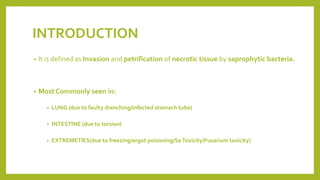
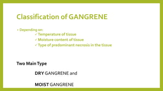
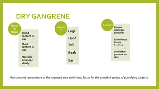
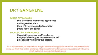
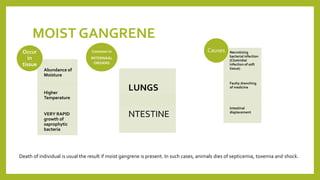
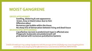
GANGRENE
UNIT-I
VETERINARY PATHOLOGY (PAPER-I)
DR. KAUSHAL KUMAR
Assistant Professor & Head
Department ofVeterinary Pathology
BiharVeterinary College, BASU, Patna, Bihar
INTRODUCTION
• It is defined as Invasion and petrification of necrotic tissue by saprophytic bacteria.
• Most Commonly seen in:
• LUNG (due to faulty drenching/infected stomach tube)
• INTESTINE (due to torsion)
• EXTREMETIES(due to freezing/ergot poisoning/Se Toxicity/Fusarium toxicity)
Classification of GANGRENE
• Depending on:
 Temperature of tissue
 Moisture content of tissue
 Type of predominant necrosis in the tissue
Two MainType
DRY GANGRENE and
MOIST GANGRENE
DRY GANGRENE
Blood
content is
less
Fluid
content is
less
Necrosis
develops
slowly
Occur
in
tissue Legs
Hoof
Tail
Beak
Ear
Common
in
Fungus
(calviceps
purpuria)
Seleniferous
Plants
Feeding
Consistent
pressure on
skin
Causes
Moisture and temperature of the necrosed area are limiting factor for the growth & spread of putrefying bacteria.
DRY GANGRENE
GROSS APPEARANCE
Dry, shriveled & mummified appearance
Colour green to black
Zone of hyperemia and inflammation
putrid odour due to H2S
MICROSCOPIC APPEARANCE
Coagulative necrosis in affected area
Phagocytic leukocytes are predominant cell
pink stained with numerous bacteria
GIT is mostly involved, because intestinal wall gets necrosed easily by putrefactive bacteria and thereby rupture
occurs, and finally fecal content discharged in to peritoneal cavity and microorganisms spread rapidly into viscera.
Finally septicemia , toxemia shock & death occurs.
MOIST GANGRENE
Abundance of
Moisture
Higher
Temperature
VERY RAPID
growth of
saprophytic
bacteria
Occur
in
tissue
LUNGS
NTESTINE
Common in
INTERNAAL
ORGANS
Necrotizing
bacterial infection
(Clostridial
infection of soft
tissue)
Faulty drenching
of medicine
Intestinal
displacement
Causes
Death of individual is usual the result if moist gangrene is present. In such cases, animals dies of septicemia, toxemia and shock.
MOIST GANGRENE
GROSS APPEARANCE
Swelling, blistering & wet appearance
Green, Gray or black Colour due to H2S
Offensive odour
Numerous gas bubble within the tissue
No sharp line of demarcation between living and dead tissue
MICROSCOPIC APPEARANCE
Liquefactive necrosis is predominant type in affected area
Phagocytic leukocytes are predominant cell
pink stained with numerous putrefactive bacteria
Dried & shriveled part may undergo liquefaction with the passage of time and finally may cause sloughing of time
and finally cause sloughing of the affected tissue or area termed asVERTUAL AMPUTATION
THANKS

Gangrene

  • 1.
    GANGRENE UNIT-I VETERINARY PATHOLOGY (PAPER-I) DR.KAUSHAL KUMAR Assistant Professor & Head Department ofVeterinary Pathology BiharVeterinary College, BASU, Patna, Bihar
  • 2.
    INTRODUCTION • It isdefined as Invasion and petrification of necrotic tissue by saprophytic bacteria. • Most Commonly seen in: • LUNG (due to faulty drenching/infected stomach tube) • INTESTINE (due to torsion) • EXTREMETIES(due to freezing/ergot poisoning/Se Toxicity/Fusarium toxicity)
  • 3.
    Classification of GANGRENE •Depending on:  Temperature of tissue  Moisture content of tissue  Type of predominant necrosis in the tissue Two MainType DRY GANGRENE and MOIST GANGRENE
  • 4.
    DRY GANGRENE Blood content is less Fluid contentis less Necrosis develops slowly Occur in tissue Legs Hoof Tail Beak Ear Common in Fungus (calviceps purpuria) Seleniferous Plants Feeding Consistent pressure on skin Causes Moisture and temperature of the necrosed area are limiting factor for the growth & spread of putrefying bacteria.
  • 5.
    DRY GANGRENE GROSS APPEARANCE Dry,shriveled & mummified appearance Colour green to black Zone of hyperemia and inflammation putrid odour due to H2S MICROSCOPIC APPEARANCE Coagulative necrosis in affected area Phagocytic leukocytes are predominant cell pink stained with numerous bacteria GIT is mostly involved, because intestinal wall gets necrosed easily by putrefactive bacteria and thereby rupture occurs, and finally fecal content discharged in to peritoneal cavity and microorganisms spread rapidly into viscera. Finally septicemia , toxemia shock & death occurs.
  • 6.
    MOIST GANGRENE Abundance of Moisture Higher Temperature VERYRAPID growth of saprophytic bacteria Occur in tissue LUNGS NTESTINE Common in INTERNAAL ORGANS Necrotizing bacterial infection (Clostridial infection of soft tissue) Faulty drenching of medicine Intestinal displacement Causes Death of individual is usual the result if moist gangrene is present. In such cases, animals dies of septicemia, toxemia and shock.
  • 7.
    MOIST GANGRENE GROSS APPEARANCE Swelling,blistering & wet appearance Green, Gray or black Colour due to H2S Offensive odour Numerous gas bubble within the tissue No sharp line of demarcation between living and dead tissue MICROSCOPIC APPEARANCE Liquefactive necrosis is predominant type in affected area Phagocytic leukocytes are predominant cell pink stained with numerous putrefactive bacteria Dried & shriveled part may undergo liquefaction with the passage of time and finally may cause sloughing of time and finally cause sloughing of the affected tissue or area termed asVERTUAL AMPUTATION
  • 8.