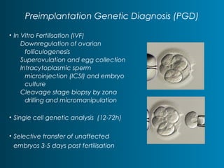
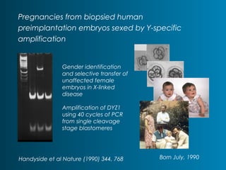
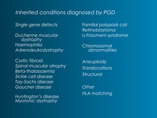
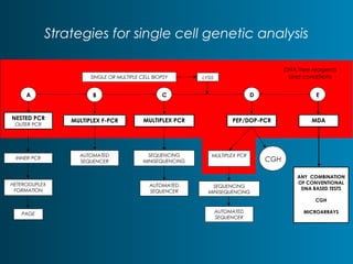
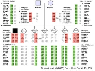
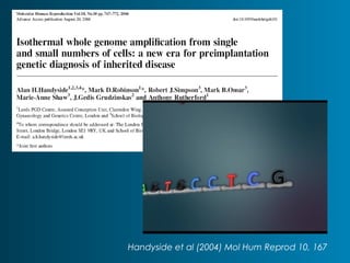
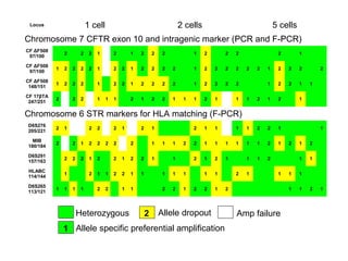
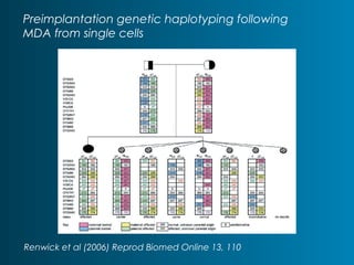
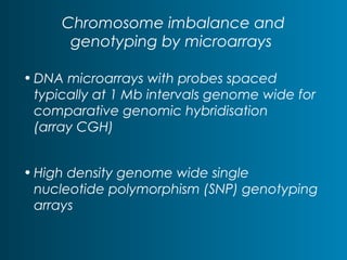
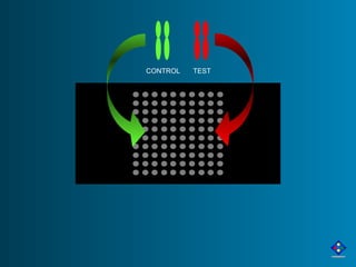
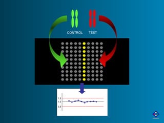
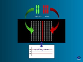
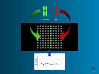
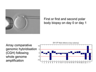
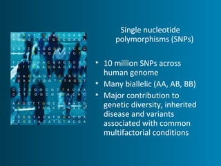
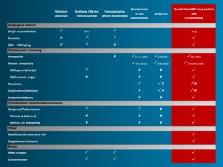
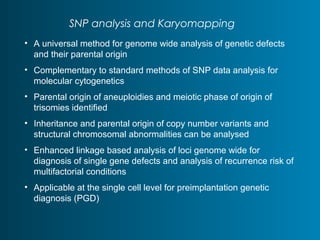
Gabbay Award Lecture 
Brandeis University 
November 16th, 2009 
‘Designer babies’: 
twenty years on 
Alan H Handyside 
London Bridge Fertility, Gynaecology 
And Genetics Centre 
London 
United Kingdom
Preimplantation Genetic Diagnosis (PGD) 
• In Vitro Fertilisation (IVF) 
Downregulation of ovarian 
folliculogenesis 
Superovulation and egg collection 
Intracytoplasmic sperm 
microinjection (ICSI) and embryo 
culture 
Cleavage stage biopsy by zona 
drilling and micromanipulation 
• Single cell genetic analysis (12-72h) 
• Selective transfer of unaffected 
embryos 3-5 days post fertilisation
Pregnancies from biopsied human 
preimplantation embryos sexed by Y-specific 
amplification 
Handyside et al Nature (1990) 344, 768 
Born July, 1990 
Gender identification 
and selective transfer of 
unaffected female 
embryos in X-linked 
disease 
Amplification of DYZ1 
using 40 cycles of PCR 
from single cleavage 
stage blastomeres
Inherited conditions diagnosed by PGD 
Familial polyposis coli 
Retinoblastoma 
Li-Fraumeni syndrome 
Chromosomal 
abnormalities 
Aneuploidy 
Translocations 
Structural 
Other 
HLA matching 
Single gene defects 
Duchenne muscular 
dystrophy 
Haemophilia 
Adrenoleukodystrophy 
Cystic fibrosis 
Spinal muscular atrophy 
Beta-thalassaemia 
Sickle cell disease 
Tay-Sachs disease 
Gaucher disease 
Huntington’s disease 
Myotonic dystrophy
Strategies for single cell genetic analysis 
SINGLE OR MULTIPLE CELL BIOPSY LYSIS 
A B C D E 
NESTED PCR 
OUTER PCR MULTIPLEX F-PCR MULTIPLEX PCR PEP/DOP-PCR 
INNER PCR 
HETERODUPLEX 
FORMATION 
PAGE 
MDA 
AUTOMATED 
SEQUENCER 
SEQUENCING 
MINISEQUENCING 
AUTOMATED 
SEQUENCER 
MULTIPLEX PCR 
SEQUENCING 
MINISEQUENCING 
AUTOMATED 
SEQUENCER 
ANY COMBINATION 
OF CONVENTIONAL 
DNA BASED TESTS 
CGH 
MICROARRAYS 
CGH 
DNA free reagents 
and conditions
Fiorentino et al (2005) Eur J Hum Genet 13, 953
Handyside et al (2004) Mol Hum Reprod 10, 167
Locus 1 cell 2 cells 5 cells 
Chromosome 7 CFTR exon 10 and intragenic marker (PCR and F-PCR) 
CF ΔF508 
97/100 2 2 2 1 2 1 2 2 2 1 2 2 2 2 1 
CF ΔF508 
97/100 1 2 2 2 2 1 2 2 1 2 2 2 2 1 2 2 2 2 2 2 1 2 2 2 2 
CF ΔF508 
148/151 1 2 2 2 1 2 2 1 2 2 2 2 1 2 2 2 2 1 2 2 1 1 
CF 17βTA 
247/251 2 2 2 1 1 1 2 1 2 2 1 1 1 2 1 1 1 2 1 2 1 
Chromosome 6 STR markers for HLA matching (F-PCR) 
D6S276 
205/221 2 1 2 2 2 1 2 1 2 1 1 1 1 2 2 1 1 
MIB 
180/184 2 2 1 2 2 2 2 2 1 1 1 2 2 1 1 1 1 1 1 2 1 2 1 2 
D6S291 
157/163 2 2 2 1 2 2 1 2 2 1 1 2 1 2 1 1 1 2 1 1 
HLABC 
114/144 1 2 1 1 2 2 1 1 1 1 1 1 1 2 1 1 1 1 
D6S265 
113/121 1 1 1 1 2 2 1 1 2 2 1 2 2 1 2 1 1 2 1 
Heterozygous Allele dropout Amp failure 
1 
2 
Allele specific preferential amplification
Preimplantation genetic haplotyping following 
MDA from single cells 
Renwick et al (2006) Reprod Biomed Online 13, 110
Embryo 2 
Monosomy 16, 21 
Trisomy 22
Chromosome imbalance and 
genotyping by microarrays 
•DNA microarrays with probes spaced 
typically at 1 Mb intervals genome wide for 
comparative genomic hybridisation 
(array CGH) 
• High density genome wide single 
nucleotide polymorphism (SNP) genotyping 
arrays
CONTROL TEST
CONTROL TEST 
1.5 
1.0 
0.5
1.5 
1.0 
0.5 
CONTROL TEST
1.5 
1.0 
0.5 
CONTROL TEST
First or first and second polar 
body biopsy on day 0 or day 1 
Array comparative 
genomic hybridisation 
(CGH) following 
whole genome 
amplification
Single nucleotide 
polymorphisms (SNPs) 
• 10 million SNPs across 
human genome 
• Many biallelic (AA, AB, BB) 
• Major contribution to 
genetic diversity, inherited 
disease and variants 
associated with common 
multifactorial conditions
Karyomapping 
• A universal method for genome wide analysis of 
genetic disease based on mapping crossovers 
between parental haplotypes 
• High density genome wide single nucleotide 
polymorphisms (SNP) genotyping of proband, parents and 
appropriate family member(s) to establish phase (Illumina 
Human CNV 370 Infinium-II Quad and Duo >300K SNP loci) 
• Mendelian analysis and karyomapping of the parental and 
grandparental haplotypes for each chromosome or 
chromosome segment in recombinant chromosomes 
Handyside et al (2009) J Med Genet Online First
♂ ♀ 
BAA 
ABA 
AAB 
BBA 
AAB 
ABA 
AAB 
AAB
A ♂ ♀ 
B 
C D 
Karyomapping combines 
genome wide linkage based 
detection of single gene defects 
(A) with chromosomal 
aneuploidy including 
monosomy/deletions (B) and 
trisomies involving inheritance of 
two different meiotic 
chromosomes from one parent 
(D). Chromosome duplication is 
not detected (C).
Family 1
Family 2 
Preimplantation genetic diagnosis 
for cystic fibrosis 
Whole genome amplification by 
isothermal multiple displacement 
amplification of 2-10 cells in each 
biopsy/embryo
Embryo 7 
13, 16, 18, 21, 22 
Monosomy 18, 21, 22 
X, Y, 21 
Trisomy X 
Monosomy 21 
Array CGH 
Karyomap
Mutation 
detection 
Multiplex PCR and 
minisequencing 
Preimplantation 
genetic haplotyping 
Fluorescence 
in situ 
hybridisation 
Array CGH 
Quantitative SNP array analysis 
and 
Karyomapping 
Single gene defects 
Single or combination  Any  Any 
Exclusion     
SGD + HLA typing     
Chromosome screening 
Aneuploidy  (5-12 chr) (24 chr) (24 chr) 
Meiotic aneuploidy (PB only) (PB only)  Trisomy only 
With parental origin     
With meiotic origin     
Mosaicism    
Duplications/deletions    
Uniparental disomy    
Translocation chromosome imbalance 
Reciprocal/Robertsonian     
Normal vs balanced     
With 24 chr aneuploidy     
Other 
Multifactorial recurrence risk  
Copy Number Variants  
Errors 
Allele dropout    
Contamination   
SNP analysis and Karyomapping 
• A universal method for genome wide analysis of genetic defects 
and their parental origin 
• Complementary to standard methods of SNP data analysis for 
molecular cytogenetics 
• Parental origin of aneuploidies and meiotic phase of origin of 
trisomies identified 
• Inheritance and parental origin of copy number variants and 
structural chromosomal abnormalities can be analysed 
• Enhanced linkage based analysis of loci genome wide for 
diagnosis of single gene defects and analysis of recurrence risk of 
multifactorial conditions 
• Applicable at the single cell level for preimplantation genetic 
diagnosis (PGD)
Acknowledgements 
Genoma Laboratory, Rome 
Anil Biricik 
Francesco Fiorentino 
Bridge Genoma, London 
Helen Tempest 
Mira Grigorova 
Alan Thornhill 
Department of Biosciences, 
University of Kent 
Alem Gebrasselasi 
Darren Griffin 
BlueGnome, Cambridge 
Anthony Brown 
Tony Gordon 
Graham Snudden 
Nick Haan 
Department of Pathology, 
University of Cambridge 
Ben Dunmore 
Nabeel Affara 
Genetics and IVF Institute, 
Fairfax, Virginia 
Gary Harton 
Brian Mariani

Gabbay81109pre07 12588919989864-phpapp02

  • 1.
    Gabbay Award Lecture Brandeis University November 16th, 2009 ‘Designer babies’: twenty years on Alan H Handyside London Bridge Fertility, Gynaecology And Genetics Centre London United Kingdom
  • 3.
    Preimplantation Genetic Diagnosis(PGD) • In Vitro Fertilisation (IVF) Downregulation of ovarian folliculogenesis Superovulation and egg collection Intracytoplasmic sperm microinjection (ICSI) and embryo culture Cleavage stage biopsy by zona drilling and micromanipulation • Single cell genetic analysis (12-72h) • Selective transfer of unaffected embryos 3-5 days post fertilisation
  • 4.
    Pregnancies from biopsiedhuman preimplantation embryos sexed by Y-specific amplification Handyside et al Nature (1990) 344, 768 Born July, 1990 Gender identification and selective transfer of unaffected female embryos in X-linked disease Amplification of DYZ1 using 40 cycles of PCR from single cleavage stage blastomeres
  • 5.
    Inherited conditions diagnosedby PGD Familial polyposis coli Retinoblastoma Li-Fraumeni syndrome Chromosomal abnormalities Aneuploidy Translocations Structural Other HLA matching Single gene defects Duchenne muscular dystrophy Haemophilia Adrenoleukodystrophy Cystic fibrosis Spinal muscular atrophy Beta-thalassaemia Sickle cell disease Tay-Sachs disease Gaucher disease Huntington’s disease Myotonic dystrophy
  • 6.
    Strategies for singlecell genetic analysis SINGLE OR MULTIPLE CELL BIOPSY LYSIS A B C D E NESTED PCR OUTER PCR MULTIPLEX F-PCR MULTIPLEX PCR PEP/DOP-PCR INNER PCR HETERODUPLEX FORMATION PAGE MDA AUTOMATED SEQUENCER SEQUENCING MINISEQUENCING AUTOMATED SEQUENCER MULTIPLEX PCR SEQUENCING MINISEQUENCING AUTOMATED SEQUENCER ANY COMBINATION OF CONVENTIONAL DNA BASED TESTS CGH MICROARRAYS CGH DNA free reagents and conditions
  • 7.
    Fiorentino et al(2005) Eur J Hum Genet 13, 953
  • 8.
    Handyside et al(2004) Mol Hum Reprod 10, 167
  • 9.
    Locus 1 cell2 cells 5 cells Chromosome 7 CFTR exon 10 and intragenic marker (PCR and F-PCR) CF ΔF508 97/100 2 2 2 1 2 1 2 2 2 1 2 2 2 2 1 CF ΔF508 97/100 1 2 2 2 2 1 2 2 1 2 2 2 2 1 2 2 2 2 2 2 1 2 2 2 2 CF ΔF508 148/151 1 2 2 2 1 2 2 1 2 2 2 2 1 2 2 2 2 1 2 2 1 1 CF 17βTA 247/251 2 2 2 1 1 1 2 1 2 2 1 1 1 2 1 1 1 2 1 2 1 Chromosome 6 STR markers for HLA matching (F-PCR) D6S276 205/221 2 1 2 2 2 1 2 1 2 1 1 1 1 2 2 1 1 MIB 180/184 2 2 1 2 2 2 2 2 1 1 1 2 2 1 1 1 1 1 1 2 1 2 1 2 D6S291 157/163 2 2 2 1 2 2 1 2 2 1 1 2 1 2 1 1 1 2 1 1 HLABC 114/144 1 2 1 1 2 2 1 1 1 1 1 1 1 2 1 1 1 1 D6S265 113/121 1 1 1 1 2 2 1 1 2 2 1 2 2 1 2 1 1 2 1 Heterozygous Allele dropout Amp failure 1 2 Allele specific preferential amplification
  • 10.
    Preimplantation genetic haplotypingfollowing MDA from single cells Renwick et al (2006) Reprod Biomed Online 13, 110
  • 11.
    Embryo 2 Monosomy16, 21 Trisomy 22
  • 12.
    Chromosome imbalance and genotyping by microarrays •DNA microarrays with probes spaced typically at 1 Mb intervals genome wide for comparative genomic hybridisation (array CGH) • High density genome wide single nucleotide polymorphism (SNP) genotyping arrays
  • 13.
  • 14.
  • 15.
    1.5 1.0 0.5 CONTROL TEST
  • 16.
    1.5 1.0 0.5 CONTROL TEST
  • 17.
    First or firstand second polar body biopsy on day 0 or day 1 Array comparative genomic hybridisation (CGH) following whole genome amplification
  • 18.
    Single nucleotide polymorphisms(SNPs) • 10 million SNPs across human genome • Many biallelic (AA, AB, BB) • Major contribution to genetic diversity, inherited disease and variants associated with common multifactorial conditions
  • 19.
    Karyomapping • Auniversal method for genome wide analysis of genetic disease based on mapping crossovers between parental haplotypes • High density genome wide single nucleotide polymorphisms (SNP) genotyping of proband, parents and appropriate family member(s) to establish phase (Illumina Human CNV 370 Infinium-II Quad and Duo >300K SNP loci) • Mendelian analysis and karyomapping of the parental and grandparental haplotypes for each chromosome or chromosome segment in recombinant chromosomes Handyside et al (2009) J Med Genet Online First
  • 20.
    ♂ ♀ BAA ABA AAB BBA AAB ABA AAB AAB
  • 21.
    A ♂ ♀ B C D Karyomapping combines genome wide linkage based detection of single gene defects (A) with chromosomal aneuploidy including monosomy/deletions (B) and trisomies involving inheritance of two different meiotic chromosomes from one parent (D). Chromosome duplication is not detected (C).
  • 22.
  • 23.
    Family 2 Preimplantationgenetic diagnosis for cystic fibrosis Whole genome amplification by isothermal multiple displacement amplification of 2-10 cells in each biopsy/embryo
  • 24.
    Embryo 7 13,16, 18, 21, 22 Monosomy 18, 21, 22 X, Y, 21 Trisomy X Monosomy 21 Array CGH Karyomap
  • 25.
    Mutation detection MultiplexPCR and minisequencing Preimplantation genetic haplotyping Fluorescence in situ hybridisation Array CGH Quantitative SNP array analysis and Karyomapping Single gene defects Single or combination  Any  Any Exclusion     SGD + HLA typing     Chromosome screening Aneuploidy  (5-12 chr) (24 chr) (24 chr) Meiotic aneuploidy (PB only) (PB only)  Trisomy only With parental origin     With meiotic origin     Mosaicism    Duplications/deletions    Uniparental disomy    Translocation chromosome imbalance Reciprocal/Robertsonian     Normal vs balanced     With 24 chr aneuploidy     Other Multifactorial recurrence risk  Copy Number Variants  Errors Allele dropout    Contamination   
  • 26.
    SNP analysis andKaryomapping • A universal method for genome wide analysis of genetic defects and their parental origin • Complementary to standard methods of SNP data analysis for molecular cytogenetics • Parental origin of aneuploidies and meiotic phase of origin of trisomies identified • Inheritance and parental origin of copy number variants and structural chromosomal abnormalities can be analysed • Enhanced linkage based analysis of loci genome wide for diagnosis of single gene defects and analysis of recurrence risk of multifactorial conditions • Applicable at the single cell level for preimplantation genetic diagnosis (PGD)
  • 27.
    Acknowledgements Genoma Laboratory,Rome Anil Biricik Francesco Fiorentino Bridge Genoma, London Helen Tempest Mira Grigorova Alan Thornhill Department of Biosciences, University of Kent Alem Gebrasselasi Darren Griffin BlueGnome, Cambridge Anthony Brown Tony Gordon Graham Snudden Nick Haan Department of Pathology, University of Cambridge Ben Dunmore Nabeel Affara Genetics and IVF Institute, Fairfax, Virginia Gary Harton Brian Mariani