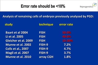
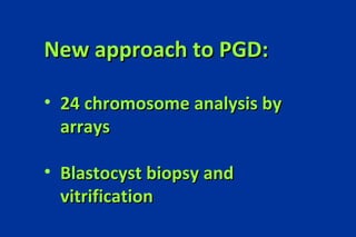
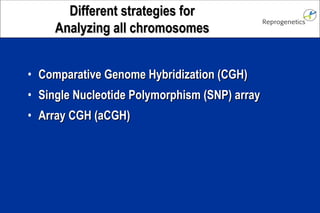
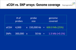
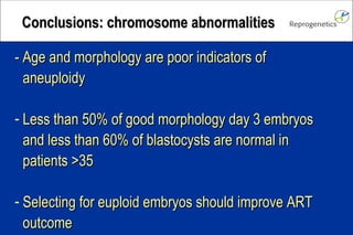
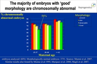
USA: Livingston, NJ Europe: Barcelona, Spain Oxford, UK Hamburg, Germany Asia:  Kobe, Japan South America: Lima, Peru Santiago Munné Preimplantation Genetic  Diagnosis
CLIA and NYDH approved laboratory.  Approximately 1/3 (150 centers) of US PGD cases >20,000 PGD cycles performed PGD Services:  - 24-chromosome analysis by array CGH - translocations by FISH or array CGH - gene defects by PCR or SNP array - Blasotcyst biopsy and vitrification teaching PGD portal for the electronic medical records of  e IVF Reprogenetics services
High rates of  Chromosome abnormalities
embryos analyzed: 6054. Morphologically normal embryos: 3751. Source: Munn é  et al. 2007. Similar results also found by Munne et al 1995, Marquez et al. 2000, Magli et al. 2007. % chromosomally  abnormal embryos 56% Maternal age Morphology: The majority of embryos with ‘good’ morphology are chromosomally abnormal
PGD Hypothesis PGD  may  improve ART outcome in women of advanced maternal age Munné et al. (1993)
Despite large studies indicating the advantages of aneuploidy screening, the notion that PGS for infertility is beneficial is not yet shared uniformly.
Positive effect Gianaroli et al. 1999 Munne et al 1999 Gianaroli et al 2001a Gianaroli et al. 2001b Munne et al. 2003 Gianaroli et al. 2004 Munne et al. 2005 Munne et al 2006 Verlinsky et al. 2005 Colls et al. 2007 Garrisi et al. 2009 Rubio et al. 2009 No effect (small)  Werlin et al. 2003 Jansen et al. 2008 Mersereau et al. 2008 Scholcraft et al. 2009 No effect (Large) Staessen et al. 2004 Platteau et al. 2005 Negative effect  Mastenbroek et al. 2007 Hardarson et al. 2008 Contradicting PGD results  using day 3 biopsy and FISH
Proposed explanations: Mosaicism Self-correction of abnormal embryos Sub-optimal PGD and biopsy methods Contradicting PGD results  using day 3 biopsy and FISH
Mosaicism
592 embryos found abnormal by PGD were reanalyzed and found to be:  normal    13 mosaic  <49% abnormal   27 mosaic  50-99% abnormal  124 mosaic  100% abnormal  297 homogeneously abnormal  131 Colls et al. (2007) Mosaicism produces <7% misdiagnosis 6.8% 1[13]1[16]2[18]2[21]1[22] 2[13]1[16]2[18]2[21]2[22] 1[13]1[16]2[18]2[21]1[22] 1[16] 2[13]2[16]2[18]2[21]2[22] 2[13]1[16]2[18]1[21]1[22] 2[13]3[18]1[21]1[22] 3[13]1[16]2[18]1[21]3[22]  1[13]1[16]1[18]1[21]1[22] 3[13]1[16]2[18]1[21]3[22]
The FISH error of the assay translates into a  clinical misdiagnosis rate of 0.5% NEGATIVE PREDICTIVE VALUE:  99.5% Data from >150 IVF centers referring to Reprogenetics 10 aneuploid abortions of 2148 implanted fetuses  - Average maternal age = 37 years Clinical misdiagnosis
self-correction myths
UNIPARENTAL DISOMY:  a trisomy correction creates a zygote with 2 chromosomes of one parent, none of the other (trisomy rescue). Theoretical chance : 1/3  EXAMPLE  TRISOMY 15: Prognosis is ethal because of imprinting Trisomy 15 in cleavage stage embryos: 1.874% a UPD-15 in newborns:  0.001% b Estimated correction of trisomy 15 to UDP:   1/3  c Trisomy 15 day 3 embryos that self-corrected:  1.874 x 0.001 / 3 =  0.56% a: Munne et al. (2004), b: From: OMIM, c: 3 X UPD   Trisomy correction is rare: UPD evidence
Fetus seldom self correct:  it’s the placenta that becomes abnormal This work questions the assumption that placental confined mosaicism is the result of fetal self-correction. At the contrary, it suggests that  normal fetuses may develop abnormal placenta.
Sub-optimizal methods (FISH studies, day 3 biopsy)
Optimal PGS Questionable PGS Biopsy media with aminoacids   simple media Biopsy time / embryo 1 min > 5 min # cells biopsied one two Fixation method Carnoy’s Tween 20 # chromosomes tested ≥ 8  6 # analysts / case 2 1 Use of NRR* yes no Large experience yes no Error rate <10% 10-50% Number of zygotes >5     5 *NRR: No result rescue, or re-testing of dubious chromosome with different probes. Optimal PGS methods
CONTROL  2 -CELLS   BIOPSIED  Staessen et al (2004) 11.5% 17.1% Implantation Staessen et al. (2004): No significant differences But 2 cells biopsied PGD for AMA: randomized studies
“  The data presented here clearly indicates that two cell biopsy significantly impacts clinical outcome. Our previous report providing no arguments in favor of PGS (Staessen et al., 2004) was criticized by others arguing that PGS might have been beneficial if only one cell had been removed (Cohen et al., 2007). In respect to the present findings, this criticism seems justified ”. P < 0.001 Two cell biopsy is detrimental
20% of  cycles  undiagnosed (literature: 1-3% of  embryos  *) 2) 59% implantation reduction due to biopsy: 3)  Average number of embryos analyzed was only 5 4)  Chromosomes 15 and 22 (21% abnormalities) not analyzed 59% reduction implantation Control    14.7% Biopsied, no PGD   6.0% Biopsied and PGD   16.8% * 1% Gianaroli et al. (2004), 3.1% Colls et al. (2007)   PGD for AMA: randomized studies Mastenbroek et al. (2007)
Analysis of remaining cells of embryos previously analyzed by PGD: study   technique error rate   Baart et al 2004  FISH   50.0%   Li et al. 2005  FISH   40.0%   Gleicher et al. 2009 FISH   15-20%   Munne et al. 2002  FISH-9   7.2%    Colls et al., 2007  FISH-9   4.7%   Magli et al. 2007  FISH-9   3.7%   Munne et al. 2010 array CGH   1.8% Error rate should be <10%
New approach to PGD: 24 chromosome analysis by arrays Blastocyst biopsy and vitrification
Comparative Genome Hybridization (CGH) Single Nucleotide Polymorphism (SNP) array Array CGH (aCGH) Different strategies for Analyzing all chromosomes
CGH and array CGH
Kallioniemi et al. (1992), applied to single cells by Wells et al. (1999) Normal DNA Test DNA Comparative Genome  Hybridization (CGH) Normal Trisomy Monosomy
Array CGH Test  DNA Normal  DNA 2700 probes Same band resolution as karyotype
46,XY
47,XY+2
44,XY-9-17
aCGH advantages All  24 chromosome  type of aneuploidies detected Results in  24  hours; allows for PB or day 3 biopsy Parental DNA  not  required:  ad hoc decisions possible Detects trisomy originating from mitotic errors or MII meiotic errors without crossing-over (SNP array may not) Used in  >15,000  patients with mental retardation
46,XY-10 +16   aCGH detected  50%  more abnormalities than FISH-12 and  20%  more abnormal embryos (Colls et al. 2009) Detection of abnormalities:  aCGH vs FISH-12 Detectable by FISH
aCGH validation: no results Embryos undiagnosed: biopsy on day  3 :  2%  (16/724) biopsy on day  5 :  >2%  (0/54) Method: Bluegnome array, Sureplex amplification.
aCGH validation: error rate Validation method 1: single cells from cell lines analyzed*  Error rate in euploid cell lines: 0/9 Error rate in aneuploid cell lines: 0/42 Validation method 2: Reanalysis of the rest of the embryo by FISH with 19 chromosomes probes**  Error rate from day 3 biopsies:  1.8%  (1/56)   * Mamas et al (submitted), ** Gutierrez et al. (submitted)
aCGH results on day 3: validation data Cells from the same embryo: X O , + 2,  - 4p, + 11, - 13,  - 18,  - 22 XX,   - 2,  - 4p, + 4q31, - 13,  - 18,  - 22 XX X , + 2,  - 4p,   - 4q31, + 11, - 13,  - 18,  - 22 X O , + 2,  + 4p,   - 4q31,   - 11, - 13,  - 18,  - 22 Who said mosaicism was a FISH artifact?
Day 3 biopsy, day 5 transfer and array CGH Cycles performed:    151 Maternal age (av.)  37.2   Pregnancy Rate Ongoing Pregnancy Rate Per Cycle Per ET Per Cycle Per ET Control 38% 38% 31% 31% PGD 46% 59% 42% 54% NS < 0.001 NS < 0.001 * Expected from each center SART data, controlled by age Data from 24 centers. Munné et al. (2010) PGDIS, and unpublished data
CGH on blastocyst biopsies: Why? Advantages: More DNA: More robust diagnosis Eliminates some mosaic embryos Reduces error rate Reduced impact of embryo biopsy Less embryos to process Facilitates single embryo transfer Uterine environment optimized after thaw
Effect of embryo vitrification and Replacement on next cycle
Average age 37.7 Av. Previous failed cycles: 2.4 Average # blastocysts: 6.4 Normal blastocysts: 52% 99.3% survival after freeze/thaw Schoolcraft et al. (in press) CGH on blastocyst biopsies: Clinical results (2)
Cycles Mat. Prev. embryos  implant. ongoing age failed replaced   (+ sac) preg.   cycles CGH :    45 37.7   2.4    2.0   67% 79%   control :  113 37.1   1.2    2.7   28% 60%     p=0.0003 Schoolcraft et al. (in press) CGH on blastocyst biopsies: Preliminary clinical results
CGH on blastocyst biopsies: Implantation is independent of age Patients <43 who are eligible for blastocyst transfer have a >95% change of having normal embryos available for transfer
Pregnancy rates were above those for controls for all age groups  However, pregnancy rates were lower for older patients due to the increased frequency of cycles with no euploid embryos.  Spontaneous abortion rates were reduced for all patients, including those with a history of multiple miscarriage Chromosomally normal embryos from older patients have a similar chance of producing a child as those derived from young patients. CGH on blastocyst biopsies: Conclusions on pilot study
What patient groups will benefit the most from this type of screening? Young patients may benefit, particularly in countries where single embryo transfer is mandatory Can the results obtained in the current study be replicated in a randomized controlled trial? How much of the observed benefit is due to transfer in a subsequent cycle? An RCT is currently being contemplated in the UK Aneuploidy explains ‘some’ of the decline in IVF success with advancing maternal age. What explains the remainder? Uterine receptivity factor may be involved CGH on blastocyst biopsies: Remaining questions
PGD for translocations and 24 chromosome abnormalities
aCGH for translocations and 24 chromosome aneuploidy  Patient:  46,XY,t(3;11)(q22.2;q23.3)
Comparison of different approaches
aCGH vs. SNP arrays: Genome coverage   # of   probe   genome probes   size   covered aCGH 4,000 x 150,000 kb = 600.0 Mb (25%) SNPs 300,000  x   50 kb  =  1.5 Mb (>0.1%)
aCGH vs. SNP arrays: Resolution More resolution, more counseling nightmares: In pre-natal diagnosis the minimum resolution reported is 20 MB: Smaller abnormalities are not well understood and difficult to council At 1.5 Kb resolution there are >300,000 CNPs described: Difficult to differentiate a new polymorphism from an abnormality
Embryos analyzed with 9-12 probes: 91,473 Polyploid or haploid:   6,609 (8%) Abnormal embryos fully reanalyzed:   1,416 polyploid or haploid by PGD and  polyploid or haploid after reanalysis:   140 Of the above, that had only trisomies:   27 (24%) Not diagnosed by aCGH or CGH:   1.8% aCGH vs SNPs: polyploidy
aCGH vs SNPs: mitotic abnormalities Embryos analyzed with 9-12 probes: 91,473 Complex abnormal: 26,609 (29%) Abnormal embryos fully reanalyzed:   1,416 Complex abnormal by PGD and mosaic and not aneuploid after reanalysis:   330 (52%) Of the above, that had only trisomies:   53 (16%) Not diagnosed by qualitative SNP array:   3%
aCGH vs SNPs Other SNP advantages: Usefulness? Origin of aneuploidy detected 95% are maternal anyway Embryo fingerprinting None in SET. aCGH also doable Differentiates normal / balanced balanced are fine.  >80% abnormalities Other SNP disadvantages: Problem: Requires parental testing Adds costs, cancellation fees, planning in advance, prevents add oc prescription, 20% patients cancel
Comparison of platforms FISH-12  CGH   a array SNP   probes  a   CGH   a arrays Day 3 biopsy/ day 5 results yes no yes yes Parental DNA needed no no no yes Aneuploid embryos detected 89% 100% 100% 97% Detect gene defects no no no yes Detect polyploidy (errors) yes (1.8%) (1.8%  ) yes Detect MII trisomies w/o crossover yes yes yes no  if qual. Detect mitotic trisomies (errors) yes yes yes (3%) Error rate (cell lines analysis) 2% 1% 0%  d 4%   b Error rate (embryo reanalysis)  7% 8%   2% unk Embryos with no results 3% 6% 2% 8%   b Increased implantation rate (day 3)    + n/a   ++     unk   Increased implantation rate (day 5) unk   +++  +++ +++  c a Reprogenetics data,  b  Jonhson et al. (2010),  c  Scott et al. (2010, PCRS),  d  Mamas et al (submitted)
PGD for gene defects  and 24 chromosome abnormalities
PGD for gene disorders Disease tested:   Acetil Co Oxidase type I defficiency, Adrenoleucodistrophy, Alpha-thalassemia, Alport syndrome, Autosomal Dominant Polycystic Kidney Disease (ADPKD), Autosomal Recesive Polycystic Kidney Disease (ARPKD), Beta-thalassemia,  Branchio-Oto-Renal syndrome (BOR),  BRCA1 breast cancer predisposition, BRCA2 breast cancer predisposition, CanavanCharcot-Marie-Tooth type IA (CMT1a), Choroideremia, Congenital adrenal hyperplasia (CAH), Congenital neutropenia, Connexin 26 hearing loss, Cystic fibrosis, Duchenne/Becker  Muscular Dystrophy (DMD), Ectrodactyly, Ectodermal dysplasia, and Cleft lip/palate syndrome (EEC1), Fabry Disease, Familial adenomatous poliposis coli (FAP), Familial dysautonomia, Familial intrahepatic cholestasis 2, Fanconi anemia, Fragile site mental retardation , Gangliosidosis type 1 (GM1), Gaucher disease, Glomuvenous malformations (GVM), Glycogen-storage disease type I  (GSD1), Glycosylation type 1C, Hemoglobin SC disease, Hemophilia A, Hemophilia B, Hereditary nonpolyposis colon cancer  (HNPCC), Hereditary pancreatitis, HLA matching Huntington disease, Hurler syndrome, Hypophosphatasia, Incontinential pigmenti, Krabbe disease (Globoid cell leukodystrophy), Long QT syndrome, Marfan syndrome, Meckle gruber, Metachromatic leukodystrophy (MLD), Methylmalonic aciduria cblC type (MMACHC), Myotonic Dystrophy 1, Myotubular myopathy, Neurofibromatosis 1, Neurofibromatosis 2, Niemann-Pick Disease, Noonan syndrome, Oculocutaneous albinism 1 (OCA1), Ornithine carbamoyltransferase deficiency (OTC), Osteogenesis Imperfecta 1, Rapp Hodgkin ectodermal dysplasia, Retinitis pigmentosa, Retinoblastoma, Sickle Cell Anemia, Smith-Lemli-Opitz syndrome (SLOS), Spinal bulbar muscular atrophy (SBMA), Spinal Muscular Atrophy Type 1 (SMA1), Tay Sachs, Tuberous sclerosis 1 (TSC1), Tuberous sclerosis 2 (TSC2),  Von Hippel-Lindau Syndrome (vHL),  X-linked dominant Charcot–Marie–Tooth (CMTX), etc……  (see review Gutierrez et al. (2008)) We can do PGD for any disease with known mutation
Mutation   site Polymorphic site Conventional approach to PGD of gene  defects to avoid Allele Drop Out (ADO) accurate amplification of both loci ADO affecting normal allele ADO affecting mutation site
PGD of gene defects using SNP array Karyomapping Δ 508 Father: Δ 508 Mother: Slide adapted from  A.Handyside Monosomy  11 Trisomy  21 Carrier  Δ 508
Example:   First Clinical Application of Karyomapping for PGD  of Gaucher Disease Combined with 24 Chromosome Screening Handyside A, Grifo, J, Gabriel A, Thornhill A, Griffin D, Ketterson K, Prates R,  Tormasi S, Fischer J, Munné S (2010) PGDIS Embryo # Gene defects Chromosome defects 5 Affected normal 7 Affected monosomy 22 8 Carrier monosomy 10, 21,22 9 Affected monosomy 22 2 unaffected Monosomy 22 10 Affected Monosomy 9,17,19,22, trisomy 14 Results confirmed by a-CGH and by PCR
Conclusions
- Age and morphology are poor indicators of aneuploidy  Less than 50% of good morphology day 3 embryos and less than 60% of blastocysts are normal in patients >35 Selecting for euploid embryos should improve ART outcome  Conclusions: chromosome abnormalities
Studies with improved results differ from those that show no improvement in that: 1) Reduce biopsy damage (1 cell, experience, blast?) 2) Low error rate (fixation, NRR, 2 analyzers) 3) Analyze 16,15,21,22 chromosomes + ≥4 more 4) Extensive experience 5) Appropriate population (≥5 embryos,  ≥   35 y. old) Conclusions: FISH studies
Blastocyst biopsy + CGH + vitrification shows very high implantation rates (72%, av. Age 38). Array CGH and day 5 biopsy will produce same results Array CGH and day 3 biopsy improves results when normal embryos are available.  Additional vitirifcation step may still be advantageous Conclusions: array CGH
Single gene defects and 24 chromosome analysis can be detected simultaneously for couples carriers of gene defects.  It requires parental testing. Conclusions: gene defects and 24 chromosome analysis
Integration with e IVF
Reduce time of form-filling to a few clicks:  patient demographics, cycle progress, PGD results, follow up, automatically accessed by IVF center and Reprogenetics Advantages (1) e IVF
Minimize the risk of mismanaging patients: Translocations and gene defects preparations, billing, …  Advantages (2) e IVF
Access to the aggregate data of Reprogenetics, de-identified by patient and center, with standard queries and access to the raw data. Example: Out of the 20,000 PGD cycles performed by Reprogenetics, what is the proportion of normal embryos in a patient 43 years old?  17% (n = 345) … with an E2 between 8 and 10…. etc Advantages (3) e IVF
Fields to choose:  Age by  1 year or by SART groups (drop list) stim protocol (drop list) ICSI or IVF pick E2 level (chose range) number of eggs (chose range) number of zygotes (chose range) zygote pm stage (chose range) number of blastocysts(chose range) motphology grade on day 3 (chose range) motphology grade on day 5 (chose range) number of day 3 embryos of grade X (chose range) number of day 5 embryos of grade X (chose range) ICM grade (chose range) trophectoderm grade (chose range) # of previous miscarriages (chose range) # of previous failed cycles (chose range) # of previous conceptions that were chromosomally abnormal (0, >0) PGD indication (drop list) PGD test (drop list) pregnancy rate of centers included in the query (chose range) Outcome: % of cycles with these characteristics out of the total % normal embryos % aneuploid embryos % other abnormal embryos implantation rate per pick up implantation rate per transfer pregnancy rate per pick up pregnacy rate per transfer miscarriage rate % miscarraiges chromosomally abnormal take home baby rate center ranking compared to other non identified centers e IVF
Santiago Munné, PhD, Director Jacques Cohen, PhD, Director munne@reprogenetics.com  www.reprogenetics.com   Pere Colls, Ph.D Dagan Wells, Ph.D George Pieczenik, Ph.D   Cristina Gutiérrez, Ph.D   Jorge Sanchez, PhD   John Zheng, MD   Tomas Escudero, MS Kelly Ketterson, MS Jill Fischer, MS Jessica Vega, MS   Tim Schimmel Sasha Sadowy Sophia Tormasi N-neka Goodall Renata Prates   Piedad Garzon   Laurie Ferrara Bekka Sellon-Wright Maria Feldhaus USA Spain Mireia Sandalinas, MS Carles Giménez, PhD César Arjona, MS Ana Jiménez, PhD Elena Garcia, MS  Japan Tetsuo Otani, MD Muriel Roche Miho Mizuike UK Dagan Wells, PhD Elpida Fragouli, PhD Samer Alfarawati, MS Michalis Konstantinidis South America Paul Lopez Luis Alberto Guzman Germany Karsten Held, MD

Munne Array Cgh Update 2010 05

  • 1.
    USA: Livingston, NJEurope: Barcelona, Spain Oxford, UK Hamburg, Germany Asia: Kobe, Japan South America: Lima, Peru Santiago Munné Preimplantation Genetic Diagnosis
  • 2.
    CLIA and NYDHapproved laboratory. Approximately 1/3 (150 centers) of US PGD cases >20,000 PGD cycles performed PGD Services: - 24-chromosome analysis by array CGH - translocations by FISH or array CGH - gene defects by PCR or SNP array - Blasotcyst biopsy and vitrification teaching PGD portal for the electronic medical records of e IVF Reprogenetics services
  • 3.
    High rates of Chromosome abnormalities
  • 4.
    embryos analyzed: 6054.Morphologically normal embryos: 3751. Source: Munn é et al. 2007. Similar results also found by Munne et al 1995, Marquez et al. 2000, Magli et al. 2007. % chromosomally abnormal embryos 56% Maternal age Morphology: The majority of embryos with ‘good’ morphology are chromosomally abnormal
  • 5.
    PGD Hypothesis PGD may improve ART outcome in women of advanced maternal age Munné et al. (1993)
  • 6.
    Despite large studiesindicating the advantages of aneuploidy screening, the notion that PGS for infertility is beneficial is not yet shared uniformly.
  • 7.
    Positive effect Gianaroliet al. 1999 Munne et al 1999 Gianaroli et al 2001a Gianaroli et al. 2001b Munne et al. 2003 Gianaroli et al. 2004 Munne et al. 2005 Munne et al 2006 Verlinsky et al. 2005 Colls et al. 2007 Garrisi et al. 2009 Rubio et al. 2009 No effect (small) Werlin et al. 2003 Jansen et al. 2008 Mersereau et al. 2008 Scholcraft et al. 2009 No effect (Large) Staessen et al. 2004 Platteau et al. 2005 Negative effect Mastenbroek et al. 2007 Hardarson et al. 2008 Contradicting PGD results using day 3 biopsy and FISH
  • 8.
    Proposed explanations: MosaicismSelf-correction of abnormal embryos Sub-optimal PGD and biopsy methods Contradicting PGD results using day 3 biopsy and FISH
  • 9.
  • 10.
    592 embryos foundabnormal by PGD were reanalyzed and found to be: normal 13 mosaic <49% abnormal 27 mosaic 50-99% abnormal 124 mosaic 100% abnormal 297 homogeneously abnormal 131 Colls et al. (2007) Mosaicism produces <7% misdiagnosis 6.8% 1[13]1[16]2[18]2[21]1[22] 2[13]1[16]2[18]2[21]2[22] 1[13]1[16]2[18]2[21]1[22] 1[16] 2[13]2[16]2[18]2[21]2[22] 2[13]1[16]2[18]1[21]1[22] 2[13]3[18]1[21]1[22] 3[13]1[16]2[18]1[21]3[22] 1[13]1[16]1[18]1[21]1[22] 3[13]1[16]2[18]1[21]3[22]
  • 11.
    The FISH errorof the assay translates into a clinical misdiagnosis rate of 0.5% NEGATIVE PREDICTIVE VALUE: 99.5% Data from >150 IVF centers referring to Reprogenetics 10 aneuploid abortions of 2148 implanted fetuses - Average maternal age = 37 years Clinical misdiagnosis
  • 12.
  • 13.
    UNIPARENTAL DISOMY: a trisomy correction creates a zygote with 2 chromosomes of one parent, none of the other (trisomy rescue). Theoretical chance : 1/3 EXAMPLE TRISOMY 15: Prognosis is ethal because of imprinting Trisomy 15 in cleavage stage embryos: 1.874% a UPD-15 in newborns: 0.001% b Estimated correction of trisomy 15 to UDP: 1/3 c Trisomy 15 day 3 embryos that self-corrected: 1.874 x 0.001 / 3 = 0.56% a: Munne et al. (2004), b: From: OMIM, c: 3 X UPD Trisomy correction is rare: UPD evidence
  • 14.
    Fetus seldom selfcorrect: it’s the placenta that becomes abnormal This work questions the assumption that placental confined mosaicism is the result of fetal self-correction. At the contrary, it suggests that normal fetuses may develop abnormal placenta.
  • 15.
    Sub-optimizal methods (FISHstudies, day 3 biopsy)
  • 16.
    Optimal PGS QuestionablePGS Biopsy media with aminoacids simple media Biopsy time / embryo 1 min > 5 min # cells biopsied one two Fixation method Carnoy’s Tween 20 # chromosomes tested ≥ 8  6 # analysts / case 2 1 Use of NRR* yes no Large experience yes no Error rate <10% 10-50% Number of zygotes >5  5 *NRR: No result rescue, or re-testing of dubious chromosome with different probes. Optimal PGS methods
  • 17.
    CONTROL 2-CELLS BIOPSIED Staessen et al (2004) 11.5% 17.1% Implantation Staessen et al. (2004): No significant differences But 2 cells biopsied PGD for AMA: randomized studies
  • 18.
    “ Thedata presented here clearly indicates that two cell biopsy significantly impacts clinical outcome. Our previous report providing no arguments in favor of PGS (Staessen et al., 2004) was criticized by others arguing that PGS might have been beneficial if only one cell had been removed (Cohen et al., 2007). In respect to the present findings, this criticism seems justified ”. P < 0.001 Two cell biopsy is detrimental
  • 19.
    20% of cycles undiagnosed (literature: 1-3% of embryos *) 2) 59% implantation reduction due to biopsy: 3) Average number of embryos analyzed was only 5 4) Chromosomes 15 and 22 (21% abnormalities) not analyzed 59% reduction implantation Control 14.7% Biopsied, no PGD 6.0% Biopsied and PGD 16.8% * 1% Gianaroli et al. (2004), 3.1% Colls et al. (2007) PGD for AMA: randomized studies Mastenbroek et al. (2007)
  • 20.
    Analysis of remainingcells of embryos previously analyzed by PGD: study technique error rate Baart et al 2004 FISH 50.0% Li et al. 2005 FISH 40.0% Gleicher et al. 2009 FISH 15-20% Munne et al. 2002 FISH-9 7.2% Colls et al., 2007 FISH-9 4.7% Magli et al. 2007 FISH-9 3.7% Munne et al. 2010 array CGH 1.8% Error rate should be <10%
  • 21.
    New approach toPGD: 24 chromosome analysis by arrays Blastocyst biopsy and vitrification
  • 22.
    Comparative Genome Hybridization(CGH) Single Nucleotide Polymorphism (SNP) array Array CGH (aCGH) Different strategies for Analyzing all chromosomes
  • 23.
  • 24.
    Kallioniemi et al.(1992), applied to single cells by Wells et al. (1999) Normal DNA Test DNA Comparative Genome Hybridization (CGH) Normal Trisomy Monosomy
  • 25.
    Array CGH Test DNA Normal DNA 2700 probes Same band resolution as karyotype
  • 26.
  • 27.
  • 28.
  • 29.
    aCGH advantages All 24 chromosome type of aneuploidies detected Results in 24 hours; allows for PB or day 3 biopsy Parental DNA not required: ad hoc decisions possible Detects trisomy originating from mitotic errors or MII meiotic errors without crossing-over (SNP array may not) Used in >15,000 patients with mental retardation
  • 30.
    46,XY-10 +16 aCGH detected 50% more abnormalities than FISH-12 and 20% more abnormal embryos (Colls et al. 2009) Detection of abnormalities: aCGH vs FISH-12 Detectable by FISH
  • 31.
    aCGH validation: noresults Embryos undiagnosed: biopsy on day 3 : 2% (16/724) biopsy on day 5 : >2% (0/54) Method: Bluegnome array, Sureplex amplification.
  • 32.
    aCGH validation: errorrate Validation method 1: single cells from cell lines analyzed* Error rate in euploid cell lines: 0/9 Error rate in aneuploid cell lines: 0/42 Validation method 2: Reanalysis of the rest of the embryo by FISH with 19 chromosomes probes** Error rate from day 3 biopsies: 1.8% (1/56) * Mamas et al (submitted), ** Gutierrez et al. (submitted)
  • 33.
    aCGH results onday 3: validation data Cells from the same embryo: X O , + 2, - 4p, + 11, - 13, - 18, - 22 XX, - 2, - 4p, + 4q31, - 13, - 18, - 22 XX X , + 2, - 4p, - 4q31, + 11, - 13, - 18, - 22 X O , + 2, + 4p, - 4q31, - 11, - 13, - 18, - 22 Who said mosaicism was a FISH artifact?
  • 34.
    Day 3 biopsy,day 5 transfer and array CGH Cycles performed: 151 Maternal age (av.) 37.2 Pregnancy Rate Ongoing Pregnancy Rate Per Cycle Per ET Per Cycle Per ET Control 38% 38% 31% 31% PGD 46% 59% 42% 54% NS < 0.001 NS < 0.001 * Expected from each center SART data, controlled by age Data from 24 centers. Munné et al. (2010) PGDIS, and unpublished data
  • 35.
    CGH on blastocystbiopsies: Why? Advantages: More DNA: More robust diagnosis Eliminates some mosaic embryos Reduces error rate Reduced impact of embryo biopsy Less embryos to process Facilitates single embryo transfer Uterine environment optimized after thaw
  • 36.
    Effect of embryovitrification and Replacement on next cycle
  • 37.
    Average age 37.7Av. Previous failed cycles: 2.4 Average # blastocysts: 6.4 Normal blastocysts: 52% 99.3% survival after freeze/thaw Schoolcraft et al. (in press) CGH on blastocyst biopsies: Clinical results (2)
  • 38.
    Cycles Mat. Prev.embryos implant. ongoing age failed replaced (+ sac) preg. cycles CGH : 45 37.7 2.4 2.0 67% 79% control : 113 37.1 1.2 2.7 28% 60% p=0.0003 Schoolcraft et al. (in press) CGH on blastocyst biopsies: Preliminary clinical results
  • 39.
    CGH on blastocystbiopsies: Implantation is independent of age Patients <43 who are eligible for blastocyst transfer have a >95% change of having normal embryos available for transfer
  • 40.
    Pregnancy rates wereabove those for controls for all age groups However, pregnancy rates were lower for older patients due to the increased frequency of cycles with no euploid embryos. Spontaneous abortion rates were reduced for all patients, including those with a history of multiple miscarriage Chromosomally normal embryos from older patients have a similar chance of producing a child as those derived from young patients. CGH on blastocyst biopsies: Conclusions on pilot study
  • 41.
    What patient groupswill benefit the most from this type of screening? Young patients may benefit, particularly in countries where single embryo transfer is mandatory Can the results obtained in the current study be replicated in a randomized controlled trial? How much of the observed benefit is due to transfer in a subsequent cycle? An RCT is currently being contemplated in the UK Aneuploidy explains ‘some’ of the decline in IVF success with advancing maternal age. What explains the remainder? Uterine receptivity factor may be involved CGH on blastocyst biopsies: Remaining questions
  • 42.
    PGD for translocationsand 24 chromosome abnormalities
  • 43.
    aCGH for translocationsand 24 chromosome aneuploidy Patient: 46,XY,t(3;11)(q22.2;q23.3)
  • 44.
  • 45.
    aCGH vs. SNParrays: Genome coverage # of probe genome probes size covered aCGH 4,000 x 150,000 kb = 600.0 Mb (25%) SNPs 300,000 x 50 kb = 1.5 Mb (>0.1%)
  • 46.
    aCGH vs. SNParrays: Resolution More resolution, more counseling nightmares: In pre-natal diagnosis the minimum resolution reported is 20 MB: Smaller abnormalities are not well understood and difficult to council At 1.5 Kb resolution there are >300,000 CNPs described: Difficult to differentiate a new polymorphism from an abnormality
  • 47.
    Embryos analyzed with9-12 probes: 91,473 Polyploid or haploid: 6,609 (8%) Abnormal embryos fully reanalyzed: 1,416 polyploid or haploid by PGD and polyploid or haploid after reanalysis: 140 Of the above, that had only trisomies: 27 (24%) Not diagnosed by aCGH or CGH: 1.8% aCGH vs SNPs: polyploidy
  • 48.
    aCGH vs SNPs:mitotic abnormalities Embryos analyzed with 9-12 probes: 91,473 Complex abnormal: 26,609 (29%) Abnormal embryos fully reanalyzed: 1,416 Complex abnormal by PGD and mosaic and not aneuploid after reanalysis: 330 (52%) Of the above, that had only trisomies: 53 (16%) Not diagnosed by qualitative SNP array: 3%
  • 49.
    aCGH vs SNPsOther SNP advantages: Usefulness? Origin of aneuploidy detected 95% are maternal anyway Embryo fingerprinting None in SET. aCGH also doable Differentiates normal / balanced balanced are fine. >80% abnormalities Other SNP disadvantages: Problem: Requires parental testing Adds costs, cancellation fees, planning in advance, prevents add oc prescription, 20% patients cancel
  • 50.
    Comparison of platformsFISH-12 CGH a array SNP probes a CGH a arrays Day 3 biopsy/ day 5 results yes no yes yes Parental DNA needed no no no yes Aneuploid embryos detected 89% 100% 100% 97% Detect gene defects no no no yes Detect polyploidy (errors) yes (1.8%) (1.8% ) yes Detect MII trisomies w/o crossover yes yes yes no if qual. Detect mitotic trisomies (errors) yes yes yes (3%) Error rate (cell lines analysis) 2% 1% 0% d 4% b Error rate (embryo reanalysis) 7% 8% 2% unk Embryos with no results 3% 6% 2% 8% b Increased implantation rate (day 3) + n/a ++ unk Increased implantation rate (day 5) unk +++ +++ +++ c a Reprogenetics data, b Jonhson et al. (2010), c Scott et al. (2010, PCRS), d Mamas et al (submitted)
  • 51.
    PGD for genedefects and 24 chromosome abnormalities
  • 52.
    PGD for genedisorders Disease tested: Acetil Co Oxidase type I defficiency, Adrenoleucodistrophy, Alpha-thalassemia, Alport syndrome, Autosomal Dominant Polycystic Kidney Disease (ADPKD), Autosomal Recesive Polycystic Kidney Disease (ARPKD), Beta-thalassemia, Branchio-Oto-Renal syndrome (BOR), BRCA1 breast cancer predisposition, BRCA2 breast cancer predisposition, CanavanCharcot-Marie-Tooth type IA (CMT1a), Choroideremia, Congenital adrenal hyperplasia (CAH), Congenital neutropenia, Connexin 26 hearing loss, Cystic fibrosis, Duchenne/Becker Muscular Dystrophy (DMD), Ectrodactyly, Ectodermal dysplasia, and Cleft lip/palate syndrome (EEC1), Fabry Disease, Familial adenomatous poliposis coli (FAP), Familial dysautonomia, Familial intrahepatic cholestasis 2, Fanconi anemia, Fragile site mental retardation , Gangliosidosis type 1 (GM1), Gaucher disease, Glomuvenous malformations (GVM), Glycogen-storage disease type I (GSD1), Glycosylation type 1C, Hemoglobin SC disease, Hemophilia A, Hemophilia B, Hereditary nonpolyposis colon cancer (HNPCC), Hereditary pancreatitis, HLA matching Huntington disease, Hurler syndrome, Hypophosphatasia, Incontinential pigmenti, Krabbe disease (Globoid cell leukodystrophy), Long QT syndrome, Marfan syndrome, Meckle gruber, Metachromatic leukodystrophy (MLD), Methylmalonic aciduria cblC type (MMACHC), Myotonic Dystrophy 1, Myotubular myopathy, Neurofibromatosis 1, Neurofibromatosis 2, Niemann-Pick Disease, Noonan syndrome, Oculocutaneous albinism 1 (OCA1), Ornithine carbamoyltransferase deficiency (OTC), Osteogenesis Imperfecta 1, Rapp Hodgkin ectodermal dysplasia, Retinitis pigmentosa, Retinoblastoma, Sickle Cell Anemia, Smith-Lemli-Opitz syndrome (SLOS), Spinal bulbar muscular atrophy (SBMA), Spinal Muscular Atrophy Type 1 (SMA1), Tay Sachs, Tuberous sclerosis 1 (TSC1), Tuberous sclerosis 2 (TSC2), Von Hippel-Lindau Syndrome (vHL), X-linked dominant Charcot–Marie–Tooth (CMTX), etc…… (see review Gutierrez et al. (2008)) We can do PGD for any disease with known mutation
  • 53.
    Mutation site Polymorphic site Conventional approach to PGD of gene defects to avoid Allele Drop Out (ADO) accurate amplification of both loci ADO affecting normal allele ADO affecting mutation site
  • 54.
    PGD of genedefects using SNP array Karyomapping Δ 508 Father: Δ 508 Mother: Slide adapted from A.Handyside Monosomy 11 Trisomy 21 Carrier Δ 508
  • 55.
    Example: First Clinical Application of Karyomapping for PGD of Gaucher Disease Combined with 24 Chromosome Screening Handyside A, Grifo, J, Gabriel A, Thornhill A, Griffin D, Ketterson K, Prates R, Tormasi S, Fischer J, Munné S (2010) PGDIS Embryo # Gene defects Chromosome defects 5 Affected normal 7 Affected monosomy 22 8 Carrier monosomy 10, 21,22 9 Affected monosomy 22 2 unaffected Monosomy 22 10 Affected Monosomy 9,17,19,22, trisomy 14 Results confirmed by a-CGH and by PCR
  • 56.
  • 57.
    - Age andmorphology are poor indicators of aneuploidy Less than 50% of good morphology day 3 embryos and less than 60% of blastocysts are normal in patients >35 Selecting for euploid embryos should improve ART outcome Conclusions: chromosome abnormalities
  • 58.
    Studies with improvedresults differ from those that show no improvement in that: 1) Reduce biopsy damage (1 cell, experience, blast?) 2) Low error rate (fixation, NRR, 2 analyzers) 3) Analyze 16,15,21,22 chromosomes + ≥4 more 4) Extensive experience 5) Appropriate population (≥5 embryos, ≥ 35 y. old) Conclusions: FISH studies
  • 59.
    Blastocyst biopsy +CGH + vitrification shows very high implantation rates (72%, av. Age 38). Array CGH and day 5 biopsy will produce same results Array CGH and day 3 biopsy improves results when normal embryos are available. Additional vitirifcation step may still be advantageous Conclusions: array CGH
  • 60.
    Single gene defectsand 24 chromosome analysis can be detected simultaneously for couples carriers of gene defects. It requires parental testing. Conclusions: gene defects and 24 chromosome analysis
  • 61.
  • 62.
    Reduce time ofform-filling to a few clicks: patient demographics, cycle progress, PGD results, follow up, automatically accessed by IVF center and Reprogenetics Advantages (1) e IVF
  • 63.
    Minimize the riskof mismanaging patients: Translocations and gene defects preparations, billing, … Advantages (2) e IVF
  • 64.
    Access to theaggregate data of Reprogenetics, de-identified by patient and center, with standard queries and access to the raw data. Example: Out of the 20,000 PGD cycles performed by Reprogenetics, what is the proportion of normal embryos in a patient 43 years old? 17% (n = 345) … with an E2 between 8 and 10…. etc Advantages (3) e IVF
  • 65.
    Fields to choose: Age by 1 year or by SART groups (drop list) stim protocol (drop list) ICSI or IVF pick E2 level (chose range) number of eggs (chose range) number of zygotes (chose range) zygote pm stage (chose range) number of blastocysts(chose range) motphology grade on day 3 (chose range) motphology grade on day 5 (chose range) number of day 3 embryos of grade X (chose range) number of day 5 embryos of grade X (chose range) ICM grade (chose range) trophectoderm grade (chose range) # of previous miscarriages (chose range) # of previous failed cycles (chose range) # of previous conceptions that were chromosomally abnormal (0, >0) PGD indication (drop list) PGD test (drop list) pregnancy rate of centers included in the query (chose range) Outcome: % of cycles with these characteristics out of the total % normal embryos % aneuploid embryos % other abnormal embryos implantation rate per pick up implantation rate per transfer pregnancy rate per pick up pregnacy rate per transfer miscarriage rate % miscarraiges chromosomally abnormal take home baby rate center ranking compared to other non identified centers e IVF
  • 66.
    Santiago Munné, PhD,Director Jacques Cohen, PhD, Director munne@reprogenetics.com www.reprogenetics.com Pere Colls, Ph.D Dagan Wells, Ph.D George Pieczenik, Ph.D Cristina Gutiérrez, Ph.D Jorge Sanchez, PhD John Zheng, MD Tomas Escudero, MS Kelly Ketterson, MS Jill Fischer, MS Jessica Vega, MS Tim Schimmel Sasha Sadowy Sophia Tormasi N-neka Goodall Renata Prates Piedad Garzon Laurie Ferrara Bekka Sellon-Wright Maria Feldhaus USA Spain Mireia Sandalinas, MS Carles Giménez, PhD César Arjona, MS Ana Jiménez, PhD Elena Garcia, MS Japan Tetsuo Otani, MD Muriel Roche Miho Mizuike UK Dagan Wells, PhD Elpida Fragouli, PhD Samer Alfarawati, MS Michalis Konstantinidis South America Paul Lopez Luis Alberto Guzman Germany Karsten Held, MD