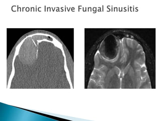
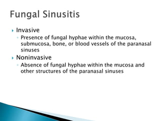
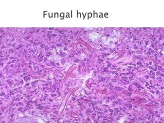
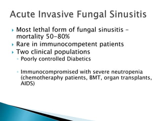
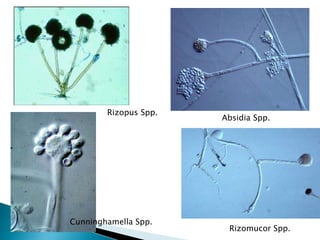
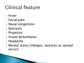
By
Lt Col Saeed Ullah MBBS, FCPS
Classified ENT specialist
CMH Quetta
 Inflammation of the sinuses due to a fungus.
 Direct effect of the fungus or indirect.
 400,000 known fungal species or which 400
are human pathogens and 50 of which cause
systemic or CNS infection
 Clinical presentation, imaging features, and
treatment differ based on type of fungal
sinusitis
 Broadly categorized into invasive and
noninvasive
 Invasive
◦ Presence of fungal hyphae within the mucosa,
submucosa, bone, or blood vessels of the paranasal
sinuses
 Noninvasive
◦ Absence of fungal hyphae within the mucosa and
other structures of the paranasal sinuses
 Most lethal form of fungal sinusitis –
mortality 50-80%
 Rare in immunocompetent patients
 Two clinical populations
◦ Poorly controlled Diabetics
◦ Immunocompromised with severe neutropenia
(chemotheraphy patients, BMT, organ transplants,
AIDS)
Absidia Spp.
Rizomucor Spp.
Rizopus Spp.
Cunninghamella Spp.
 Fever
 Facial pain
 Nasal congestion
 Epistaxis
 Proptosis
 Visual disturbance
 Headache
 Mental status changes, seizures as spread
occurs
 Necrotic nasal septum ulcer (eschar)
 Nasal discharge due to Sinusitis
 Hypertelorism due to rapid orbital spread
 Meningitis due to intracranial spread
 Angioinvasion and hematogenous
dissemination common
 73% of patients with intracranial spread die
 General
 Specific
 Severe nasal cavity soft tissue thickening
 Hypoattenuating mucosal thickening
within lumen of paranasal sinus
 Rapid aggressive bone destruction of
sinus walls
 Fungi can also spread along vessels with
spread beyond the sinus with intact bony
walls
 Intracranial extension
 cavernous sinus thrombosis
 carotid artery invasion or occlusion
 Unilateral ethmoid involvement with bone
destruction, intraorbital spread and proptosis
 Better for evaluating intracranial and
intraorbital extension
◦ Evaluate for inflammatory change in orbital fat and
extraocular muscles
◦ Obliteration of periantral fat is a subtle sign of
extension
◦ Leptomeningeal enhancement progressing to
cerebritis and abscess
Aspergillus involving the sphenoid sinus with invasion of the left cavernous
sinus, thrombosis, extension to the left sylvian fissure and infratemporal
fossa with cerebral infarctions.
Aspergillus in left maxillary sinus with extension anterior and posterior to
the retroantral space. There is diffuse involvement of the muscles of
mastication.
 Aggressive surgical debridement and
systemic antifungal therapy
 Reversal of underlying cause of
immunosuppression if possible
 Recovery from neutropenia is most predictive
of survival
 Intracranial spread is most predictive of
mortality
 Inhaled fungal organisms deposited in nasal
passageways and paranasal sinuses
 Progression over months to years with fungal
organisms invading mucosa, submucosa,
blood vessels, and bony walls
 Organisms – Mucor, Rhizopus, Aspergillus,
Bipolaris, and Candida
 Usually immunocompetent
 History of chronic rhinosinusitis
 Usually persistent and recurrent disease
 Maxillofacial soft tissue swelling
◦ Orbital invasion with proptosis,
◦ Cranial neuropathies
◦ Decreased vision
◦ Invade cribiform plate causing headaches,
seizures, decreased mental status
 Noncontrast CT – soft tissue mass within
one or more of paranasal sinuses, bone
involvement often gives mottled appearance
with or without sclerosis
◦ May mimic malignancy with masslike appearance
and extension beyond sinus confines
 MRI – decreased signal on T1, markedly
decreased signal on T2 weighted images
 Surgical exenteneratin of affected tissues and
systemic antifungal
 Needs aggressive treatment
 Most common form of fungal sinusitis
 Common in warm, humid climates
 Hypersensitivity reaction to inhaled
fungal organisms
 IgE type I immediate hypersensitivity and
type III hypersensitivity are involved
 Common organisms implicated – Bipolaris,
Curvularia, Alternaria, Aspergillus, and
Fusarium
 “Allergic mucin” within affected sinus which is
inspissated mucous the consistency of peanut
butter with eosinophils on histology
 Younger individuals, third decade,
immunocompetent
 Often associated history of atopy with allergic
rhinitis or asthma
 Chronic headaches, nasal congestion, and
chronic sinusitis for years
 Bilateral with multiple sinuses involved.
 Often has a nasal component
 Noncontrast CT – high attenuation allergic
mucin within lumen of sinuses – can mimic
a mucocele
 MRI
 Surgical removal of allergic mucin with
restoration of normal sinus drainage is goal
 Longterm use of topical nasal steroids helps
suppress the immune response and minimize
recurrence
 Topical or systemic antifungals are not
indicated
 Older individuals, female>male
 Immunocompetent
 Asymptomatic or minimal symptoms with
chronic pressure or nasal discharge
 Cacosmia (perception of foul odor when no
such odor exists)
 Mass within the lumen of paranasal sinus
 Frontal sinus most common followed by
sphenoid sinus
 Noncontrast CT – hyperattenuating mass
often with punctate calcifications
 High density material with thickened walls of
the maxillary sinus due to chronic
inflammation
 Surgical Removal with restoration of drainage
of the sinus
 Antifungal medications usually unnecessary
 Recurrence is rare
 It is the failed containment of the
cerebrospinal fluid in the subarachnoid
compartment.
 It indicates a communication with the
subarachnoid space & therefore an opening
of the arachnoid, the dura and the bone to
permit exit of the CSF through the nose.
 The actual loss of CSF is of no particular
consequence
 The persistent dural fistula represents a
persistent hazard for a potentially fatal
purulent meningitis
 Persistent CSF rhinorrhea is therefore an
absolute indication for a surgical repair of the
leak.
 Anterior
 Middle
 Posterior
 Normal CSF
pressure is 40 mm
in infants & 140
mm in adults.
Traumatic
 Accidental
◦ Acute
◦ Delayed
 Iatrogenic
◦ Acute
◦ Delayed
Non traumatic
 High pressure
 Tumours (direct/ indirect effect )
 Hydrocephalus
 Normal pressure
 Congenital anomalies
 Focal atrophy of olfactory/sellar area
 Osteomyelitic erosion
 Idiopathic
 80 % secondary to head trauma with
associated skull base fractures
 16% operations on nose, paranasal sinuses
and skull base.
 Mostly occur through anterior cranial fossa.
Anterior cranial fossa
 Cribriform plate (commonest )
 Fovea ethmoidalis
 Posterior wall of frontal sinus
Middle cranial fossa fractures
 Sphenoid sinus
 Eustachian tube
Posterior fossa
 Clivus
 Petrous temporal bone
 Uncommon
 Mostly in adults
 4th decade
 ♂ : ♀ ratio is 1:2
 May occur after an episode of coughing,
sneezing or straining.
 Arise from the cribriform area in 75 % of
cases
 They act as a safety valve to decrease the
raised ICP
 84% are associated with slow growing intra
cranial tumours (Pituitary neoplasms are the
commonest)
 16 % are related to hydrocephalus
 Mostly are from the cribriform area and the
sella turcica but may be from the middle
fossa.
 90 % are due to potential congenital pathways
 10 % are due to direct erosion of skull base
 Is the fluid CSF?
 Cause of leakage
 Site of leakage
Any persistant rhinorrhea should be
considered CSF until proved otherwise.
Patient with recurrent pneumococcal
meningitis
Bending the head forward will increase the
rate of flow
Headache
Salty taste
Anosmia
Associated Symptoms
Reservoir sign:
After being supine for sometime the patient
is brought in an upright position, with the
neck flexed. A sudden rush of clear fluid is
indicative of CSF fistulae.
 Hankerchief Test:
◦ Fluid in rhinitis contains mucous which stifins while
CSF doesnot.
 Halo Sign:
◦ When CSF rhinorrhea is blood stained it dries out
with a central blood stain surrounded by a clear
ring.
 Nasal endoscopy
◦ with or without intrathecal floresein for leak
presence or localization
 Biochemistry/ Immunochemistry
◦ Estimation of glucose, proteins and electrolytes can
be done.
◦ A concentration of 30mg/dl or 1.6mmol/l of
glucose is considered confirmatory of CSF
 β-2 Transferrin
 Radiology plays the key role
 Bone defects, air fluid levels and erosions can
be seen.
 Plain X rays
◦ Pneumocephalus/ air fluid levels
 CT scan in axial/ coronal views
◦ Skull Base fractures, CSF fistulae
 Isotope studies
In case of inactive, intermittent, small or
doubtful leak, CT scan with contrast will not
reveal the leak. In such cases radio nuclied
cisternography is more effective. Indium III-
DPTA is generally used.
Intrathecal dyes
◦ Intrathecal floreciene with nasal endoscopes are
used for anterior fossa leaks
 Management consists of cooperation between
Neurosurgeon
Neuroradiologist
Otolaryngologist
depending upon severity, etiology, extent of
injury & anatomical site of leak.
 Treatment can be divided into
Medical &
Surgical
 Majority of acute traumatic leaks heal
spontaneously.
 Bed rest in head up position
 Avoiding coughing, sneezing, nose blowing &
straining.
 Drugs to decrease spinal fluid production like
acetazolamide and frusemide.
 Repeated removal of CSF via lumbar taps or
an indwelling lumbar subarachnoid drain.
 Antibiotics
 Intracranial approach
 Extra cranial approach
 Endoscopic repair
Fungal rhinosinusitis, Qims

Fungal rhinosinusitis, Qims

  • 3.
    By Lt Col SaeedUllah MBBS, FCPS Classified ENT specialist CMH Quetta
  • 5.
     Inflammation ofthe sinuses due to a fungus.  Direct effect of the fungus or indirect.
  • 6.
     400,000 knownfungal species or which 400 are human pathogens and 50 of which cause systemic or CNS infection  Clinical presentation, imaging features, and treatment differ based on type of fungal sinusitis  Broadly categorized into invasive and noninvasive
  • 7.
     Invasive ◦ Presenceof fungal hyphae within the mucosa, submucosa, bone, or blood vessels of the paranasal sinuses  Noninvasive ◦ Absence of fungal hyphae within the mucosa and other structures of the paranasal sinuses
  • 9.
     Most lethalform of fungal sinusitis – mortality 50-80%  Rare in immunocompetent patients  Two clinical populations ◦ Poorly controlled Diabetics ◦ Immunocompromised with severe neutropenia (chemotheraphy patients, BMT, organ transplants, AIDS)
  • 10.
    Absidia Spp. Rizomucor Spp. RizopusSpp. Cunninghamella Spp.
  • 11.
     Fever  Facialpain  Nasal congestion  Epistaxis  Proptosis  Visual disturbance  Headache  Mental status changes, seizures as spread occurs
  • 12.
     Necrotic nasalseptum ulcer (eschar)  Nasal discharge due to Sinusitis  Hypertelorism due to rapid orbital spread  Meningitis due to intracranial spread  Angioinvasion and hematogenous dissemination common  73% of patients with intracranial spread die
  • 16.
  • 17.
     Severe nasalcavity soft tissue thickening  Hypoattenuating mucosal thickening within lumen of paranasal sinus  Rapid aggressive bone destruction of sinus walls  Fungi can also spread along vessels with spread beyond the sinus with intact bony walls  Intracranial extension  cavernous sinus thrombosis  carotid artery invasion or occlusion
  • 18.
     Unilateral ethmoidinvolvement with bone destruction, intraorbital spread and proptosis
  • 19.
     Better forevaluating intracranial and intraorbital extension ◦ Evaluate for inflammatory change in orbital fat and extraocular muscles ◦ Obliteration of periantral fat is a subtle sign of extension ◦ Leptomeningeal enhancement progressing to cerebritis and abscess
  • 20.
    Aspergillus involving thesphenoid sinus with invasion of the left cavernous sinus, thrombosis, extension to the left sylvian fissure and infratemporal fossa with cerebral infarctions.
  • 21.
    Aspergillus in leftmaxillary sinus with extension anterior and posterior to the retroantral space. There is diffuse involvement of the muscles of mastication.
  • 22.
     Aggressive surgicaldebridement and systemic antifungal therapy  Reversal of underlying cause of immunosuppression if possible  Recovery from neutropenia is most predictive of survival  Intracranial spread is most predictive of mortality
  • 23.
     Inhaled fungalorganisms deposited in nasal passageways and paranasal sinuses  Progression over months to years with fungal organisms invading mucosa, submucosa, blood vessels, and bony walls  Organisms – Mucor, Rhizopus, Aspergillus, Bipolaris, and Candida
  • 24.
     Usually immunocompetent History of chronic rhinosinusitis  Usually persistent and recurrent disease  Maxillofacial soft tissue swelling ◦ Orbital invasion with proptosis, ◦ Cranial neuropathies ◦ Decreased vision ◦ Invade cribiform plate causing headaches, seizures, decreased mental status
  • 25.
     Noncontrast CT– soft tissue mass within one or more of paranasal sinuses, bone involvement often gives mottled appearance with or without sclerosis ◦ May mimic malignancy with masslike appearance and extension beyond sinus confines  MRI – decreased signal on T1, markedly decreased signal on T2 weighted images
  • 27.
     Surgical exenteneratinof affected tissues and systemic antifungal  Needs aggressive treatment
  • 28.
     Most commonform of fungal sinusitis  Common in warm, humid climates  Hypersensitivity reaction to inhaled fungal organisms  IgE type I immediate hypersensitivity and type III hypersensitivity are involved
  • 29.
     Common organismsimplicated – Bipolaris, Curvularia, Alternaria, Aspergillus, and Fusarium  “Allergic mucin” within affected sinus which is inspissated mucous the consistency of peanut butter with eosinophils on histology
  • 30.
     Younger individuals,third decade, immunocompetent  Often associated history of atopy with allergic rhinitis or asthma  Chronic headaches, nasal congestion, and chronic sinusitis for years
  • 31.
     Bilateral withmultiple sinuses involved.  Often has a nasal component  Noncontrast CT – high attenuation allergic mucin within lumen of sinuses – can mimic a mucocele  MRI
  • 33.
     Surgical removalof allergic mucin with restoration of normal sinus drainage is goal  Longterm use of topical nasal steroids helps suppress the immune response and minimize recurrence  Topical or systemic antifungals are not indicated
  • 34.
     Older individuals,female>male  Immunocompetent  Asymptomatic or minimal symptoms with chronic pressure or nasal discharge  Cacosmia (perception of foul odor when no such odor exists)
  • 35.
     Mass withinthe lumen of paranasal sinus  Frontal sinus most common followed by sphenoid sinus  Noncontrast CT – hyperattenuating mass often with punctate calcifications
  • 36.
     High densitymaterial with thickened walls of the maxillary sinus due to chronic inflammation
  • 37.
     Surgical Removalwith restoration of drainage of the sinus  Antifungal medications usually unnecessary  Recurrence is rare
  • 40.
     It isthe failed containment of the cerebrospinal fluid in the subarachnoid compartment.  It indicates a communication with the subarachnoid space & therefore an opening of the arachnoid, the dura and the bone to permit exit of the CSF through the nose.
  • 42.
     The actualloss of CSF is of no particular consequence  The persistent dural fistula represents a persistent hazard for a potentially fatal purulent meningitis  Persistent CSF rhinorrhea is therefore an absolute indication for a surgical repair of the leak.
  • 43.
  • 45.
     Normal CSF pressureis 40 mm in infants & 140 mm in adults.
  • 46.
    Traumatic  Accidental ◦ Acute ◦Delayed  Iatrogenic ◦ Acute ◦ Delayed
  • 47.
    Non traumatic  Highpressure  Tumours (direct/ indirect effect )  Hydrocephalus  Normal pressure  Congenital anomalies  Focal atrophy of olfactory/sellar area  Osteomyelitic erosion  Idiopathic
  • 48.
     80 %secondary to head trauma with associated skull base fractures  16% operations on nose, paranasal sinuses and skull base.  Mostly occur through anterior cranial fossa.
  • 49.
    Anterior cranial fossa Cribriform plate (commonest )  Fovea ethmoidalis  Posterior wall of frontal sinus
  • 50.
    Middle cranial fossafractures  Sphenoid sinus  Eustachian tube
  • 51.
    Posterior fossa  Clivus Petrous temporal bone
  • 52.
     Uncommon  Mostlyin adults  4th decade  ♂ : ♀ ratio is 1:2  May occur after an episode of coughing, sneezing or straining.
  • 53.
     Arise fromthe cribriform area in 75 % of cases  They act as a safety valve to decrease the raised ICP  84% are associated with slow growing intra cranial tumours (Pituitary neoplasms are the commonest)  16 % are related to hydrocephalus
  • 54.
     Mostly arefrom the cribriform area and the sella turcica but may be from the middle fossa.  90 % are due to potential congenital pathways  10 % are due to direct erosion of skull base
  • 55.
     Is thefluid CSF?  Cause of leakage  Site of leakage
  • 56.
    Any persistant rhinorrheashould be considered CSF until proved otherwise. Patient with recurrent pneumococcal meningitis Bending the head forward will increase the rate of flow Headache Salty taste Anosmia Associated Symptoms
  • 57.
    Reservoir sign: After beingsupine for sometime the patient is brought in an upright position, with the neck flexed. A sudden rush of clear fluid is indicative of CSF fistulae.
  • 58.
     Hankerchief Test: ◦Fluid in rhinitis contains mucous which stifins while CSF doesnot.  Halo Sign: ◦ When CSF rhinorrhea is blood stained it dries out with a central blood stain surrounded by a clear ring.  Nasal endoscopy ◦ with or without intrathecal floresein for leak presence or localization
  • 59.
     Biochemistry/ Immunochemistry ◦Estimation of glucose, proteins and electrolytes can be done. ◦ A concentration of 30mg/dl or 1.6mmol/l of glucose is considered confirmatory of CSF  β-2 Transferrin
  • 60.
     Radiology playsthe key role  Bone defects, air fluid levels and erosions can be seen.  Plain X rays ◦ Pneumocephalus/ air fluid levels  CT scan in axial/ coronal views ◦ Skull Base fractures, CSF fistulae
  • 62.
     Isotope studies Incase of inactive, intermittent, small or doubtful leak, CT scan with contrast will not reveal the leak. In such cases radio nuclied cisternography is more effective. Indium III- DPTA is generally used.
  • 64.
    Intrathecal dyes ◦ Intrathecalfloreciene with nasal endoscopes are used for anterior fossa leaks
  • 66.
     Management consistsof cooperation between Neurosurgeon Neuroradiologist Otolaryngologist depending upon severity, etiology, extent of injury & anatomical site of leak.
  • 67.
     Treatment canbe divided into Medical & Surgical
  • 68.
     Majority ofacute traumatic leaks heal spontaneously.  Bed rest in head up position  Avoiding coughing, sneezing, nose blowing & straining.  Drugs to decrease spinal fluid production like acetazolamide and frusemide.  Repeated removal of CSF via lumbar taps or an indwelling lumbar subarachnoid drain.  Antibiotics
  • 69.
     Intracranial approach Extra cranial approach  Endoscopic repair