
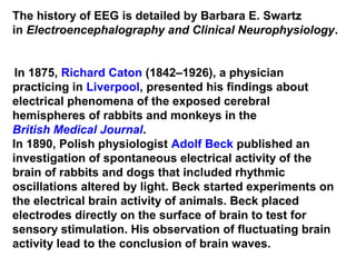
![German physiologist and psychiatrist Hans Berger
(1873–1941) recorded the first human EEG in 1924.
[7] Expanding on work previously conducted on
animals by Richard Caton and others,
Berger also invented the electroencephalogram
(giving the device its name), an invention described
"as one of the most surprising, remarkable, and
momentous developments in the history of clinical
neurology“
His discoveries were first confirmed by British
scientists Edgar Douglas Adrian and B. H. C.
Matthews in 1934 and developed by them.](https://image.slidesharecdn.com/functional-brainwavesfinal-160728165318/85/Functional-brainwaves-final-11-320.jpg)
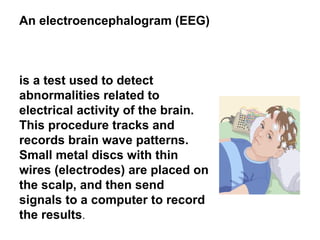
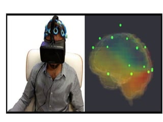
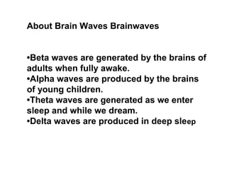
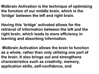


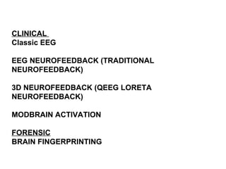
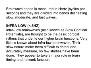
This document discusses brain waves and neurofeedback. It describes the different types of brain waves (delta, theta, alpha, beta, gamma) and what states each is associated with. It then discusses classic EEG which is used to diagnose neurological issues like seizures. Next, it covers neurofeedback (EEG biofeedback) which is used for diagnostics and therapy by teaching patients to regulate their brain waves. It also discusses 3D neurofeedback using LORETA which provides more advanced brain imaging and targeting of networks during neurofeedback training. Midbrain activation and the Shichida method of right brain training is briefly covered.


![PERI-PROSTHETIC FRACTURE NAIL-PLATE CONSTRUCT [NPC].pptx](https://cdn.slidesharecdn.com/ss_thumbnails/drarunkumardrmohamedashrafperiprostheticfrasturenail-plateconstructnpc-260209164459-7e9d15a1-thumbnail.jpg?width=640&height=640&fit=bounds)


