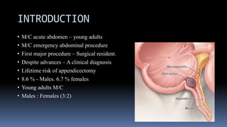
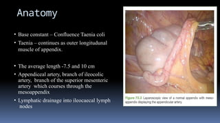
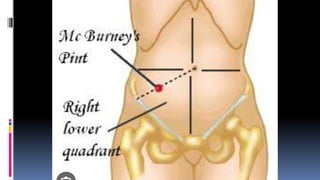
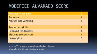
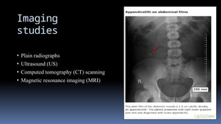
ACUTE APPENDICITIS
DR. S.P. GAYATHRE
PROF AND HEAD
DEPARTMENT OF GENERAL SURGERY
SMCH
INTRODUCTION
• M/C acute abdomen – young adults
• M/C emergency abdominal procedure
• First major procedure – Surgical resident.
• Despite advances – A clinical diagnosis
• Lifetime risk of appendicectomy
• 8.6 % - Males. 6.7 % females
• Young adults M/C
• Males : Females (3:2)
Anatomy
• Base constant – Confluence Taenia coli
• Taenia – continues as outer longitudunal
muscle of appendix.
• The average length -7.5 and 10 cm
• Appendiceal artery, branch of ileocolic
artery, branch of the superior mesenteric
artery which courses through the
mesoappendix
• Lymphatic drainage into ileocaecal lymph
nodes
Laboratory
studies
• Routine Full blood count:
leukocytosis with left shift - 90% of the cases
Higher leukocytosis - with gangrenous and perforated appendicitis
(∼17,000 cells/mm3)
• Urinalysis- to rule out nephrolithiasis
• Pregnancy test- mandatory in child bearing age
• Urea and electrolytes
• C-reactive protein
Migratory RIF pain 1
Anorexia 1
Nausea and vomiting 1
Tenderness (RIF) 2
Rebound tenderness 1
Elevated temperature 1
Leukocytosis 2
MODIFIED ALVARADO SCORE
score of 7 or more: strongly predictive of acute
appendicitis. (5–6): equivocal score
Imaging
studies
• Plain radiographs
• Ultrasound (US)
• Computed tomography (CT) scanning
• Magnetic resonance imaging (MRI)
USG
• An easily compressible appendix <5 mm in diameter generally rules out
appendicitis.
• Features on an ultrasound that suggest appendicitis include
Diameter of greater than 6 mm
Pain with compression,
Presence of an appendicolith
Increased echogenicity of the fat,
Periappendiceal fluid
CT-
scan
• Features on a CT scan that suggest appendicitis include
Enlarged lumen and double wall thickness (greater than 6 mm)
Wall thickening (greater than 2 mm)
Periappendiceal fat stranding
Appendiceal wall thickening and/or
An appendicolith
Management of
appendicitis
• Non-operative management
• Operative management
Non-operative management
– Uncomplicated
appendicitis
• Patients with uncomplicated (absence of appendicolith, perforation or
abscess) appendicitis.
• Bowel rest and intravenous antibiotics, often metronidazole and 3rd
generation
cephalosporin.
• One-quarter of patients initially treated conservatively -
require surgery within 1 year
>40 yrs – followed up – Malignancy risk.
Operative
management
• Open appendectomy -supine position.
• The choice of incision :
Oblique muscle-splitting incision (McArthur-McBurney)
Transverse incision (Rockey-Davis) or
Conservative midline incision
TREATMENT
Appendicectomy
APPROACHES
1. Gridiron incision.
2. Rutherford Morison’s incision.
3. Lanz crease incision.
4.Right lower paramedian incision/lower midline incision.
5. Laparoscopic.
Appendicectom
y
THANK YOU

finalacplendicitissjsjsususisnhruurususjsjeue.pptx

  • 1.
    ACUTE APPENDICITIS DR. S.P.GAYATHRE PROF AND HEAD DEPARTMENT OF GENERAL SURGERY SMCH
  • 2.
    INTRODUCTION • M/C acuteabdomen – young adults • M/C emergency abdominal procedure • First major procedure – Surgical resident. • Despite advances – A clinical diagnosis • Lifetime risk of appendicectomy • 8.6 % - Males. 6.7 % females • Young adults M/C • Males : Females (3:2)
  • 3.
    Anatomy • Base constant– Confluence Taenia coli • Taenia – continues as outer longitudunal muscle of appendix. • The average length -7.5 and 10 cm • Appendiceal artery, branch of ileocolic artery, branch of the superior mesenteric artery which courses through the mesoappendix • Lymphatic drainage into ileocaecal lymph nodes
  • 6.
    Laboratory studies • Routine Fullblood count: leukocytosis with left shift - 90% of the cases Higher leukocytosis - with gangrenous and perforated appendicitis (∼17,000 cells/mm3) • Urinalysis- to rule out nephrolithiasis • Pregnancy test- mandatory in child bearing age • Urea and electrolytes • C-reactive protein
  • 7.
    Migratory RIF pain1 Anorexia 1 Nausea and vomiting 1 Tenderness (RIF) 2 Rebound tenderness 1 Elevated temperature 1 Leukocytosis 2 MODIFIED ALVARADO SCORE score of 7 or more: strongly predictive of acute appendicitis. (5–6): equivocal score
  • 8.
    Imaging studies • Plain radiographs •Ultrasound (US) • Computed tomography (CT) scanning • Magnetic resonance imaging (MRI)
  • 9.
    USG • An easilycompressible appendix <5 mm in diameter generally rules out appendicitis. • Features on an ultrasound that suggest appendicitis include Diameter of greater than 6 mm Pain with compression, Presence of an appendicolith Increased echogenicity of the fat, Periappendiceal fluid
  • 10.
    CT- scan • Features ona CT scan that suggest appendicitis include Enlarged lumen and double wall thickness (greater than 6 mm) Wall thickening (greater than 2 mm) Periappendiceal fat stranding Appendiceal wall thickening and/or An appendicolith
  • 12.
    Management of appendicitis • Non-operativemanagement • Operative management
  • 13.
    Non-operative management – Uncomplicated appendicitis •Patients with uncomplicated (absence of appendicolith, perforation or abscess) appendicitis. • Bowel rest and intravenous antibiotics, often metronidazole and 3rd generation cephalosporin. • One-quarter of patients initially treated conservatively - require surgery within 1 year >40 yrs – followed up – Malignancy risk.
  • 14.
    Operative management • Open appendectomy-supine position. • The choice of incision : Oblique muscle-splitting incision (McArthur-McBurney) Transverse incision (Rockey-Davis) or Conservative midline incision
  • 15.
  • 16.
    APPROACHES 1. Gridiron incision. 2.Rutherford Morison’s incision. 3. Lanz crease incision. 4.Right lower paramedian incision/lower midline incision. 5. Laparoscopic.
  • 17.
  • 18.