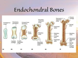
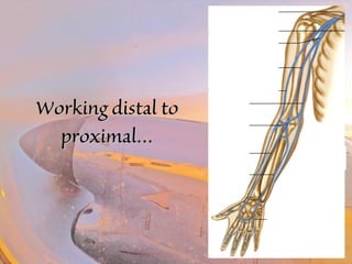
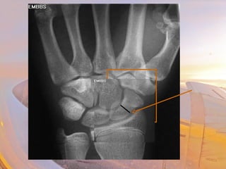
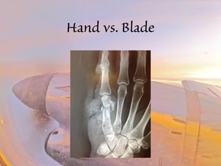
Orthopedics 
101 
101 
V. Bonales, M.D. 
Emergency Medicine Physician 
RMH PCMD
OObbjjeeccttiivveess 
• Physiology of the skeletal system 
• Skeletal anatomy 
• Bone trauma 
• FCA (2 cases)
SSkkeelleettaall SSyysstteemm
DDiivviissiioonnss 
• 206 bones 
–Axial Skeleton 
• Skull, vertebral column, thoracic cage 
–Appendicular Skeleton 
• Clavicle & scapula, upper limbs, pelvic girdle, 
lower limbs
SSkkeelleettaall SSyysstteemm 
• Functions 
–Support and protection 
–Body movement 
–Blood cell formation (hematopoiesis) 
–Storage of salts (CaPO4)
BBoonneess 
• Bone is a connective tissue 
–Made up of calcium phosphate & 
calcium carbonate 
–Cells are called “osteocytes”
OOsstteeooccyyttee
BBoonnee oorrggaanniizzaattiioonn 
• Osteocytes sit in lacunae 
– Communicate via 
canaliculi 
• Lacunae form concentric 
circles around central 
(Haversian) canals 
• Groupings form long 
cylinder groups called 
osteons (Haversian system)
BBoonnee oorrggaanniizzaattiioonn 
• Compact bone 
• Spongy bone
BBoonnee oorrggaanniizzaattiioonn 
• Periosteum 
• Compact bone 
• Spongy bone 
• Endosteum 
• Medullary cavity 
• Marrow 
• N/A/V
BBoonnee oorrggaanniizzaattiioonn 
• Perforating canals 
connect central canals 
–Connect to medullary 
cavity 
• Periosteum covers the 
entire bone
BBoonnee DDeevveellooppmmeenntt -- 
CCaarrttiillaaggee 
• Cells = chondrocytes 
• Covered by perichondrium 
–Provides blood & nutrients to 
avascular cartilage
FFiibbrrooccaarrttiillaaggee 
• Contains collagen 
–Very tough 
• Need vitamin C 
• Shock absorber 
• Cushions the 
vertebrae (discs) & 
knees
EEllaassttiicc CCaarrttiillaaggee 
• Contains elastic 
fibers 
• Flexible 
• Found in ears & 
larynx - epiglottis
Hyaline CCaarrttiillaaggee 
• Most common 
–Ends of bones 
–Tip of nose 
–Rings of the 
respiratory tract 
–Important in bone 
growth
BBoonnee DDeevveellooppmmeenntt 
• Two types 
– Intramembranous – flat bones 
– Endochondral – long bones
EEnnddoocchhoonnddrraall BBoonneess
EEppiipphhyysseeaall PPllaatteess
Bone Strength 
• 120 lbs of pressure to break the femur 
• 15 lbs of pressure to break the humerus 
• 7 lbs of pressure to break a collar bone or rib 
• 5 lbs of pressure to break a wrist
TThhee UUppppeerr EExxttrreemmiittyy
WWoorrkkiinngg ddiissttaall ttoo 
pprrooxxiimmaall……
HHaanndd BBoonneess 
• Phalanges 
• Metacarpals 
• Carpals 
– Scaphoid 
– Lunate 
– Triquetrum 
– Pisiform 
– Trapezoid 
– Trapezium 
– Capitate 
– Hamate
CCaarrppaallss 
• Scaphoid 
–Anatomical snuff box 
–Most often broken with fall (FOOSH) 
–Needs to be cast even if no evidence of a fx 
• Fx will show up 10 days later
HHaanndd CCiirrccuullaattiioonn 
• Redundant circulation 
• Volar arch (deep & superficial) 
– Half from radial artery, half from ulnar artery
Hand vs. Blade
FFoorreeaarrmm BBoonneess 
• Radius 
– Styloid process 
– Head of the radius 
• Ulna 
– Styloid process 
– Olecranon & 
coronoid processes 
– Trochlear notch 
• Olecranon process 
• Coronoid process 
• Trochlear notch
MMeeddiiaann NNeerrvvee 
• Crosses under flexor 
retinaculum & carpal 
ligament to get to hand 
• Irritation of tendons 
w/in this area leads to 
carpal tunnel syndrome
FFoorreeaarrmm CCiirrccuullaattiioonn 
• Radial Artery & Vein 
• Ulnar Artery & Vein 
• Cubital fossa
UUppppeerr AArrmm -- BBoonnee 
• Humerus 
– Trochlea & capitulum 
– Lateral & medial 
epicondyles 
– Coronoid & olecranon 
fossas 
–Greater & lesser tubercles 
– Head
UUppppeerr AArrmm -- CCiirrccuullaattiioonn
UUppppeerr EExxttrreemmiittyy NNeerrvveess
BBrraacchhiiaall PPlleexxuuss 
• Musculocutaneous 
• Axillary 
• Radial 
– Extensors of forearm 
– Sensation to dorsum of arm 
• Ulnar 
– ½ Hand muscles 
– Sensation lateral 1/3 hand 
• Median 
– Flexors of forearm 
– ½ Hand muscles 
– Sensation medial 1/3 hand
??? Questions ???
Spine 
Cervical Spine 
7 vertebrae 
Axis 
Atlas 
Ligaments 
Anterior 
Posterior
Spine Trauma 
Axial Load Injuries
Spine Trauma 
Hyperextension injuries 
Hyperflexion injuries
Spine Trauma
Spine Trauma
Spine Trauma
Spine Trauma 
Hangman’s Fracture 
- Bilateral pedicle Fx 
- Distraction C2-C3 
- Due to complete disruption
Spine Trauma
Spine Trauma
Spine Trauma 
C spine precautions placed when: 
– Any neck discomfort or numbness in extremities 
– Not fully alert or AMS 
– Other painful injuries
Spine Trauma 
Rigid collars, such as the Philadelphia and the extrication collars, 
reduce flexion and extension to about 30% normal and 
rotation and lateral movement to about 50%.
Spine Trauma 
best immobilization method 
– secure patient to a hard board 
– place sandbags at either side of head 
– rigid collar around the neck 
– decreases movement to about 5% of normal.
Spine Trauma 
Thoracic Spine 
12 vertebrae 
Attached to rib cage 
Ligaments 
Anterior 
Posterior
Spine Trauma 
• Axial loading 
– Burst fracture 
– Nerve findings common
Spine Trauma 
• Hyperextension injuries 
– Very unstable fracture
Spine Trauma
Spine Trauma 
• Lumbar Spine 
– 5 vertebrae 
– Fractures require 
significant mechanism
Spine Trauma 
Flexion injuries 
Wedge compression fracture 
Requires a high energy process
Head and Spine Injuries 
Look for motor or sensory 
deficits 
Deficit will point to location of 
injury 
Remember dual innervation of 
cervical cape 
Log roll & keep spine in 
neutral position
???? QQuueessttiioonnss ????
Chest Trauma 
Sternal Fracture 
Indicates worse mechanism of injury 
Not an indicator of cardiac injury
Chest Trauma 
Rib Fractures 
9 - 11 watch for intra-abdominal injuries 
Flail Chest 
Three or more consecutive ribs 
Broken in 2 or more places 
Same side of chest 
Paradoxical Chest Movement
Chest Trauma 
Lung contusions 
More common in children 
Serious cause of morbidity 
Elderly 
COPD
??? Questions ???
Blunt Force Trauma 
Medial Malleolus
Iliac 
Artery & vein 
Femoral 
Artery & vein 
Popliteal 
Artery & vein 
Anterior & posterior 
Tibial Artery & Vein 
Dorsalis pedis artery
• Sciatic nerve 
• Knee flexion 
• Leads to 
• Common peroneal nerve 
• Ankle & toes dorsiflexion 
• “foot drop” 
• Tibial nerve 
• Ankle plantar flexion
• Femur 
• Head 
• Inserts into pelvic bone 
• Neck 
• Most common site of Fx 
• Greater trocanter 
• Lesser trocanter 
• Shaft 
• Medial epicondyle 
• Lateral epicondyle 
• Medial condyle 
• Lateral condyle
• Femoral nerve 
• Knee extension, hip flexion 
• Knee jerk response 
• Femoral triangle 
• Nerve 
• Artery 
• Vein 
• lYmph 
“The NAVY goes toward the water”
• Cartilage 
• Hyaline 
• Fibrocartilage 
• Meniscus 
• Medial & lateral 
• Ligaments 
• Medial collateral 
• Lateral collateral 
• Anterior cruciate 
• Posterior cruciate 
• Patellar
“Unhappy trio” 
• Medial meniscus & ligament attached 
• Anterior cruciate
• Tibia & fibula 
• Medial & lateral condyles 
• Head of fibula 
• Tibial tuberosity 
• Lateral malleolus 
• Medial malleolus 
L 
M
• Mortise joint 
• Medial & lateral 
ligaments 
• Lateral ligament & 
fibular fx
• Tarsals 
• Calcaneus 
• Talus 
• Metatarsals 
• Phalanges
Crush Injury 
occurs when body part subjected to high degree of force 
or pressure
Blunt Force Trauma 
Bleeding 
Bruising 
Fracture 
Laceration 
Compartment Syndrome
Compartment Syndrome 
Not an acute occurrence 
Symptoms: 
Pain out of proportion/passive 
motion 
Parasthesias/Paralysis 
Pallor 
Pulselessness 
Poiklothermia
Blunt Force Trauma 
Compartment Pressures 
Normal about zero; usually less than 10 mm Hg 
Capillary blood flow compromised at >20 mm Hg 
Muscles/nerves at risk for ischemic necrosis >30 to 40 mm Hg. 
Nerve is most sensitive, followed by muscle tissue.
Questions?? 
Thank You!!

Field Care Audit 10/2014

Editor's Notes

  • #46 Type I fracture A type I fracture (less than 5% of cases) is an oblique fracture through the upper part of the odontoid process. This type of fracture occasionally is associated with gross instability due to traction forces applied to, and subsequent injury of, the apical and/or alar ligaments. This is an avulsion injury to the tip of the odontoid and usually is stable. Type II fracture: A type II fracture (more than 60% of cases) is a fracture occurring at the base of the odontoid as it attaches to the body of C2 Type III fracture A type III fracture (30% of cases) occurs when the fracture line extends through the body of the axis. The fracture line can extend laterally into the superior articular facet for the atlas. Another type of odontoid process fracture is a vertical fracture through the odontoid process and body of the axis (less than 5% of cases).
  • #48 traumatic spondylolisthesis for the more common injury pattern usually seen as a result of falls and MVAs. The injury pattern seen secondary to judicial hangings is a fracture-dislocation of C2. More precisely, it is a bilateral pedicle fracture of C2, along with distraction of C2 from C3 secondary to complete disruption of the disk and ligaments between C2 and C3.
  • #56 T10 – T11 fracture dislocation secondary to logging accident
  • #57 50% chance of associated intra-abdominal injury.
  • #59 Wearing seatbelt with no shoulder belt
  • #95 Although not an acute event, should be aware of if managing crush injuries