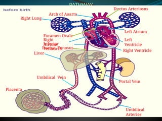
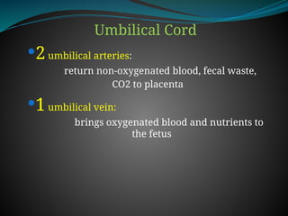
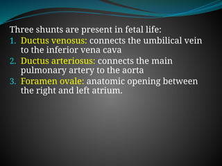
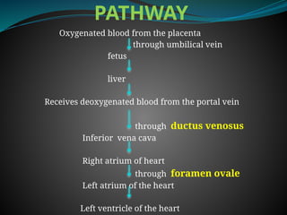
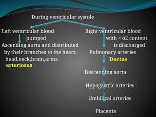
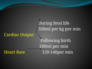
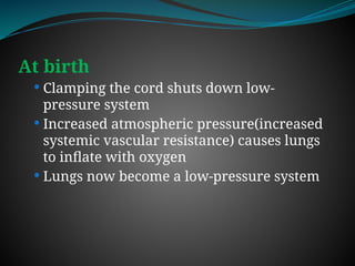
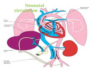
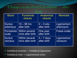
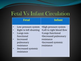
The document discusses fetal circulation, detailing the circulatory system of a human fetus, including the roles of the umbilical cord and placenta. It describes key pathways and structures such as the umbilical veins and arteries, ductus venosus, ductus arteriosus, and foramen ovale, along with the changes that occur in circulation after birth. The transition from a low-pressure to a high-pressure system, as well as the closures of fetal shunts, are also highlighted.


![MANAGEMNT OF JAUNDICE FOR NURSING STUDENTSt[1].ppt](https://cdn.slidesharecdn.com/ss_thumbnails/jaundiceeditedfor2024cohort1-250318173231-642cf8e8-thumbnail.jpg?width=640&height=640&fit=bounds)
![Simulation based learning _Rev[1].ppt JOAB.ppt](https://cdn.slidesharecdn.com/ss_thumbnails/emamatiyafinalrev1-250309104337-a8877e76-thumbnail.jpg?width=640&height=640&fit=bounds)
![INTER-PERSONAL COMMUNICATION IN NURSING [1].pptx](https://cdn.slidesharecdn.com/ss_thumbnails/inter-communication1-250309103945-e946794c-thumbnail.jpg?width=640&height=640&fit=bounds)
![Upper_resp_tract_edited_for_2024_cohort[1].pptx](https://cdn.slidesharecdn.com/ss_thumbnails/upperresptracteditedfor2024cohort1-250225134620-84a86c8f-thumbnail.jpg?width=640&height=640&fit=bounds)
![20971INTRODUCTION_TO_HIV&AIDS for nursing students[1].ppt](https://cdn.slidesharecdn.com/ss_thumbnails/20971introductiontohivaids1-250225134220-578a1188-thumbnail.jpg?width=640&height=640&fit=bounds)

![4. EDUCATIONAL PSYCHOLOGY EDITED[1].pptx](https://cdn.slidesharecdn.com/ss_thumbnails/4-250224085157-3c676047-thumbnail.jpg?width=640&height=640&fit=bounds)
![The Immune system edited for 2024 cohort[1].pptx](https://cdn.slidesharecdn.com/ss_thumbnails/theimmunesystemeditedfor2024cohort1-250224085036-4b3e6a28-thumbnail.jpg?width=640&height=640&fit=bounds)


![Pneumocystis_jirovecii_pneumonia[1].pptx](https://cdn.slidesharecdn.com/ss_thumbnails/malamulo-pneumocystisjiroveciipneumonia1-241127111242-957cc6eb-thumbnail.jpg?width=640&height=640&fit=bounds)
![management of Chest_trauma for nursing [1].ppt](https://cdn.slidesharecdn.com/ss_thumbnails/malamulochesttrauma1-241127110255-71befbaa-thumbnail.jpg?width=640&height=640&fit=bounds)
![Chest_trauma types and management[1].ppt](https://cdn.slidesharecdn.com/ss_thumbnails/malamulochesttrauma1-241126062258-4b388e87-thumbnail.jpg?width=640&height=640&fit=bounds)
![ASTHMA_IN_CHILDREN for NURSING STUDENT[1].pptx](https://cdn.slidesharecdn.com/ss_thumbnails/malamuloasthmainchildren1-241125202930-914bc525-thumbnail.jpg?width=640&height=640&fit=bounds)
![Acute Respiratory Infection Guidelines(ARI)__PRESENTATION[1].pptx](https://cdn.slidesharecdn.com/ss_thumbnails/aripresentation1-241114171345-63d1411f-thumbnail.jpg?width=640&height=640&fit=bounds)
![Acute Respiratory Infections(ARI)__PRESENTATION[1].pptx](https://cdn.slidesharecdn.com/ss_thumbnails/aripresentation1-241020112302-e350d353-thumbnail.jpg?width=640&height=640&fit=bounds)
![ECTOPIC_PREGNANCY_disorders of reproductive organs_1_(3)[1].ppt](https://cdn.slidesharecdn.com/ss_thumbnails/ectopicpregnancyupg131-241020092422-a0e8252f-thumbnail.jpg?width=640&height=640&fit=bounds)
![BURNS assessment and management_GRP_1_PPT[1].pptx](https://cdn.slidesharecdn.com/ss_thumbnails/burnsgrp1ppt1-241020072724-5845af38-thumbnail.jpg?width=640&height=640&fit=bounds)
![Integrated Management -IMNCI_case_management_process[1].pptx](https://cdn.slidesharecdn.com/ss_thumbnails/imncicasemanagementprocess1-241018200546-4cf422ad-thumbnail.jpg?width=640&height=640&fit=bounds)


