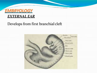
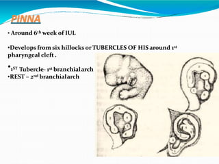
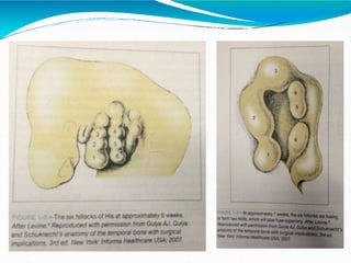
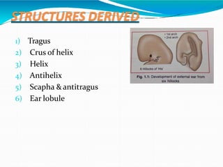
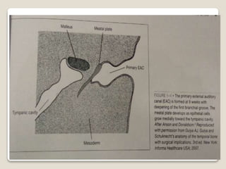
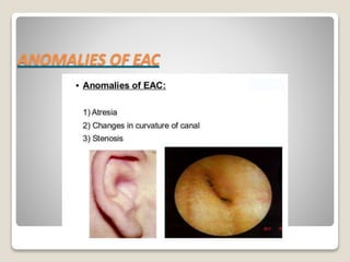
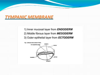
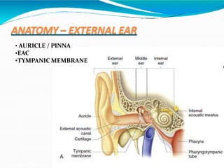
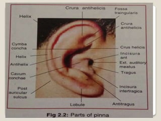
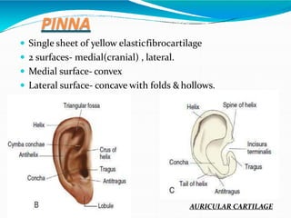
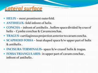
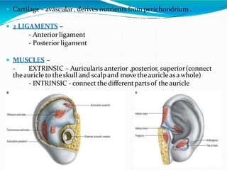
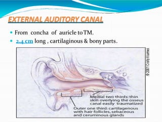
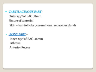
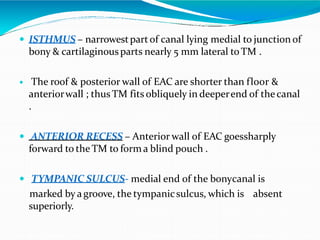
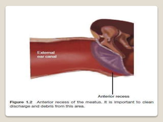
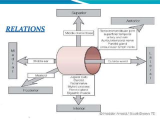
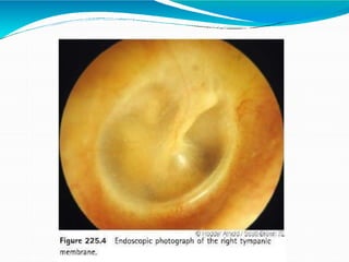
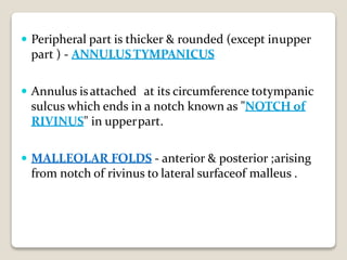
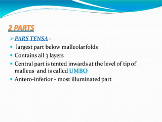
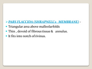
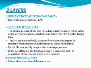
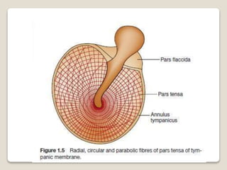
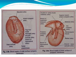
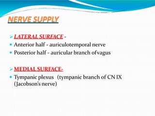
DR MOHAMMED NISHAD N
CONTENTS
 EMBRYOLOGY
 ANOMALIES
 STRUCTURE
 BLOOD SUPPLY
 NERVE SUPPLY
 LYMPHATIC DRAINAGE
 CLINICAL & SURGICAL IMPORTANCE
EMBRYOLOGY
EXTERNAL EAR
Develops from first branchialcleft
PINNA
• Around 6th week of IUL
•Developsfrom six hillocksorTUBERCLES OF HIS around 1st
pharyngeal cleft.
•1ST Tubercle- 1st branchialarch
•REST – 2nd branchialarch
STRUCTURES DERIVED
1) Tragus
2) Crus of helix
3) Helix
4) Antihelix
5) Scapha &antitragus
6) Ear lobule
ANOMALIES OF PINNA
 PRE-AURICULAR SINUS- Defective fusion of tubercles.
 ANOTIA – Failure of development of hillocks.
 BAT EAR DEFORMITY – Defective development of4th
tubercle causing absence of antihelix.
 CRYPTOTIA (hidden or pocket ear) - an abnormality
of the auricle where the upper pole is buried beneath the
temporal skin.
 MICROTIA (diminutive ear)- is usually an isolated
congenital abnormality, but is sometimes associated with
recognized syndromes, e.g. fetal alcohol syndrome,
maternal diabetic syndrome, thalidomide and
isotretinoin exposure.
 POLYOTIA (mirror ear) - is caused by persistence of pre-
auricular tissue that would normally be included in the
pinna, but instead lies in front of the tragus in the
posterior aspect of thecheek.
 STAHL’S EAR - helix is flattened and the upper crus of
the antihelix is duplicated, producing a ridge of cartilage
running from the antihelix to the rim of the helix.
This causes a pointing of the ear and a reversal of the
normal concavity of the scaphoid fossa. Occasionally, the
upper part of the pinna flops over to produce an
appearance known as ‘lopear
CRYPTOTIA
MICROTIA STAHL’S BAR
POLYOTIA LOP EAR
EXTERNAL AUDITORY CANAL
 Develops around the 1st branchial cleft as an
invagination into funnel shaped pit to form primary
EAC.
 Subsequent medial growthwith solid coreof ectoderm
leads to formation of a meatal plate called secondary
EAC.
 Between 8th-10th week of IUL solid core ofepithelium
undergoes canalization form in definitive EAC.
ANOMALIES OF EAC
TYMPANIC MEMBRANE
ANATOMY – EXTERNAL EAR
• AURICLE / PINNA
•EAC
•TYMPANIC MEMBRANE
PINNA
 Single sheet of yellow elasticfibrocartilage
 2 surfaces- medial(cranial) , lateral.
 Medial surface- convex
 Lateral surface- concave with folds &hollows.
AURICULAR CARTILAGE
Lateral surface
 HELIX – most prominent outerfold.
 ANTIHELIX- fold infront of helix.
 CONCHA – infrontof antihelix , hollow spacedivided bycrus of
helix – Cymba conchae & Cavumconchae.
 TRAGUS- cartilaginousprojectionanteriortocavumconcha.
 SCAPHOID FOSSA – boatshaped space b/w upper partof helix
& antihelix .
 INCISURA TERMINALIS- space b/w crusof helix & tragus.
 FOSSATRIANGULARIS- in upperpart of cavum conchae ,
infront of antihelix.
 Cartilage – avascular , derives nutrients from perichondrium .
 2 LIGAMENTS –
- Anterior ligament
- Posterior ligament
 MUSCLES –
- EXTRINSIC – Auricularis anterior ,posterior, superior(connect
theauricle to the skull and scalpand move theauricleasawhole)
- INTRINSIC - connect thedifferentpartsof theauricle
IMPORTANCE
 INCISURA TERMINALIS- This area is devoid of
cartilage , can be used for giving incision for
procedures in ear to avoid post- op
perichondritis .
 LATERAL SURFACE – Skin is firmly
adherent to perichondrium ; so
more prone for frost bite.
 MEDIAL SURFACE – More of subcutaneous
tissue , skin is loosely adherent to
underlying cartilage ; so cysts like
sebaceous cyst are common .
IMPORTANCE
 Stripping the perichondrium from thecartilage, as occurs
following injuries that cause haematoma, can lead to
cartilage necrosis with crumpled up'boxer's ears'.
 Small pieces of skin from the lobule of the pinna are
commonly used for demonstration of lepra bacilli to
confirm the diagnosis of leprosy.
BLOOD SUPPLY
 ARTERIAL SUPPLY – External carotid artery
 Posterior auricularartery
 Anterior auricular branches of superficial temporal
artery
 Superior auricularartery
 VENOUS DRAINAGE- Auricular veins correspond to
the arteries of the auricle. Arteriovenous anastomoses
are numerous in the skin of the auricle and are thought
to be important in the regulation of coretemperature
NERVE SUPPLY
LYMPHATIC DRAINAGE
 The posterioraspectof the pinnadrains to nodesat
the mastoid tip.
 The tragus and upperpartof the pinnadrain into pre-
auricular nodes
 The remainderof the pinnadrains toupperdeep
cervical lymph nodes.
EXTERNAL AUDITORY CANAL
 From concha of auricle toTM.
 2.4 cm long , cartilaginous & bony parts.
 “S” shaped - itsouterpart isdirected upwards, backwards
and medially while its inner part is directed downwards,
forwards and medially.
 Therefore, to see the tympanic membrane, the pinna has to
be pulled upwards, backwardsand laterally soas to bring the
two parts inalignment.
 In the neonate, there is virtually no bony external meatusas
the tympanic bone is not yet developed, and the tympanic
membrane is more horizontally placed so that the auricle
must be gentlydrawn downwardsand backwards for the best
view of the tympanicmembrane.
 CARTILAGINOUS PART -
Outer 1/3rd of EAC , 8mm
Fissure of santorini
Skin – hair follicles , ceruminous , sebaceousglands
• BONY PART–
Inner 2/3rd of EAC ,16mm
Isthmus
Anterior Recess
 ISTHMUS – narrowestpartof canal lying medial to junction of
bony & cartilaginousparts nearly 5 mm lateral toTM .
 The roof & posterior wall of EAC are shorter than floor &
anteriorwall ; thus TM fits obliquely in deeperend of thecanal
.
 ANTERIOR RECESS – Anterior wall of EAC goessharply
forward to the TM to forma blind pouch .
 TYMPANIC SULCUS- medial end of the bonycanal is
marked by agroove, the tympanicsulcus, which is absent
superiorly.
IMPORTANCE
• ANTERIOR RECESS- cmn site for foreign body impaction
lodgement.
• FURUNCULOSIS – outer cartilaginouscanal
• WAX - impaction (deafness,irritation , itching , otalgiaetc)
IMPORTANCE
 Skin lining TM & Bony canal has self cleansing
property due to migrationof keratin layerof
epithelium from drum towards cartilaginousportion
Loss of this property – keratosisobturans
IMPORTANCE
 Irritation of the auricular branch of the vagus in the
external ear by ear wax or syringing may reflexly
produce persistent cough, vomiting or even death due
to sudden cardiac inhibition. On theother hand, mild
stimulationof this nerve may reflexly produce
increased appetite.
 Accumulation of wax in theexternal acoustic meatus is
often a source of excessive itching, although fungal
infection and foreign bodies should be excluded.
Troublesome impaction of large foreign bodies like
seeds, grains, insects is common.
RELATIONS
BLOOD SUPPLY
 ARTERIAL SUPPLY – derived from branches of theexternal
carotid.
 Theauricular branchesof the superficial temporal artery supply
the roof and anteriorportion of thecanal.
 Thedeepauricular branch of the first partof the maxillaryartery
supplies the anterior meatal wall skin and the epithelium of the
outer surface of the tympanicmembrane.
 Theauricular branchesof the posteriorauricularartery pierce the
cartilage of the auricle and supply the posterior portions of the
canal.
 VENOUS DRAINAGE - Theveinsdrain into theexternal jugular
vein, the maxillaryveinsand the pterygoid plexus.
NERVE SUPPLY
 Anterior wall and roof: auriculotemporal (V3).
 Posterior wall and floor: auricular branch of vagus
(CN X).
 Posterior wall of the auditory canal also receives
sensory fibres of CN VII through auricular branch
ofvagus.
 IMP- In herpes zoster oticus, lesions are seen in the
distribution of facial nerve, i.e. concha,
posterior partof tympanic membrane and
postauricularregion.
LYMPHATIC DRAINAGE
 Anterior wall – pre-auricularLN
 Posteriorwall – postauricular LN
 Floor – retroauricularLN
TYMPANIC MEMBRANE
 Thin semi-translucent membrane , pearlywhite in
colour, oval in shape.
 Lies obliquelyatan angleof 55°.
 VD- 10mm ; HD- 9mm
 Inner surface isconvex
 Forms majorityof lateral wall of middleearcavity
 Peripheral part is thicker & rounded (except inupper
part ) - ANNULUSTYMPANICUS
 Annulus isattached at its circumference totympanic
sulcus which ends in a notch known as "NOTCH of
RIVINUS" in upperpart.
 MALLEOLAR FOLDS - anterior & posterior ;arising
from notch of rivinus to lateral surfaceof malleus .
2 PARTS
PARS TENSA -
 largest part below malleolarfolds
 Contains all 3 layers
 Central part is tented inwards at the level of tipof
malleus and is called UMBO
 Antero-inferior - most illuminatedpart
PARS FLACCIDA (SHRAPNELL's MEMBRANE) -
 Triangular area above malleolarfolds
 Thin , devoid of fibroustissue & annulus.
 It fits into notch ofrivinus.
3 LAYERS
1) OUTER CUTICULAR/EPITHELIAL LAYER -
 It iscontinuouswith skin of EAC
2) MIDDLE FIBROUS LAYER -
 The lamina propria of the pars tensa has radially oriented fibres in the
outer layers and circular, parabolic and transverse fibres in the deeper
layer.
 Thisarrangement probablyaccounts forthecomplex pattern of
tympanic membrane displacementduring sound stimulation.
 Radial fibres normally merge with annulustympanicus
 In the pars flaccida, the lamina propria is less marked and the
orientationof thecollagen fibres seems random.
3) INNER MUCOSAL LAYER -
 It is continuouswith middleear mucosa
BLOOD SUPPLY
 ARTERIAL SUPPLY–
 OUTER SURFACE - deepauricular branch of maxillary
artery
 INNER SURFACE -
 Anterior tympanic branch of maxillaryartery
 Posterior tympanic branch of stylomastoidartery
 Inferior tympanicartery , branch of ascending pharyngeal
artery
 Arteria nutricia incudomallea , a twig from middle meningeal
artery
VENOUS DRAINAGE –
 OUTER SURFACE - external jugularvein
 INNER SURFACE - Transverse sinus & venousplexus
around Eustachian tube
NERVE SUPPLY
LATERAL SURFACE -
 Anterior half - auriculotemporal nerve
 Posterior half - auricular branch ofvagus
MEDIAL SURFACE-
 Tympanic plexus (tympanic branch of CN IX
(Jacobson’s nerve)
ReferencEs
 Scott Brown’s otorhinolarynology , head & neck
surgery
 Glasscock-Shambaugh surgery of theear
 Gray’s Anatomy
 Diseases of ENT & HNS – PL Dhingra, Shruti Dhingra
 Textbook of ENT & HNS -P Hazarika, D.R.Nayak,
R.Balakrishnan
THANK YOU

ANATOMY OF EXTERNAL EAR

  • 1.
  • 2.
    CONTENTS  EMBRYOLOGY  ANOMALIES STRUCTURE  BLOOD SUPPLY  NERVE SUPPLY  LYMPHATIC DRAINAGE  CLINICAL & SURGICAL IMPORTANCE
  • 3.
  • 4.
    PINNA • Around 6thweek of IUL •Developsfrom six hillocksorTUBERCLES OF HIS around 1st pharyngeal cleft. •1ST Tubercle- 1st branchialarch •REST – 2nd branchialarch
  • 6.
    STRUCTURES DERIVED 1) Tragus 2)Crus of helix 3) Helix 4) Antihelix 5) Scapha &antitragus 6) Ear lobule
  • 7.
  • 8.
     PRE-AURICULAR SINUS-Defective fusion of tubercles.  ANOTIA – Failure of development of hillocks.  BAT EAR DEFORMITY – Defective development of4th tubercle causing absence of antihelix.  CRYPTOTIA (hidden or pocket ear) - an abnormality of the auricle where the upper pole is buried beneath the temporal skin.  MICROTIA (diminutive ear)- is usually an isolated congenital abnormality, but is sometimes associated with recognized syndromes, e.g. fetal alcohol syndrome, maternal diabetic syndrome, thalidomide and isotretinoin exposure.
  • 9.
     POLYOTIA (mirrorear) - is caused by persistence of pre- auricular tissue that would normally be included in the pinna, but instead lies in front of the tragus in the posterior aspect of thecheek.  STAHL’S EAR - helix is flattened and the upper crus of the antihelix is duplicated, producing a ridge of cartilage running from the antihelix to the rim of the helix. This causes a pointing of the ear and a reversal of the normal concavity of the scaphoid fossa. Occasionally, the upper part of the pinna flops over to produce an appearance known as ‘lopear
  • 10.
  • 11.
    EXTERNAL AUDITORY CANAL Develops around the 1st branchial cleft as an invagination into funnel shaped pit to form primary EAC.  Subsequent medial growthwith solid coreof ectoderm leads to formation of a meatal plate called secondary EAC.  Between 8th-10th week of IUL solid core ofepithelium undergoes canalization form in definitive EAC.
  • 13.
  • 14.
  • 15.
    ANATOMY – EXTERNALEAR • AURICLE / PINNA •EAC •TYMPANIC MEMBRANE
  • 17.
    PINNA  Single sheetof yellow elasticfibrocartilage  2 surfaces- medial(cranial) , lateral.  Medial surface- convex  Lateral surface- concave with folds &hollows. AURICULAR CARTILAGE
  • 18.
    Lateral surface  HELIX– most prominent outerfold.  ANTIHELIX- fold infront of helix.  CONCHA – infrontof antihelix , hollow spacedivided bycrus of helix – Cymba conchae & Cavumconchae.  TRAGUS- cartilaginousprojectionanteriortocavumconcha.  SCAPHOID FOSSA – boatshaped space b/w upper partof helix & antihelix .  INCISURA TERMINALIS- space b/w crusof helix & tragus.  FOSSATRIANGULARIS- in upperpart of cavum conchae , infront of antihelix.
  • 19.
     Cartilage –avascular , derives nutrients from perichondrium .  2 LIGAMENTS – - Anterior ligament - Posterior ligament  MUSCLES – - EXTRINSIC – Auricularis anterior ,posterior, superior(connect theauricle to the skull and scalpand move theauricleasawhole) - INTRINSIC - connect thedifferentpartsof theauricle
  • 20.
    IMPORTANCE  INCISURA TERMINALIS-This area is devoid of cartilage , can be used for giving incision for procedures in ear to avoid post- op perichondritis .  LATERAL SURFACE – Skin is firmly adherent to perichondrium ; so more prone for frost bite.  MEDIAL SURFACE – More of subcutaneous tissue , skin is loosely adherent to underlying cartilage ; so cysts like sebaceous cyst are common .
  • 21.
    IMPORTANCE  Stripping theperichondrium from thecartilage, as occurs following injuries that cause haematoma, can lead to cartilage necrosis with crumpled up'boxer's ears'.  Small pieces of skin from the lobule of the pinna are commonly used for demonstration of lepra bacilli to confirm the diagnosis of leprosy.
  • 22.
    BLOOD SUPPLY  ARTERIALSUPPLY – External carotid artery  Posterior auricularartery  Anterior auricular branches of superficial temporal artery  Superior auricularartery  VENOUS DRAINAGE- Auricular veins correspond to the arteries of the auricle. Arteriovenous anastomoses are numerous in the skin of the auricle and are thought to be important in the regulation of coretemperature
  • 23.
  • 25.
    LYMPHATIC DRAINAGE  Theposterioraspectof the pinnadrains to nodesat the mastoid tip.  The tragus and upperpartof the pinnadrain into pre- auricular nodes  The remainderof the pinnadrains toupperdeep cervical lymph nodes.
  • 27.
    EXTERNAL AUDITORY CANAL From concha of auricle toTM.  2.4 cm long , cartilaginous & bony parts.
  • 28.
     “S” shaped- itsouterpart isdirected upwards, backwards and medially while its inner part is directed downwards, forwards and medially.  Therefore, to see the tympanic membrane, the pinna has to be pulled upwards, backwardsand laterally soas to bring the two parts inalignment.  In the neonate, there is virtually no bony external meatusas the tympanic bone is not yet developed, and the tympanic membrane is more horizontally placed so that the auricle must be gentlydrawn downwardsand backwards for the best view of the tympanicmembrane.
  • 29.
     CARTILAGINOUS PART- Outer 1/3rd of EAC , 8mm Fissure of santorini Skin – hair follicles , ceruminous , sebaceousglands • BONY PART– Inner 2/3rd of EAC ,16mm Isthmus Anterior Recess
  • 30.
     ISTHMUS –narrowestpartof canal lying medial to junction of bony & cartilaginousparts nearly 5 mm lateral toTM .  The roof & posterior wall of EAC are shorter than floor & anteriorwall ; thus TM fits obliquely in deeperend of thecanal .  ANTERIOR RECESS – Anterior wall of EAC goessharply forward to the TM to forma blind pouch .  TYMPANIC SULCUS- medial end of the bonycanal is marked by agroove, the tympanicsulcus, which is absent superiorly.
  • 32.
    IMPORTANCE • ANTERIOR RECESS-cmn site for foreign body impaction lodgement. • FURUNCULOSIS – outer cartilaginouscanal • WAX - impaction (deafness,irritation , itching , otalgiaetc)
  • 33.
    IMPORTANCE  Skin liningTM & Bony canal has self cleansing property due to migrationof keratin layerof epithelium from drum towards cartilaginousportion Loss of this property – keratosisobturans
  • 34.
    IMPORTANCE  Irritation ofthe auricular branch of the vagus in the external ear by ear wax or syringing may reflexly produce persistent cough, vomiting or even death due to sudden cardiac inhibition. On theother hand, mild stimulationof this nerve may reflexly produce increased appetite.  Accumulation of wax in theexternal acoustic meatus is often a source of excessive itching, although fungal infection and foreign bodies should be excluded. Troublesome impaction of large foreign bodies like seeds, grains, insects is common.
  • 35.
  • 36.
    BLOOD SUPPLY  ARTERIALSUPPLY – derived from branches of theexternal carotid.  Theauricular branchesof the superficial temporal artery supply the roof and anteriorportion of thecanal.  Thedeepauricular branch of the first partof the maxillaryartery supplies the anterior meatal wall skin and the epithelium of the outer surface of the tympanicmembrane.  Theauricular branchesof the posteriorauricularartery pierce the cartilage of the auricle and supply the posterior portions of the canal.  VENOUS DRAINAGE - Theveinsdrain into theexternal jugular vein, the maxillaryveinsand the pterygoid plexus.
  • 37.
    NERVE SUPPLY  Anteriorwall and roof: auriculotemporal (V3).  Posterior wall and floor: auricular branch of vagus (CN X).  Posterior wall of the auditory canal also receives sensory fibres of CN VII through auricular branch ofvagus.  IMP- In herpes zoster oticus, lesions are seen in the distribution of facial nerve, i.e. concha, posterior partof tympanic membrane and postauricularregion.
  • 38.
    LYMPHATIC DRAINAGE  Anteriorwall – pre-auricularLN  Posteriorwall – postauricular LN  Floor – retroauricularLN
  • 39.
    TYMPANIC MEMBRANE  Thinsemi-translucent membrane , pearlywhite in colour, oval in shape.  Lies obliquelyatan angleof 55°.  VD- 10mm ; HD- 9mm  Inner surface isconvex  Forms majorityof lateral wall of middleearcavity
  • 43.
     Peripheral partis thicker & rounded (except inupper part ) - ANNULUSTYMPANICUS  Annulus isattached at its circumference totympanic sulcus which ends in a notch known as "NOTCH of RIVINUS" in upperpart.  MALLEOLAR FOLDS - anterior & posterior ;arising from notch of rivinus to lateral surfaceof malleus .
  • 44.
    2 PARTS PARS TENSA-  largest part below malleolarfolds  Contains all 3 layers  Central part is tented inwards at the level of tipof malleus and is called UMBO  Antero-inferior - most illuminatedpart
  • 45.
    PARS FLACCIDA (SHRAPNELL'sMEMBRANE) -  Triangular area above malleolarfolds  Thin , devoid of fibroustissue & annulus.  It fits into notch ofrivinus.
  • 46.
    3 LAYERS 1) OUTERCUTICULAR/EPITHELIAL LAYER -  It iscontinuouswith skin of EAC 2) MIDDLE FIBROUS LAYER -  The lamina propria of the pars tensa has radially oriented fibres in the outer layers and circular, parabolic and transverse fibres in the deeper layer.  Thisarrangement probablyaccounts forthecomplex pattern of tympanic membrane displacementduring sound stimulation.  Radial fibres normally merge with annulustympanicus  In the pars flaccida, the lamina propria is less marked and the orientationof thecollagen fibres seems random. 3) INNER MUCOSAL LAYER -  It is continuouswith middleear mucosa
  • 48.
    BLOOD SUPPLY  ARTERIALSUPPLY–  OUTER SURFACE - deepauricular branch of maxillary artery  INNER SURFACE -  Anterior tympanic branch of maxillaryartery  Posterior tympanic branch of stylomastoidartery  Inferior tympanicartery , branch of ascending pharyngeal artery  Arteria nutricia incudomallea , a twig from middle meningeal artery
  • 50.
    VENOUS DRAINAGE – OUTER SURFACE - external jugularvein  INNER SURFACE - Transverse sinus & venousplexus around Eustachian tube
  • 51.
    NERVE SUPPLY LATERAL SURFACE-  Anterior half - auriculotemporal nerve  Posterior half - auricular branch ofvagus MEDIAL SURFACE-  Tympanic plexus (tympanic branch of CN IX (Jacobson’s nerve)
  • 52.
    ReferencEs  Scott Brown’sotorhinolarynology , head & neck surgery  Glasscock-Shambaugh surgery of theear  Gray’s Anatomy  Diseases of ENT & HNS – PL Dhingra, Shruti Dhingra  Textbook of ENT & HNS -P Hazarika, D.R.Nayak, R.Balakrishnan
  • 53.