VIP Call Girls Tirunelveli Aaradhya 8250192130 Independent Escort Service Tir...
Exotic viral infections and the liver
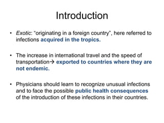
1. Introduction
• Exotic: “originating in a foreign country”, here referred to
infections acquired in the tropics.
• The increase in international travel and the speed of
transportation exported to countries where they are
not endemic.
• Physicians should learn to recognize unusual infections
and to face the possible public health consequences
of the introduction of these infections in their countries.
2. Liver involvement in infections
by exotic viruses
• The liver is often affected:
– Primary target organ
– Only marginally affected
– In very few cases the infection is only
hepatotropic.
3. Exotic viruses and the liver
•
•
•
•
•
•
•
Dengue fever
Yellow fever
Rift Valley fever
Crimean/Congo haemorrhagic fever
Lassa fever
Marburg and Ebola virus diseases
Others
5. Epidemiology of Dengue Fever
The most prevalent mosquito-borne viral
disease. Transmitted by mosquito bite
(usually Aedes aegypti, but trasmission by Aedes
albopictus and Aedes polynesiensis also described).
•
Annual incidence:
• 100 million cases of Dengue fever
• 250.000 cases of Dengue
haemorrhagic fever
• mortality rate of 24,000-25.000
(W vs developing countries)
CDC 2009
7. Epidemiology of Dengue Fever
Population growth, urbanization
and air travel closely tied with
resurgence of epidemic DF and
emergence of DHF in the 20th
century.
•
10.4 % of post-travel systemic febrile illnesses
(second to malaria).
Freedman. N Engl J Med 2006
8. Dengue virus
• RNA virus. Flaviviridae family.
• Four serotypes (DENV 1-4).
– People living in an area of endemic dengue
can be infected with three or four dengue
serotypes during their lifetime.
– Types 2 and 4 more likely to cause inapparent
infections
9. Clinical presentation of Dengue Fever
Incubation period : 3-14 days it can be excluded in a
traveler developing an illness more than 14 days after returning from a
dengue-endemic country.
• Asymptomatic infection
• Self-limited dengue fever
• Dengue haemorrhagic
fever with shock syndrome
Risk of severe
disease is much
higher in
sequential rather
than primary
dengue infection
10. Clinical presentation of Dengue Fever
Classic dengue fever (DF):
– Acute febrile illness rash,
headache and marked
muscle and joint pains.
“break-bone fever”
– Haemorrhagic
manifestations are common
but in rare cases can be
life-threatening.
Dengue haemorrhagic fever (DHF):
Increased
(the most specific and life-threatening
feature of DHF)
Marked
thrombocytopaenia
(<100,000 cells/mm3)
Fever
Haemorrhagic
+/-
World Health Organization
vascular permeability
shock
tendency
Dengue Shock
syndrome (DSS)
11. Controversy regarding classification
system of Dengue Fever
Currently, WHO have adopted a new and easier revised classification:
• Dengue
– Without warning signs.
– With “warning signs”: abdominal pain persistent vomiting,
clinical fluid accumulation, mucosal bleeding, lethargy, liver
enlargement >2 cm or increase in hematocrit and rapid decrease
in platelet count.
• Severe dengue:
– severe plasma leakage,
– severe haemorrhage or
– severe organ impairment (defined as AST or ALT >1000,
impaired consciousness, or severe involvement of the
heart or other organs).
World Health Organization. Geneva 2009
12. Liver involvement in Dengue Fever
• The liver is not the main target organ
hepatic disease has been considered as
unusual manifestation.
• However, clinical and experimental
observations suggest that liver
involvement occurs during dengue
infections.
13. Evidence of liver involvement in
Dengue Fever
Kuo (1992)
270 pt
Souza (2004)
1585 pt
% abnormal AST
% abnormal AST
% abnormal ALT
% abnormal ALT
ALT/AST>10 times
Nimmannitya (1987)
145 pt
Mohan (2000)
37 children
% normal AST
% slightly elevated
ALT
% significantly
elevated ALT
14. Liver involvement in Dengue Fever
Elevation AST is greater than ALT
Nguyen (1997)
45 pt
Souza (2004)
1585 pt
Parkash (2010)
699 pt
Kuo (1992)
270 pt
% abnormal AST
% abnormal ALT
15. Liver involvement in Dengue Fever
Elevation AST is greater than ALT
Differs from viral hepatitis: useful for differential
diagnosis.
The exact significance of this pattern is
uncertain.
It has been suggested that may be due to excess release of AST
form damaged myocytes (Chung 1992).
16. Liver involvement in Dengue Fever
• Aminotransferases peak 9th day after first
symptoms and go back to normal within 3
weeks.
• Liver failure has been documented after
resuscitation from shock, and, in some
cases, may be caused by prolonged
hypotension rather than a direct viral effect
hypoxic hepatitis
17. Liver involvement in Dengue Fever
• Dengue viral antigens within hepatocytes,virus appears
to replicate in both hepatocytes and Kupffer cells
(Huerre 2001).
• Liver cellular apoptosis in vivo and in vitro (Couvelard
1999; Marianneau 1996, 1997).
• Mid-zone and centrilobular necrosis, fatty
alterations, hyperplasia of the Kupffer cells, acidophil
bodies and monocyte infiltrations of the portal tract
with DHF.
18. Liver involvement in Dengue Fever
Mild
and moderate hepatitis are more frequent than severe hepatitis
However,
severe hepatitis appear to be higher in DHF/DSS
Parkash (2010)
Souza (2004)
19. Liver involvement in Dengue Fever
Mild
and moderate hepatitis are more frequent than severe hepatitis
However,
severe hepatitis appear to be higher in DHF/DSS
Wahid (2000)
DF
DF
DHF
DHF
P>0.05
Elevation AST/ALT
Mean levels ALT
20. Liver involvement in Dengue Fever
• Fulminant hepatic failure complicating
severe DHF/DSS has also been
documented, and is associated with a
poor prognosis.
Alvarez and Ramirez-Ronda 1985
Lawn 2003
Munasinghe and Rajasuriya 1967
Nguyen 1997
Subramanian 2005
21. Liver involvement in Dengue Fever
Severe liver dysfunction associated with worst prognosis
Also higher rate of:
•bleeding
•renal failure
•acalculous cholecystitis
699 patients
Parkash (2010)
22. Dengue shock syndrome in a liver
transplant recipient
– 3 weeks after transplant and died 2 days after
admission with DSS.
– Serum quantitative PCR and liver tissue
immunohistochemistry and PCR confirmed
the dengue diagnosis of type 3 dengue virus.
– No documented previous history of dengue
infection
Garcia. Transplantation. 2006
23. Diagnosis of Dengue Fever
• Based mainly on clinical signs and symptoms in
endemic countries.
• Viral isolation and/or serotyping useful for future risk
of DHF in individuals exposed to different serotypes.
• MAC-ELISA: IgM antibodies
– Rapid diagnosis
– Sensitivity and specificity much lower than the HI assay
– In countries with serologic assays available, is the
procedure of choice. If negative and the clinical suspicion
is high, testing paired acute and covalescent serum by HI
or ELISA.
Rothman. Uptodate 2010
26. Epidemiology of Yellow Fever
• First recognized in the 17th Century in Central America.
• The disease occurs in tropical regions of South America and subSaharan Africa.
• Heterologous Flaviviral (previous Dengue) immunity thought to
provide partial protection and low incidence of YF in Asia.
OMS: 200,000 cases of clinical
disease (90% in Africa) and
30,000 deaths per each year
(possibly underestimated).
OMS 1985-99
27. Epidemiology of Yellow Fever
• Flavivirus transmitted by Aedes aegypti (human-to-human
transmission in the absence of the mosquito does not occur).
• Three transmission cycles:
28. Liver involvement in Yellow Fever
• The liver is the main target organ, but other
tissues (especially kidneys) become infected.
• Kupffer cells are initially infected
• Hepatocytes subsequently infected directly via
Kupffer cells or haematogenously.
• Eosinophilic degeneration with Councilman
bodies (apoptosis), rather than by ballooning
and necrosis seen in A-E hepatitis.
29. Liver involvement in Yellow Fever
• In fatal cases, amorphous masses (Torres bodies) or
small, granular, yellow bodies (Villela bodies) can also
be seen. Not diagnostic.
• Liver cells death is due to apoptosis (Councilman
bodies).
– Hepatocytes in the midzone of the lobe express Fas
ligand and lymphocytes infiltrated the liver mediate
apoptosis.
Quaresma. Virology 2006
• There is no disruption of the reticular
architecture of the liver, even in fatal cases.
30. Clinical features in Yellow Fever
• Incubation period: 3-6 days
• Classical illness 3 stages:
– Period of infection
– Period of remission
– Period of intoxication
31. Clinical features in Yellow Fever
– Period of infection
Fever and non specific generalized
malaise, headache, pain in lower
extremities, myalgia, anorexia, vomiting, irritability
and dizzness.
Tongue is characteristically red at the tips and
sides, with white coating in the center.
• Pulse rate is slow relative to the height of the fever
Faget´s sign
• Transaminases start to rise which precede the appearance of
jaundice.
32. Clinical features in Yellow Fever
– Period of remission
• Lasting up 48 h may follow the period of
infection, characterized by abatement of fever and
symptoms.
• Patients with abortive infections recover at this
stage.
• 15% enter the third stage.
33. Clinical features in Yellow Fever
– Period of intoxication
• On the 3th to 6th day after the onset of infection
• Return fever, prostration, nausea, vomiting, epigastric
pain, jaundice, oliguria, albuminuria and a
haemorrhagic diathesis (GI
bleeding, hematuria, metrorrhagia, petechiae...).
• Viremia teminates and neutralising antibodies appear in
the blood.
• Characterized by variable dysfunction of multiple organs:
LIVER, kidneys and cardiovascular system.
• 20-30% of the patients who enter this period die.
34. Liver involvement in Yellow Fever
• AST levels > ALT, probably due to viral injury to the
myocardium and skeletal muscle.
– Useful to distinguish from other viral hepatitis.
• Levels are proportional to disease severity and might
remain elevated for up to 2 months after onset.
Oudart. Bull World Health Organ 1979
• Advanced disease encephalopathy
• Global reductions in clotting factors synthesized by the
liver contribute to bleeding. DIC may also be involved.
35. Yellow fever vs Lábrea Hepatitis
• The Brazilian Amazon Basin:
– highly endemic area: YF,HBV and delta viral hepatitis.
– an unusual type of fulminant hepatic failure Lábrea
hepatitis (LH).
•
LH courses with hepatic failure and
death . Associated with HBV and
delta virus.
Fulminant YF and LH cases may
present a similar clinical course.
36. Liver Histology in fulminant hepatic failure in
northern Brazil: YF and LH
Evaluation 42 samples from fulminant hepatic failure in
order to differentiate YF from LH.
LH
• Extensive necrosis, portal
and hepatic vein phlebitis and
morula cells + defective
pattern of hepatocellular
regeneration and fibrous
portal and acinar
proliferationpossibly
resulting from cumulative
viral cytopathic from livers
already affected by chronic
hepatitis.
YF
Extensive midzonal
apoptosis, hepatocellular
ballooning, protal phlebitis
and remarkable regeration
liver without previous
lesion.
Dias Junior. 2006
37. Liver Histology in fulminant hepatic failure in
northern Brazil: YF and LH
Dias Junior. 2006
38. Diagnosis of Yellow Fever
• Virus isolation
– From the blood or post-mortem liver tissue
• Rapid diagnostic test:
– Detection viral genome by PCR in blood o tissues
– Not widely available
• Detection IgM antibodies (ELISA)
– Simple sample provides a presumptive diagnosis. Confirmation
is made by a rise between paired acute and convalescent
samples or a fall between early and late convalescent samples.
– Cross-reactions with other flaviviruses complicate the diagnosis
particularity in Africa (multiple flaviviruses cocirculate).
39. Vaccine of Yellow Fever
• Liver attenuated virus
• Protective immunity occurs in 90% within 10 days
and in nearly 100% within 3-4 weeks.
• Immunity probably is lifelong after a single
dose, international certificate of immunizations is
valid 10 years, a booster every 10 years.
• Adverse events : 43/100,000
– >70 years 13.4 x
Department of Health and Human Services Centers for Disease Control and Prevention 2010
40. Travelers
• The risk for travelers is determined by multiple
factors: vaccination status, location of
travel, duration exposure…
• 1970-2009: nine cases of YF were reported in
unvaccinated travelers from the United States and
Europe in Africa or South America. Eight died.
• Only one case has been documented in
vaccinated traveller.
42. Rift Valley Fever Epidemiology
•
Zoonotic disease of domestic ruminants
and humans, described in Kenya in 1931.
Outbreaks have
occurred in
subSaharan
Africa and
Egypt (1976)
WHO 2009
43. Rift Valley Fever and epidemiology
• Arbovirus belonging to genus Phlebovirus
(family Bunyaviridae).
• Transmitted by mosquito bites (Aedes mcintoshi as a
main route), aerosol route or by close contact with the
blood or organs of infected animals, especially during
epizootics.
No transmission person-to-person, but
• the blood of patients during the acute
illness is highly infectious.
44. Liver and Rift Valley Fever
• The liver is the main target organ.
– Invasion of the hepatocytes, causing midzonal
hyaline degeneration leading to necrosis and
formation of Councilman bodies.
• Also, invasion of the endothelial cells
vasculitis haemorrhagic phenomena.
45. Clinical manifestations
for Rift Valley Fever
• Incubation period: 2-7 days.
• Most humans: non-specific, self-limiting febrile
illness.
• <5% disease is much more severe 3 different
clinical syndromes:
– Extensive bleeding, hepatic failure, jaundice.
– Encephalitis, confusion and coma.
– Maculo-retinitis (~10%) damage may be permanent
46. Diagnosis in Rift Valley Fever
• Differential diagnosis is simple during epidemics
and when the 3 typical clinical forms occur
together in the same area
(haemorrhagic, diathesis, encephalitis, and
ocular disease).
• Confirmed by the PCR-RNA or detection of
IgM antibodies by ELISA (exclusively in highcontainment laboratories).
• Antibodies appear to 5-10 days after the onset.
47. Treatment
• No specific treatment for either humans or
animals.
• Ribavirin may be effective in the treatment
of RVF (even more in CCHF).
48. Prevention
• During epizootics the movement of animals
from areas where the infection has been
detected should be restricted.
• Two vaccines are avaliable for use in
animals, a live-attenuated and inactivated
vaccine.
• Experimental vaccines in humans.
• Insecticide treatments.
50. Introduction
• First description of human cases in 1940s in Crimea.
In 1956 a virus named
Congo was isolated from a
child in that country. In 1969 it
was demonstrated that the
two viruses were identical:
Crimean/Congo
haemorrhagic fever virus.
•
Severe hemorrhagic fever
with a mortality rate ranging
from 10-80% in different
outbreaks.
51. Crimean/Congo virus and liver involvement
• Genus Nairovirus of the family Bunyaviridae.
• The infection is transmitted by ticks of the genus Hyalomma.
Also, contact with the blood of an infected person during the
acute phase of the disease.
• CHF resembles many other viral haemorrhagic fevers.
• Liver is often involved, with focal necrosis, often localized in
the midzonal areas and with the presence of Councilman bodes.
52. Clinical manifestations of Crimean/Congo virus
• Incubation period: 3-7days
• Mild or asymptomatic infections may occur.
• Abrupt onset with fever, headache, nausea and vomiting.
• In some patients, after 4-5 days, severe hemorrhages develop
with massive bleeding, hypotensive shock and death.
• There may be acute, icteric hepatitis.
53. Diagnosis of Crimean/Congo virus
• Antigen-capture by ELISA or RT-PCR
• Antibodies detection by ELISA.
• Isolation in cell cultures (high-containment
laboratories).
54. Treatment and prevention of
Crimean/Congo virus
• General supportive therapy is the mainstay
• A recent meta-analysis show that there is a very low
quality evidence about Ribavirin being effective but
no clear benefit from the current data.
Soares-Weiser 2010
• Although an inactivated vaccine has been
developed and used on a small scale in Eastern
Europe, there is no safe and effective vaccine
widely available for human use.
56. Introduction
• Both are among the most virulent human
pathogens, causing severe hemorrhagic fever that
resembles fulminant septic shock.
• Fatality rates in epidemics in central
Africa, including a large outbreak of Marburg
hemorrhagic fever in Angola in 2005, have reached
80-90%.
• Bioterrorism: Category A
57. Epidemiology Marburg and Ebola Fever
• Since 1967, when the importation of
infected monkeys from Uganda into
Germany and Yugoslavia resulted in
explosive outbreaks, all cases of filoviral
hemorrhagic fever have occurred in subSaharan Africa.
58. Marburg and Ebola viruses
• RNA viruses. Family Filoviridae.
• Experimentally filoviruses can initiate infection via many
routes, including ingestion, inhalation or passage through
breaks in the skin.
Mahanty. Lancet Infect Dis 2004
• Aerosolized filoviruses are highly infectious for laboratory
animals. However, epidemiologic studies have shown that
these agents rarely spread from person to person by
the respiratory route.
• No evidence that are carried by mosquitoes or other
biting arthropods.
59. Epidemiology Marburg and Ebola Fever
• Study of these virus is extremely difficult, because outbreaks are
sporadic, far from optimal analyses and clearly contagious
•
.
The reservoirs host of the filoviruses is not known (nonhuman
primates are at least as susceptible as humans to rapidly lethal
filoviral HF).
• isolation of Marburg virus from fruit bats in Uganda showed
cosiderable genetic diversity, suggesting that they had long
been present in the bat population.
Towner. PLoS Pathog 2009
60. Pathogenesis Marburg and Ebola
• Systemic inflammatory syndrome by inducing proinflammatory
mediators from infected macrophages and other cells.
• Coagulations defects (induced indirectly)
– Infected macrophages Tissue Factor triggering the extrinsic
coagulation pathway.
• Impairment of adaptive immunity
– Dendritic cells (primary responsibility for the initiation of adaptive
immune responses) major site of filoviral replication
– Lymphocytes remain uninfected, but undergo apoptosis induced by
inflammatory mediators and/or the loss of support signals from dendritic
cells. similar phenomenon is observed in septic shock.
61. Clinical manifestations Marburg and Ebola Fever
• Incubation period: 5-7 days
• Abrupt onset of fever, chills and general malaise and other non
specifics sign and syntoms.
• Conjunctival haemorrhages, easy bruising. Gross bleeding is
common only in moribund patients.
• Conjunctival injection and dark red discoloration of the soft palate
• Nonpruritic maculopapular rash on the upper body is a distinctive
sign of filoviral infection.
62. Liver involvement Marburg and Ebola Fever
• Filoviruses cause multifocal hepatic necrosis.
– Elevated serum AST > ALT
• In Marburg outbreak, transaminases rose rapidly on day 6
to 8 highest in patients who died.
– May also cause a decline in plasma levels of certain
coagulation factors.
• Death usually occurs following a shock and
hepatic failure.
63. Diagnosis Marburg and Ebola Fever
• Rapid diagnostic test: detection of viral
antigens by ELISA or RT-PCR
• Culture: confirms the diagnosis.
64. Treatment Marburg and Ebola Fever
• No specific therapy is available.
• Replacement of blood, coagulation factors, and
platelets, together with careful maintenance of
hydration, are the mainstay of therapy.
• Ribavirin, which inhibits some other RNA viruses, is
ineffective against these agents.
• Proof of concept trials have indicated that Ebola-targeted
siRNAs may be effective as post-exposure prophylaxis in
humans, with 100% of non-human primates surviving a
lethal dose of Zaire Ebola virus, the most lethal strain.
66. Epidemiology of Lassa Fever
• Zoonotic disease of rodents.
•
The first outbreak was in 1969
in Nigeria. Since then, is more
prevalent and less frequently
fatal than was supposed at first
•
Endemic in West Africa
• 300,000 infections and 5000
deaths annually
CDC 2004
67. Epidemiology of Lassa Fever
• Family Arenaviridae.
• Mastomys natalensis, a common rat around human
habitations in Africa, is the host.
• The virus spreads to humans primarily through direct
contact with infected rats or by direct contact with
water or food contaminated by rodent urine.
• Person-to-person transmission occurs.
Mastomys rodent, also
known as the
"multimammate rat".
68. Liver involvement in Lassa Fever
• Lassa virus is pantropic, causing lesions and
dysfunction in multiple organ systems.
• In experimentally infected animals, the highest
concentration of virus is found in the liver.
• There is no correlation between the titre of virus
in blood or liver tissue and the histological liver
damage.
69. Liver involvement in Lassa Fever
• Four principal alterations:
– Focal cytoplasmic degeneration of
hepatocytes
– Randomly distributed multifocal
hepatocellular necrosis with
Councilman bodies
– Monocytic reaction to confluent
necrotic hepatoytes
– Hepatocellular mitoses
70. Clinical manifestations of Lassa Fever
• Incubation period: 7-18 days.
• 80% infections are asymptomatic.
• Similar clinical manifestations to other hemorrhagic
fever viruses, with respiratory distress, hypovolemic
shock, pulmonary oedema, and, sometimes, massive
pleural effusions and ascites rapidly developing during the
2nd week in severe cases.
• The case fatality rate in pregnant women is much higher
than in non-pregnant adults.
71. Laboratory findings of Lassa Fever
• Two reliable predictors of the outcome:
– AST>150UI/L
– High viraemia
78% fatality rate
(vs 17 % in patients who do not
presents these features)
Johnson 1987
72. Diagnosis of Lassa Fever
– ELISA
• Antigen detection is very useful in the early
diagnosis.
• IgM antibodies.
– RT-PCR to detect virus this method is
primarily a research tool.
73. Treatment of Lassa Fever
• Treatment of choice: Ribavirin
– Can reduce mortality, especially when given
intravenously before day 7
– 2 g loading dose followed by 1 gr /6 h x 4
days; then 0.5 g/ 8 h x 6 days.
• Supportive care
74. Other viral haemorrhagic fevers
occasionally affecting the liver
• Chapare
• Flexal
Flavivirus
Argentina
Bolivia
Venezuela
• Sabia
Arenavirus
• Junin virus
• Machupo virus
• Guanarito virus
Brazil
Bolivia
Brazil
• Alkhurma
Egypt
75.
76.
77. Signs and Symptoms of Dengue
Fever
• High fever, up to 105 F (40.6 C)
• A rash over most of your body, can subside after a couple of days
and then reappear
• Sever headache, backache or both
• Pain behind your eyes
• Severe joint and muscle pain (breakbone fever)
• Nausea and vomiting
• Signs and symptoms usually begin about four to seven days after
being bitten by A. aegypti.
• Mild dengue fever rarely causes death, it is usually self-limiting
Mayo clinic, (2010). Dengue fever symptoms. Retrieved April 19, 2010 from
http://www.mayoclinic.com/health/dengue_fever/DS01028/DSECTION=symptoms
78. Signs and Symptoms of Dengue
Hemorrhagic fever (DHF)
• A more severe form of dengue that begins the same way
as dengue fever but become worse after several days
• Significant damage to your blood and lymph vessels
• A decrease in the number of blood cells that help your
blood clot (platelets)
• Bleeding from nose and mouth
• Bleeding under the skin, which gives the appearance of
bruising
• Death
Mayo clinic, (2010). Dengue fever symptoms. Retrieved April 19, 2010 from
http://www.mayoclinic.com/health/dengue_fever/DS01028/DSECTION=symptoms
79. Signs and Symptoms of Dengue
Shock Syndrome (DSS)
• The most severe form of the disease
• It may start with the signs and symptoms of mild dengue
plus
• Severe abdominal pain
• Frequent vomiting
• Disorientation
• Fluid (plasma) leakage from blood vessels
• Heavy bleeding
• A sudden drop in blood pressure (shock)
• Death
Mayo clinic, (2010). Dengue fever symptoms. Retrieved April 19, 2010 from
http://www.mayoclinic.com/health/dengue_fever/DS01028/DSECTION=symptoms
Editor's Notes
These are photos of some examples where water can accumulate, serving as breeding grounds for mosquitoes.
The signs and symptoms of dengue fever are the following: high fever, up to 105 F. A rash over most of your body, can subside after a couple of days and then reappear. Sever headache, backache or both, pain behind your eyes, severe joint and muscle pain, nausea, and vomiting. As mention before the incubation period is from four to seven days. Mild dengue fever rarely causes death, it is usually self-limiting
The more severe clinical manifestation of dengue fever, is known as Dengue hemorrhagic fever. The signs and symptoms begin the same way as the “mild” dengue fever. It will become worse after several days. The dengue hemorrhagic fever can cause significant damage to blood and lymph vessels. A decrease in the number of platelets (blood cells that help your blood clots), and bleeding from nose and mouth. Also bleeding under the skin, which gives the appearance of bruising. If this disease is not properly manage it can lead to death.
The most severe form of dengue is called dengue shock syndrome. It starts with the signs and symptoms of mild dengue. After several days severe abdominal pain, frequent vomiting, disorientation, plasma leakage from blood vessels, heavy bleeding and drop of blood pressure can occur. Hospitalization is necessary because of increase risk of death.