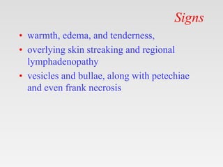
Download to read offline


This document provides tips for using a PowerPoint presentation on erysipelas: 1. The presentation can be freely downloaded, edited, and modified. Blank slides are included to engage students by asking what they know about each topic before presenting additional information. 2. The presentation follows an active learning approach, showing blank slides, asking questions, and then presenting content over three revisions to reinforce learning. 3. The presentation is also useful for self-study, with notes providing references and bibliography.


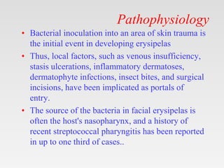
![PERI-PROSTHETIC FRACTURE NAIL-PLATE CONSTRUCT [NPC].pptx](https://cdn.slidesharecdn.com/ss_thumbnails/drarunkumardrmohamedashrafperiprostheticfrasturenail-plateconstructnpc-260209164459-7e9d15a1-thumbnail.jpg?width=640&height=640&fit=bounds)


![ONFH[AVN HIP] -TRIPLE REGIME -A NOVAL SURGICAL CONCEPT .pptx](https://cdn.slidesharecdn.com/ss_thumbnails/onfhavnhip2026koaconcalicutdrgokuldevdrmashraf-260210064517-213ec005-thumbnail.jpg?width=640&height=640&fit=bounds)