Cellulitis
SACHIN DWIVEDI
CLINICAL INSTRUCTOR
COLLEGE OF NURSING, AIIMS RISHIKESH
Introduction
Cellulitis is a common infection of the dermis and
subcutaneous tissues. Cellulitis typically presents
with pain, erythema, oedema, and warmth.
Cellulitis is not transmitted by person-to-person
contact as the epidermis is not involved.
Early diagnosis and treatment are essential to
prevent complications like abscess formation or
bloodstream infection.
Definition
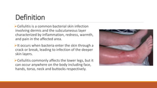
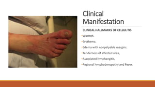
Cellulitis is a common bacterial skin infection
involving dermis and the subcutaneous layer
characterized by inflammation, redness, warmth,
and pain in the affected area.
It occurs when bacteria enter the skin through a
crack or break, leading to infection of the deeper
skin layers.
Cellulitis commonly affects the lower legs, but it
can occur anywhere on the body including face,
hands, torso, neck and buttocks respectively.
Epidemiology
Cellulitis is relatively common, occurring
often in middle-aged and older adults.
Men and women, have a similar incidence
of cellulitis.
There are approximately 50 cases per 1000
patient-years.
Etiological Factor
β hemolytic Streptococcus pyogenes.
Staphylococcus aureus (MRSA).
Risk factors include:
Traumatic injury,
Leg ulcers,
Overweight,
Lymphedema,
Diabetes mellitus,
Vasculitis,
Previous surgery,
Radiotherapy and immunocompromised states.
Pathophysiology
The skin is a protective barrier that stops normal skin flora and other
microbial pathogens reaching the subcutaneous tissue and lymphatic
system
If a skin breakage occurs (Risk factors & Causative agent)normal skin
flora and other bacteria can enter the dermis and subcutaneous tissue.
Leading potentially to an acute infection affecting the deep dermis and
subcutaneous tissue.
Affected area poorly demarcated and spreading erythema along with
pain, swelling, and warmth.
Clinical
Manifestation
CLINICAL HALLMARKS OF CELLULITIS
•Warmth.
•Erythema.
•Edema with nonpalpable margins.
•Tenderness of affected area,
•Associated lymphangitis,
•Regional lymphadenopathy and Fever.
Diagnostic
measures
History-
• Complete history of the presenting illness.
• Any trauma or injuries,
• A history of intravenous drug use, and/or have had insect or
animal bites to the affected area,
• Underline disease condition such as diabetes mellitus,
venous stasis, peripheral vascular disease, chronic tinea
pedis, and lymphedema.
Physical Examination:
• Inspected area to look for any area of skin breakdown.
• The area should be demarcated with a marker to monitor
for continuous spread.
• The area should be palpated to feel for fluctuance that
could indicate the formation of a possible abscess.
• Gently palpate the affected area, be sure to note any
presence of warmth, tenderness, or purulent drainage.
Diagnostic measures
•Blood Examination: raised CRP levels,
•ASO titres in suspected Streptococcal infections,
•X-ray to rule out gas gangrene or underlying osteomyelitis or
necrotizing fasciitis.
• Colour Doppler is done to rule out DVT/ Venous
insufficiency.
•Swab cultures are done to isolate the causative organism.
•Blood cultures to know if bacteraemia is present.
Management
Patients were managed conservatively with
glycerin magnesium-sulfate dressing.
Intravenous antibiotics (inj Cefotaxime/ inj
Gentamycin/ inj Metronidazole/ inj Amikacin)
initially and then as per culture sensitivities.
Surgical debridement with or without skin
grafting and IV antibiotics.
All patients were monitored for response to
treatment and resolution of cellulitis.
Physical Therapy Management
Rest and elevation of the affected limb is
important and can help alleviate pain.
The application of cool, wet, sterile bandages is
also recommended for pain relief, and ice can be
used as well.
Complications
Cellulitis that leads to
Bacteremia,
Endocarditis, or
Osteomyelitis will require a longer duration of antibiotics and possibly
surgery.
Severe sepsis, gangrene, or necrotizing fasciitis.
Preventative Measures:
Compression stockings. Exercise promotion and specific exercises e.g
calf pumps whilst standing in lines etc.
Educate patient about importance of
maintaining good hand hygiene and
adequately clean any future abrasions in their
skin.
References
1.https://www.researchgate.net/publication/318665012_Limb_cellulitis_in_rural_setting_in_In
dia_a_case_control_study
2. Badipatla VN, Gurugubelli SR,Chandra MR, Teja PL, Bade V, Erabati SR. A clinical study of lower
limb cellulitis. Int Surg J 2022;9:1421-5
3. Bailey E, Kroshinsky D. Cellulitis: Diagnosis and Management. Dermatologic Therapy.
2011;24:229–39.http://onlinelibrary.wiley.com/doi/10.1111/j.1529-8019.2011.01398.x/full
(accessed 10 July 2024).
Tsai C-YL, Calvin MK, Chung C, Susan Shin-Jung L, Yao-Shen C, Hung C. Development of a
prediction model for bacteremia in hospitalized adults with cellulitis to aid in the efficient use of
blood cultures: a retrospective cohort study. BMC Infectious Diseases. 2016;16(1):581.
Thank you!

Cellulitis- Causes, Diagnosis and its Management.

  • 1.
  • 2.
    Introduction Cellulitis is acommon infection of the dermis and subcutaneous tissues. Cellulitis typically presents with pain, erythema, oedema, and warmth. Cellulitis is not transmitted by person-to-person contact as the epidermis is not involved. Early diagnosis and treatment are essential to prevent complications like abscess formation or bloodstream infection.
  • 3.
    Definition Cellulitis is acommon bacterial skin infection involving dermis and the subcutaneous layer characterized by inflammation, redness, warmth, and pain in the affected area. It occurs when bacteria enter the skin through a crack or break, leading to infection of the deeper skin layers. Cellulitis commonly affects the lower legs, but it can occur anywhere on the body including face, hands, torso, neck and buttocks respectively.
  • 4.
    Epidemiology Cellulitis is relativelycommon, occurring often in middle-aged and older adults. Men and women, have a similar incidence of cellulitis. There are approximately 50 cases per 1000 patient-years.
  • 5.
    Etiological Factor β hemolyticStreptococcus pyogenes. Staphylococcus aureus (MRSA). Risk factors include: Traumatic injury, Leg ulcers, Overweight, Lymphedema, Diabetes mellitus, Vasculitis, Previous surgery, Radiotherapy and immunocompromised states.
  • 6.
    Pathophysiology The skin isa protective barrier that stops normal skin flora and other microbial pathogens reaching the subcutaneous tissue and lymphatic system If a skin breakage occurs (Risk factors & Causative agent)normal skin flora and other bacteria can enter the dermis and subcutaneous tissue. Leading potentially to an acute infection affecting the deep dermis and subcutaneous tissue. Affected area poorly demarcated and spreading erythema along with pain, swelling, and warmth.
  • 7.
    Clinical Manifestation CLINICAL HALLMARKS OFCELLULITIS •Warmth. •Erythema. •Edema with nonpalpable margins. •Tenderness of affected area, •Associated lymphangitis, •Regional lymphadenopathy and Fever.
  • 8.
    Diagnostic measures History- • Complete historyof the presenting illness. • Any trauma or injuries, • A history of intravenous drug use, and/or have had insect or animal bites to the affected area, • Underline disease condition such as diabetes mellitus, venous stasis, peripheral vascular disease, chronic tinea pedis, and lymphedema. Physical Examination: • Inspected area to look for any area of skin breakdown. • The area should be demarcated with a marker to monitor for continuous spread. • The area should be palpated to feel for fluctuance that could indicate the formation of a possible abscess. • Gently palpate the affected area, be sure to note any presence of warmth, tenderness, or purulent drainage.
  • 9.
    Diagnostic measures •Blood Examination:raised CRP levels, •ASO titres in suspected Streptococcal infections, •X-ray to rule out gas gangrene or underlying osteomyelitis or necrotizing fasciitis. • Colour Doppler is done to rule out DVT/ Venous insufficiency. •Swab cultures are done to isolate the causative organism. •Blood cultures to know if bacteraemia is present.
  • 10.
    Management Patients were managedconservatively with glycerin magnesium-sulfate dressing. Intravenous antibiotics (inj Cefotaxime/ inj Gentamycin/ inj Metronidazole/ inj Amikacin) initially and then as per culture sensitivities. Surgical debridement with or without skin grafting and IV antibiotics. All patients were monitored for response to treatment and resolution of cellulitis.
  • 11.
    Physical Therapy Management Restand elevation of the affected limb is important and can help alleviate pain. The application of cool, wet, sterile bandages is also recommended for pain relief, and ice can be used as well.
  • 12.
    Complications Cellulitis that leadsto Bacteremia, Endocarditis, or Osteomyelitis will require a longer duration of antibiotics and possibly surgery. Severe sepsis, gangrene, or necrotizing fasciitis.
  • 13.
    Preventative Measures: Compression stockings.Exercise promotion and specific exercises e.g calf pumps whilst standing in lines etc. Educate patient about importance of maintaining good hand hygiene and adequately clean any future abrasions in their skin.
  • 14.
    References 1.https://www.researchgate.net/publication/318665012_Limb_cellulitis_in_rural_setting_in_In dia_a_case_control_study 2. Badipatla VN,Gurugubelli SR,Chandra MR, Teja PL, Bade V, Erabati SR. A clinical study of lower limb cellulitis. Int Surg J 2022;9:1421-5 3. Bailey E, Kroshinsky D. Cellulitis: Diagnosis and Management. Dermatologic Therapy. 2011;24:229–39.http://onlinelibrary.wiley.com/doi/10.1111/j.1529-8019.2011.01398.x/full (accessed 10 July 2024). Tsai C-YL, Calvin MK, Chung C, Susan Shin-Jung L, Yao-Shen C, Hung C. Development of a prediction model for bacteremia in hospitalized adults with cellulitis to aid in the efficient use of blood cultures: a retrospective cohort study. BMC Infectious Diseases. 2016;16(1):581.
  • 15.