Endocrine System Pathology_ Ppt Lecture Series (5 in 1)
•
105 likes•13,549 views

1) This document discusses pathology of the endocrine system, focusing on diseases of the pituitary gland, thyroid gland, parathyroid glands, adrenal glands, and diabetes mellitus. 2) Key learning objectives include describing various endocrine tumors, thyroid disorders like Graves' disease and Hashimoto's thyroiditis, parathyroid hypo- and hyperfunction, adrenal hypo- and hyperfunction, and diabetes mellitus. 3) The topics will be presented over 5 lectures covering the major endocrine organs and their diseases.

Recommended
More Related Content
What's hot
What's hot (20)
Similar to Endocrine System Pathology_ Ppt Lecture Series (5 in 1)
Similar to Endocrine System Pathology_ Ppt Lecture Series (5 in 1) (20)
More from Imhotep Virtual Medical School
More from Imhotep Virtual Medical School (20)
Recently uploaded
Recently uploaded (20)
Endocrine System Pathology_ Ppt Lecture Series (5 in 1)
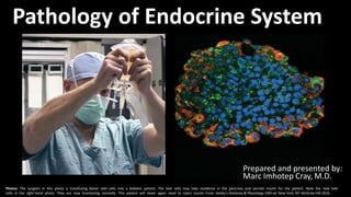
- 1. Pathology of Endocrine System Photos: The surgeon in this photo is transfusing donor islet cells into a diabetic patient. The islet cells may take residence in the pancreas and secrete insulin for the patient. Note the new islet cells in the right-hand photo. They are now functioning normally. This patient will never again need to inject insulin. From: Seeley’s Anatomy & Physiology 10th ed. New York, NY: McGraw-Hill 2010. Prepared and presented by: Marc Imhotep Cray, M.D.
- 2. Marc Imhotep Cray, MD Learning Objectives 2 1. List types of pituitary adenomas and describe their morphology. 2. List the causes of hypopituitarism. 3. Classify thyroiditis and describe the pathogenesis, complications and morphology of Hashimoto’s thyroiditis in particular. 4. Define Graves’ disease and describe its pathogenesis and morphology and correlate with the clinical features.
- 3. Marc Imhotep Cray, MD Learning Objectives cont. 3 5. Describe different types of goiters and their pathology and clinical features. 6. Classify tumors of thyroid gland and describe the morphology and clinical features of thyroid adenoma and thyroid carcinoma 7. Describe causes, pathology, clinical features and complication of parathyroid hypo and hyperfunction. 8. Describe etiology, pathophysiology, clinical features and complications of adrenocortical hyperfunction and hypofunctions.
- 4. Marc Imhotep Cray, MD Learning Objectives cont. 4 9. Describe etiology, pathophysiology , clinical features and complications of adrenomedullary lesions 10. Define Diabetes mellitus (DM), classify it and describe its pathogenesis of the different types. 11. Describe the morphological changes of blood vessels in different organs in DM. 12. List and classify the long-term complications of DM. 13. Describe the primary cause, signs, symptoms and treatment of hypoglycemia and other acute complications of DM.
- 5. Marc Imhotep Cray, MD Topics Outline 5 This sequence will cover the following topics: Lect. 1 Overview and the pituitary gland (hyperpituitarism, hypopituitarism, mass effect as related to pituitary gland lesions, and posterior pituitary gland pathology) Lect. 2 Diseases of the thyroid gland (goiter, hyperthyroidism, hypothyroidism, thyroiditis, and thyroid neoplasms) Lect. 3 Diseases of the parathyroid glands (hyperparathyroidism and hypoparathyroidism) Lect. 4 Diseases of the adrenal glands (hyperadrenalism hypoadrenalism, hyperaldosteronism, and adrenal neoplasms) Lect. 5 Diabetes mellitus (T1DM & T2DM) and complications
- 6. Marc Imhotep Cray, MD Function of Endocrine System and Overview of Endocrine Disease 6 A working knowledge of the pathways that regulate normal hormone levels helps to interpret the symptoms, signs and diagnostic studies in patients being worked up for suspected endocrine disorders.
- 7. Marc Imhotep Cray, MD Function of Endocrine System 7 Main function of endocrine system is communication orchestrates metabolic equilibrium among organs of body Although nervous and endocrine systems use some of same mediators and sometimes overlap functionally (=neuroendocrine integration) endocrine system is unique in its ability to communicate at a distance using soluble mediators= hormones Ultimately, body’s chemical messenger systems(nervous & endocrine) interact with one another to maintain homeostasis
- 8. Marc Imhotep Cray, MD Function of Endocrine System (2) 8 Term hormone (from Greek, horman, “set in motion”) applies to chemicals secreted by “ductless” (i.e., endocrine) glands into circulation carries it to target organ= classic endocrine pathway Many hormones, such as thyroid hormone, corticosteroids and pituitary hormones, fit this definition o Biological messages may also be transmitted by mechanisms other than classic endocrine pathway (see next 2 slides)
- 9. Marc Imhotep Cray, MD Function of Endocrine System (3) 9 Some hormones, such as catecholamines, are produced in multiple sites and act either locally or through circulation Other mediators function only in restricted circulation compartments: e.g., hypothalamic hormones only act on pituitary and reach it via portal tributaries without entering systemic circulation Some hormones exert their effects in very tissues that make them (autocrine), e.g., MIF (Müllerian inhibitory factor) Biological messages of endocrine system may also be transmitted by autocrine, paracrine, neuroendocrine & cytokine modes of communication
- 10. Marc Imhotep Cray, MD Mechanisms of chemically mediated cell-to-cell communication illustrated. 10 Strayer D, et al., eds. Rubin’s Pathology. Clinicopathologic Foundations of Medicine, 6th ed. Baltimore: Wolters Kluwer Health, 2012. Learn more: Molecular and Cell Biology of Endocrine System Ppt.
- 11. Marc Imhotep Cray, MD Function of Endocrine System (5) 11 NB: To qualify as a hormone, a chemical messenger must bind a receptor, whether on cell’s surface or inside (cytoplasm or nucleus) Hormones act either on final effector target or on other glands that in turn produce other hormones For example, thyroid stimulating hormone (TSH) released by pituitary promotes thyroid hormone (TH) secretion by thyroid gland TH, then, directly affects many types of peripheral cells TH will in turn down-regulate activity of pituitary TSH (as well as hypothalamic TRH)= a process known as feedback inhibition
- 12. Marc Imhotep Cray, MD Function of Endocrine System (6) At the core of endocrine system are endocrine organs, include: pituitary, adrenals, thyroid, parathyroids, pancreas & gonads Endocrine glands synthesize and secrete hormones into bloodstream hormones are carried to distant sites to exert their physiologic effects In this way endocrine glands are able to influence function of distant target organs and tissues Disorders of endocrine system are usually due to either overproduction or underproduction of a particular hormone, or mass lesions (mass effect) To aid understanding, these lectures will be presented in a similar scheme 12
- 13. Marc Imhotep Cray, MD Major Endocrine Organs 13Merali Z, Woodfine JD (eds.) Toronto Notes 2016, 33rd Ed. Toronto, Ontario, Canada, 2016.
- 14. Marc Imhotep Cray, MD Overview of Endocrine Disease/Disorders Endocrine system plays an important part in regulation of reproduction, growth and development, maintenance of internal environment, and energy production, utilization and storage Disorders of endocrine system are therefore important b/c they can have far-reaching and devastating effects in some cases can be life-threatening (e.g. thyroid storm, myxedema coma, addisonian crisis, diabetic ketoacidosis, pituitary apoplexy etc.) 14 NB: Study of endocrine diseases requires integration of morphologic findings w biochemical measurements of levels of hormones, their regulators, and other metabolites.
- 15. Marc Imhotep Cray, MD Overview of Endocrine Disease (2) 15 Several processes can disturb normal activity of endocrine system, including (3): 1) impaired synthesis or release of hormones 2) abnormal interactions betw. hormones and their target tissues 3) abnormal responses of target organs Endocrine diseases can be classified as 1) diseases of underproduction or overproduction of hormones and their resulting biochemical and clinical consequences 2) diseases associated w development of mass lesions These lesions might be nonfunctional, or assoc. w overproduction or underproduction of hormones
- 16. Marc Imhotep Cray, MD Overview of Endocrine Disease (3) Tumors (benign & malignant), hyperplasia, or inflammatory lesions of endocrine organs can cause endocrine hypofunction and hyperfunction Pathogenesis is frequently autoimmune and (or) genetic Since hypothalamus and pituitary gland control hormone secretion by many endocrine organs there is potential for lesions at this level to result in abnormal hormone secretion by downstream endocrine organs 16
- 17. Marc Imhotep Cray, MD Overview of Endocrine Disease (4) Identifying root cause of abnormal hormone production is critical for establishing correct diagnosis and managing treatment Simultaneous measurement of conc. of pituitary hormones (e.g., TSH or ACTH) and downstream hormones (e.g., thyroid hormone or cortisol) often allows localization of endocrine abnormality Assessing stimulation or inhibition of hormone release using various pharmacologic agents is also applied 17
- 18. Marc Imhotep Cray, MD Overview of Endocrine Disease (5) Stimulation tests Evaluate hypofunctioning disorders Example—adrenocorticotropic hormone (ACTH)= Cosyntropin stimulation test is used in workup of hypocortisolism Causes of hypofunction • Autoimmune destruction (most common) Examples—Addison’s disease, Hashimoto’s thyroiditis, Grave’s disease • Infarction Example—Sheehan’s postpartum necrosis, Waterhouse- Friderichsen syndrome • Decreased hormone stimulation Example—decreased thyroid- stimulating hormone in hypopituitarism • Enzyme deficiency, infection, neoplasia, congenital disorder 18
- 19. Marc Imhotep Cray, MD Overview of Endocrine Disease (6) Suppression tests Evaluate hyperfunctioning disorders Examples o dexamethasone suppression test evaluates hypercortisolism o saline infusion test evaluations of hyperaldosteronism o glucose tolerance test evaluations GH excess Most hyperfunctioning disorders cannot be suppressed • Notable exceptions prolactinoma and pituitary Cushing's syndrome (= Cushing's Disease) Causes of hyperfunction • Adenoma (most common), acute inflammation, hyperplasia, cancer 19
- 20. Marc Imhotep Cray, MD Overview of Endocrine Disease (7) 20 Also remember, it is important to understand hypothalamic-pituitary axis so you can distinguish 1° from 2° disorders primary diseases are diseases that originate within gland in question e.g., primary hyperthyroidism is due to a defect in thyroid gland), and secondary diseases represent change in one organ as a result of disease in another organ e.g., secondary hyperthyroidism may be due to a TSH-secreting pituitary adenoma
- 21. Marc Imhotep Cray, MD Overview of Endocrine Disease (8) Negative feedback loops Normally, control an increase or decrease in hormone production Example—↑ calcium, ↓ PTH; ↓ calcium, ↑ PTH Hormone synthesis and release are governed at multiple levels typically involves regulation by a pituitary hormone which itself is regulated by a hypothalamic hormone (=releasing factor) This general pathway structure is commonly referred to as a hypothalamic-pituitary-(organ) axis e.g., HPO axis, where O refers to ovary, HPA axis, where A refers to adrenal gland etc. o NB: These various axes represent examples of nervous system-endocrine system integration (or “neuroendocrine systems”) 21
- 22. Marc Imhotep Cray, MD Overview of Endocrine Disease (9) 22 As indicated above, important to understand hypothalamic–pituitary axis so you can distinguish primary from secondary disorders In primary endocrine disturbances, gland itself is malfunctioning (e.g., from tumor, inflammation, enzyme deficiency), but pituitary and hypothalamus are functioning normally and exhibit appropriate response to gland's action o For example, thyroid-stimulating hormone (TSH) is low in Graves disease b/c thyroid is overproducing thyroid hormone (TH) in response to presence of thyroid-stimulating antibody appropriate response is for pituitary to secrete less TSH b/c of feedback inhibition
- 23. Marc Imhotep Cray, MD Overview of Endocrine Disease (10) 23 In a secondary endocrine disturbance, gland is perfectly normal, but pituitary or hypothalamus is malfunctioning For example, if pituitary secretes low or normal levels of TSH in pts w low thyroid hormone levels, then pituitary is malfunctioning b/c it should be secreting higher levels of TSH in response to inadequate levels of TH
- 24. Marc Imhotep Cray, MD Neuroendocrine System (Neuroendocrinology) 24 Neuroendocrine cells receive neuronal input (NTs released by nerve cells or neurosecretory cells) and release message molecules (hormones) into blood In this way they bring about an integration betw. nervous system and endocrine system known as neuroendocrine integration o Example of a neuroendocrine cell is a cell of adrenal medulla which releases Epi & NE (=neuroendocrine hormones) into bld A major center of neuroendocrine integration is hypothalamus and pituitary gland hypothalamic neurosecretory cells release factors (=neuroendocrine hormones) in blood Some of these hormones released at hypothalamic median eminence, control secretion of anterior pituitary hormones, while others (oxytocin & vasopressin) are released directly into blood
- 25. Marc Imhotep Cray, MD Hypothalamic-pituitary axis feedback loops are neuroendocrine integration systems 25 In most cases, a hypothalamic– pituitary–target gland axis is regulated by negative feedback, whereby tropic hormone of anterior pituitary gland has negative feedback effects on hypothalamus and target gland hormone has negative feedback effects on both hypothalamus and anterior pituitary o By way of these mechanisms levels of target gland hormone are maintained within normal physiological range
- 26. 26 Hormones of hypothalamic-pituitary axis McInnis M., Mehta S. Step-up to USMLE Step 1 2015 Ed. Wolters Kluwer, 2015. Major neuroendocrine systems (Hormonal Feedback Regulatory Systems) Individual Axes: Anterior Pituitary Gland Hypothalamic-Pituitary–GH Axis Hypothalamic-Pituitary–Prolactin Axis Hypothalamic-Pituitary–Thyroid Axis Hypothalamic-Pituitary–Adrenal Axis Hypothalamic-Pituitary–Gonadal Axis Posterior Pituitary Gland Antidiuretic Hormone (ADH) & Oxytocin
- 27. Marc Imhotep Cray, MD “the negative feedback principle” It is essential to understand “the negative feedback principle” of hypothalamic /pituitary/ target organ axis A negative feedback mechanism is an example of a negative effect Negative feedback occurs when a product downstream of an axis inhibits production of a reactant by which it is regulated for example, thyroid hormone inhibition of thyroid-stimulating hormone (TSH) Solidlines=positiveeffect Dashedlines=negativeeffect Pazdernik TL, Kerecsen L. Rapid Review Pharmacology, 3rd Ed. Mosby, 2010 27
- 28. Marc Imhotep Cray, MD Example, thyroid hormone feedback loop Brown TA, Brown D. USMLE Step 1 Secrets, 3rd Ed. Saunders, 2013 28 A small reduction of TH triggers a rapid increase of TRH and TSH secretion, resulting in thyroid gland stimulation and increased thyroid hormone production When thyroid hormone reaches a normal level, it feeds back to suppress TRH and TSH, and a new steady state is attained
- 29. Marc Imhotep Cray, MD Example of no feedback loop in Endemic Goiter Iodine deficiency no TH synthesis no feedback >>↑TSH>>follicular hyperplasia Widmaier EP, Raff H & Strang KT. Vander’s Human Physiology : The Mechanisms of Body Function, 11th ed. New York, NY: McGraw-Hill, 2008. 28
- 30. Marc Imhotep Cray, MD The Pituitary Gland 30 Topics discussed Outline: Anatomy: Gross and Microscopic and Hypothalamic–Pituitary Axis Anterior Pituitary Tumors Pituitary Adenomas: General Features Functioning Adenomas and Hyperpituatarism Hypopituitarism Posterior Pituitary Syndromes
- 31. Marc Imhotep Cray, MD Pituitary Gland 31 Anatomy Pituitary gland (hypophysis) a small gland that weighs 0.5-1.0 g and measures 1.3 × 0.9 × 0.5 cm It sits at base of brain in a bony cavity called sella turcica, within sphenoid bone Anatomically, it is composed of two lobes Anterior lobe, or adenohypophysis arises from ectoderm, makes up 80% of gland is populated by epithelial cells Posterior lobe, or neurohypophysis originates from neuroectoderm as a prolongation of hypothalamus
- 32. Marc Imhotep Cray, MD Pituitary gland anatomy illustration 32 Anterior pituitary is formed from an out-pouching of pharyngeal roof and is called Rathke's pouch Posterior pituitary gland arises from an extension of hypothalamic neurons NB: pituitary gland sits in a protective bony enclosure called sella turcica (Turkish chair/saddle) Strayer D, et al., eds. Rubin’s Pathology. Clinicopathologic Foundations of Medicine, 6th ed. Baltimore: Wolters Kluwer Health, 2012.
- 33. 33 Normal pituitary gland, gross Klatt EC. Robbins and Cotran Atlas of Pathology, 3rd Ed. Philadelphia: Saunders, 2015.
- 34. 34 Normal pituitary, microscopic Klatt EC. Robbins and Cotran Atlas of Pathology, 3rd Ed. Philadelphia: Saunders, 2015.
- 35. 35 Normal pituitary, microscopic (2) Klatt EC. Robbins and Cotran Atlas of Pathology, 3rd Ed. Philadelphia: Saunders, 2015.
- 36. Marc Imhotep Cray, MD Pituitary Gland (6) 36 Histologic sections of anterior pituitary reveals cells that contain eosinophilic cytoplasm (acidophil), basophilic cytoplasm (basophil), or poorly staining cytoplasm (chromophobe) cells Note also presence of a fine reticulin network between cells Kumar V, Abbas AK, Aster JC. Robbins and Cotran Pathologic Basis of Disease, 9th ed. Philadelphia: Saunders-Elsevier, 2015. Basophils: FSH, LH, ACTH, TSH (B-FLAT) Acidophils: GH, PRL
- 37. Marc Imhotep Cray, MD Hypothalamic–Pituitary Axis 37 Endocrine function responds to feedback control Hypothalamus, pituitary stalk and pituitary gland constitute an anatomically and functionally integrated “neuroendocrine system” Neuron groups in hypothalamus secrete a number of factors that stimulate anterior pituitary secretion of hypothalamic factors, in turn, are antagonized by hormones secreted by peripheral target organs, thereby completing a feedback loop In addition, specific hypothalamic inhibitory hormones have been identified o For example, dopamine inhibits pituitary secretion of prolactin
- 38. Marc Imhotep Cray, MD Hypothalamic-pituitary axis cont. 38 Hypothalamus regulates secretion of hormones from adenohypophysis (anterior pituitary gland) by releasing stimulatory factors (corticotropin-releasing hormone, CRH; growth hormone-releasing hormone, GHRH; gonadotropin- releasing hormone, GnRH; thyrotropin-releasing hormone TRH, and inhibitory factors (growth hormone inhibitory hormone, GHIH or somatostatin; prolactin inhibitory factor, PIF or dopamine) these in turn modulate release of six hormones from anterior pituitary (next slide)
- 39. Marc Imhotep Cray, MD Anterior pituitary (6 major hormones) 39 Anterior pituitary synthesizes and releases six hormones: 1. Growth hormone (GH): regulated by growth hormone-releasing hormone (GHRH) 2. Prolactin (PRL): inhibited by dopamine from hypothalamus, stimulated by thyrotrophin-releasing hormone (TRH) 3. Adrenocorticotrophic hormone (ACTH): regulated by corticotrophin- releasing hormone (CRH) 4. Thyroid-stimulating hormone (TSH): regulated by TRH 5. Follicle-stimulating hormone (FSH): regulated by gonadotrophin- releasing hormone (GnRH) 6. luteinizing hormone (LH): regulated by GnRH FLAT PiG: FSH, LH, ACTH, TSH, PRL, GH
- 40. Marc Imhotep Cray, MD hypothalamic-hypophysial portal system 40 Hall JE. Guyton and Hall Textbook of Medical Physiology, 13e. Philadelphia: Elsevier , 2016.
- 41. Marc Imhotep Cray, MD Hypothalamic-pituitary axis illustrated 41 Production of pituitary hormones is controlled by positively and negatively acting factors from hypothalamus carried to anterior pituitary by a portal vascular system Kumar V, Abbas AK, Aster JC. Robbins and Cotran Pathologic Basis of Disease, 9th ed. Philadelphia: Saunders-Elsevier, 2015.
- 42. Marc Imhotep Cray, MD Pituitary Gland (7) 42 Posterior pituitary contains pituicytes modified glial cells w no secretory function, axon terminals and unmyelinated nerve fibers containing ADH and oxytocin Both are synthesized in neurons of hypothalamus and transported along axons to neurohypophysis (=stored and released here) o ADH promotes water resorption from distal renal tubules o oxytocin stimulates contraction of pregnant uterus at term and also of cells around lactiferous ducts in breasts
- 43. Marc Imhotep Cray, MD Pituitary Gland (8) 43 Clinical Manifestations of Pituitary Disease Manifestations of pituitary disorders are related to either excess or deficiency of pituitary hormones, or to mass effects Hyperpituitarism: Arising from excess secretion of trophic hormones o Causes include pituitary adenoma, hyperplasia & carcinomas of anterior pituitary, secretion of hormones by nonpituitary tumors, & certain hypothalamic disorders o Symptoms (Sx) of hyperpituitarism are discussed later in context of individual tumors Hypopituitarism: Arising from deficiency of trophic hormones o Caused by destructive processes, including ischemic injury, surgery or radiation, inflammatory reactions, and nonfunctional (silent) pituitary adenomas
- 44. Marc Imhotep Cray, MD Pituitary Gland (9) 44 Local mass effects: Among earliest changes referable to mass effect are radiographic abnormalities of sella turcica, including sellar expansion, bony erosion, and disruption of diaphragma sella o b/c of close proximity of optic nerves & chiasm to sella expanding pituitary lesions often compress decussating fibers in optic chiasm gives rise to visual field abnormalities, classically, defects in both lateral (temporal) visual fields called bitemporal hemianopsia (See next slide) • may also invade cavernous sinus compromising cranial nerves III, IV, and VI o Like any expanding intracranial mass pituitary adenomas can produce signs (Sn) and symptoms (Sx) of elevated intracranial pressure, including headache, vomiting, ocular palsies, altered level of consciousness, back pain and papilledema
- 45. Marc Imhotep Cray, MD Effects of Pituitary Tumors on Visual Apparatus 45 Young WF. The Netter Collection of Medical Illustrations Vol 2- The Endocrine System 2nd Edn. Philadelphia: Saunders, 2011. To learn more study: Endocrinology Tutorial 3_Anterior Pituitary Endocrine Pathology Case 2 SDL Tutorial Graphic shows compression of optic chiasm causing defects in both lateral (temporal) visual fields= bitemporal hemianopsia In addition, a variety of other visual field abnormalities may be caused by asymmetric growth of many tumors
- 46. Marc Imhotep Cray, MD Anterior Pituitary Tumors 46 Most common cause of anterior pituitary disorders are pituitary tumors most are benign adenomas Some produce hormones and result in endocrine abnormalities Others are nonfunctional, but produce mass effects o Non-functioning (or silent) adenomas progressively enlarge until they break out of sella turcica in an upwards direction, compressing optic chiasm Following, we will first discuss general features of pituitary adenomas, then specific tumors
- 47. Marc Imhotep Cray, MD Pituitary Adenomas: General Features 47 Most common cause of hyperpituitarism is a hormone producing adenoma arising in anterior lobe Other, less common, causes include hyperplasia and carcinomas of anterior pituitary; secretion of hormones by some extrapituitary tumors; hypothalamic disorders Main features of pituitary adenomas are: Classified on basis of hormone(s) produced by neoplastic cells detected by immunohistochemical stains of tissue sections (see slide 50)
- 48. Marc Imhotep Cray, MD Pituitary Adenomas: General Features (2) 48 Main features cont. Pituitary adenomas can be functional (hormone producing) or nonfunctioning (not producing hormone) , or silent (i.e., demonstration of hormone production at tissue level only, without clinical manifestations of hormone excess) o Both functional and nonfunctioning pituitary adenomas usually are composed of a single cell type and produce at most a single predominant hormone but there are exceptions • Some pituitary adenomas secrete two different hormones (GH and PRL most common combination)
- 49. Marc Imhotep Cray, MD Pituitary Adenomas: General Features (3) 49 Main features cont. Pituitary adenomas are designated as microadenomas if less than 1 cm in diameter and macroadenomas if they exceed 1 cm Nonfunctioning adenomas more likely to come to clinical attention at a later stage are, therefore, more likely to be macroadenomas o b/c of larger size, nonfunctioning adenomas may encroach upon and destroy adjacent anterior pituitary parenchyma leading to hypopituitarism
- 50. 50 Classification of Pituitary Adenomas Kumar V, Abbas AK, Aster JC. Robbins and Cotran Pathologic Basis of Disease, 9th ed. Philadelphia: Saunders-Elsevier, 2015. Note: PRL producing adenomas (prolactinoma) are most common hormone secreting tumors in both adults and children. Gonadotroph adenomas are more common in elderly.
- 51. Marc Imhotep Cray, MD 51 Kemp WL, Burns DK, Brown TG, Pathology: The Big Picture. New York:McGraw-Hill,2008. Stevens A, Lowe J, Scott I. Core Pathology, 3rd Ed. St. Louis: Mosby-Elsevier, 2009. CT scan (a) of a large pituitary adenoma (A) expanding upwards to compress optic chiasma (arrows) Large pituitary adenoma was an incidental finding at autopsy. As would be suggested by size of tumor, this pituitary adenoma did not secrete any hormones.
- 52. Marc Imhotep Cray, MD Important point regarding pituitary adenomas: Stalk effect 52 Secretion of all of AP hormones, except prolactin, is stimulated by delivery of releasing hormones including TRH, GnRH, and CRH, from hypothalamus via hypophyseal portal system Secretion of PRL, however, is tonically inhibited by delivery of dopamine via same portal system A mass pressing on stalk will prevent dopamine from reaching pituitary gland thus causing increased levels of prolactin without actually producing prolactin o However, level of PRL in “stalk effect” less than that produced by a PRL secreting adenoma This stalk effect will, simultaneously, cause inhibition of secretion of other AP hormones
- 53. Marc Imhotep Cray, MD Pituitary Adenomas: Pathogenesis 53 Several genetic abnormalities associated w pituitary adenomas have been identified: G-protein mutations are one of most common genomic alterations causing pituitary adenomas o G-proteins play a critical role in signal transduction transmitting signals from cell surface receptors e.g. growth hormone-releasing hormone (GHRH) receptor to intracellular effectors (e.g., adenyl cyclase) which then generate second messengers (e.g., cAMP) o G-proteins are heterotrimeric proteins, composed of a specific α- subunit that binds guanine nucleotides and interacts w both cell surface receptors and intracellular effectors • β- and γ-subunits are noncovalently bound to α-subunit
- 54. Marc Imhotep Cray, MD G-protein signaling in endocrine neoplasia 54 Mutations that lead to G-protein hyperactivity are seen in a variety of endocrine neoplasms, including pituitary, thyroid, and parathyroid adenomas G proteins play a critical role in signal transduction, transmitting signals from cell surface receptors (GHRH, TSH or PTH receptor) to intracellular effectors (e.g., adenyl cyclase) which generate second messengers (cAMP) Kumar V, Abbas AK, Aster JC. Robbins and Cotran Pathologic Basis of Disease, 9th ed. Philadelphia: Saunders-Elsevier, 2015. For Hormone-Receptor Interactions and Signal Transduction Mechanisms see Molecular and Cell Biology of Endocrine System : Ppt.
- 55. Marc Imhotep Cray, MD Pituitary Adenomas: Pathogenesis (3) 55 Gs is a stimulatory G protein that has a pivotal role in signal transduction in several endocrine organs, including pituitary o α-subunit of Gs (Gsα) is encoded by GNAS gene o In basal state, Gs exists in an inactive state, w guanosine diphosphate (GDP) bound to guanine nucleotide-binding site of Gsα o On interaction w ligand-bound cell surface receptor, GDP dissociates, and guanosine triphosphate (GTP) binds to Gsα, activating G protein activation of Gsα results in generation of cAMP= a potent mitogenic stimulus in several endocrine cells (e.g., pituitary somatotrophs and corticotrophs, thyroid follicular cells, parathyroid cells) promoting cellular proliferation and hormone synthesis and secretion
- 56. Marc Imhotep Cray, MD Pituitary Adenomas: Pathogenesis (4) 56 Normally, Gsα activation is transient b/c of an intrinsic GTPase activity in α-subunit hydrolyzes GTP into GDP Approx. 40% of somatotroph cell adenomas bear GNAS mutations that nullify GTPase activity of Gsα leading to continual activation of Gsα persistent generation of cAMP unchecked cellular proliferation GNAS mutations also occur in minority of corticotroph adenomas Contrastly, GNAS mutations are absent in thyrotroph, lactotroph, & gonadotroph adenomas b/c these arise from cells whose hypothalamic release hormones do not signal via cAMP-dependent pathways
- 57. 57 Pituitary adenoma, gross and microscopic Monomorphism of these cells contrasts with admixture of cells seen in normal anterior pituitary gland. Note also absence of reticulin network. Massive, nonfunctioning adenoma that has grown far beyond confines of sella turcica and has distorted overlying brain. Kumar V, Abbas AK, Aster JC. Robbins and Cotran Pathologic Basis of Disease, 9th ed. Philadelphia: Saunders-Elsevier, 2015. Two distinctive morphologic features cellular monomorphism and absence of a reticulin network.
- 58. Marc Imhotep Cray, MD Functioning Adenomas & Hyperpituatarism 58 Adenomas arising from different pituitary cells produce hormones characteristic of that cell type and cause clinical syndromes that reflect activity of hormones Lactotroph Adenomas Prolactin-secreting lactotroph adenomas are most frequent type of hyperfunctioning pituitary adenoma accounting for 30% of all clinical cases Hyperprolactinemia causes amenorrhea, galactorrhea, loss of libido, and infertility b/c manifestations of hyperprolactinemia (e.g., amenorrhea) are obvious in premenopausal women prolactinomas are diagnosed at an earlier stage in women of reproductive age than in others w these tumors
- 59. Marc Imhotep Cray, MD Functioning Adenomas & Hyperpituatarism (2) 59 Lactotroph Adenomas cont. Compared to premenopausal women effects of hyperprolactinemia are subtle in men and older women thus, tumor may reach a large size before coming to clinical attention= mass effect Hyperprolactinemia also is a feature of other conditions, including pregnancy, high-dose estrogen therapy, renal failure, hypothyroidism, hypothalamic lesions, and dopamine- inhibiting drugs (e.g., antipsychotic agents)
- 60. Marc Imhotep Cray, MD Functioning Adenomas & Hyperpituatarism (3) 60 Somatotroph Adenomas Growth hormone–secreting somatotroph adenomas are second most common type of functioning pituitary adenoma, and cause gigantism in children or acromegaly in adults b/c clinical manifestations of excess GH may be subtle somatotroph cell adenomas may be quite large by time they come to clinical attention
- 61. Marc Imhotep Cray, MD Physiologic actions of Growth hormone (GH) (somatotropin or STH) 61 Brown TA, Brown D. USMLE Step 1 Secrets, 3rd Ed. Saunders, 2013.
- 62. Marc Imhotep Cray, MD Functioning Adenomas & Hyperpituatarism (4) 62 Somatotroph Adenomas cont. Persistent GH excess stimulates hepatic secretion of insulin-like growth factor 1 (IGF1, somatomedin C), which acts in conjunction w GH to induce overgrowth of bones and muscle If a GH-secreting adenoma develops before epiphyses close, as is case in prepubertal children, result in gigantism o Characterized by a generalized increase in body size, w disproportionately long arms and legs
- 63. Marc Imhotep Cray, MD Functioning Adenomas & Hyperpituatarism (5) 63 Somatotroph Adenomas cont. If elevated levels of GH and IGF1 persist or develop after closure of epiphyses, affected individuals develop acromegaly o growth is most conspicuous in soft tissues, skin, viscera, and bones of face, hands, and feet o Enlargement of jaw results in its protrusion (prognathism), broadening of lower face, and separation of teeth o hands and feet are enlarged, and fingers are broad and sausage-like Strayer D, et al., eds. Rubin’s Pathology. Clinicopathologic Foundations of Medicine, 6th ed. Baltimore: Wolters Kluwer Health, 2012.
- 64. Marc Imhotep Cray, MD Functioning Adenomas & Hyperpituatarism (6) 64 Somatotroph Adenomas cont. Persistent GH excess also is assoc. w metabolic abnormalities most important is diabetes mellitus o DM arises b/c of GH-induced peripheral insulin resistance “blunts” body’s response to elevated glucose levels=GH is diabetogenic • Failure to suppress GH production in response to an oral load of glucose most specific tests to Dx acromegaly • IGF1 provides most sensitive lab test for the Dx of acromegaly Other manifestations of GH excess include gonadal dysfunction, generalized muscle weakness, hypertension, arthritis, congestive heart failure, increased risk for GI cancers
- 65. Marc Imhotep Cray, MD Question A 38-year-old man presents complaining of gradually enlarging hands and feet over past several years. In comparison with a photo from 15 years ago, his facial features have become obviously coarsened. Laboratory evaluation shows mildly elevated plasma glucose, and MRI of brain reveals an enlarged mass in sella turcica. Given suspected diagnosis, specialized testing is performed in which GH levels are measured following administration of an oral glucose load; no measurable decrease is seen. What is the diagnosis? Note: One good way to diagnose this disorder is to look at an old picture of pt. and compare it w patient’s current appearance. Because physical changes take place over decades, family members and friends often do not recognize them. 65
- 66. Marc Imhotep Cray, MD A. A 26-year-old attractive woman prior to acromegaly changes. B. Facial changes 20 years later in the same woman. Note the coarse facial features with large nose, lips, and chin. Protrusion of lower jaw is visible. Usatine RP etal. (Eds.) The Color Atlas of Family Medicine. New York: McGraw-Hill, 2013 66
- 67. Features of acromegaly /gigantism A 22-year-old man w gigantism due to excess GH is shown to left of his identical twin increased height and prognathism (A) and enlarged hand (B) and foot (C) of affected twin are apparent Their clinical features began to diverge at age of approx. 13 years 67
- 68. Marc Imhotep Cray, MD Functioning Adenomas & Hyperpituatarism(10) 68 Corticotroph Adenomas Excess production of ACTH by functioning corticotroph adenomas leads to adrenal hypersecretion of cortisol and development of hypercortisolism (also known as Cushing syndrome) o Cushing syndrome (discussed later w diseases of adrenal gland) may be caused by other conditions as well o When hypercortisolism is caused by excessive production of ACTH by pituitary, it is called Cushing disease after neurosurgeon Harvey Cushing who first described disorder
- 69. Marc Imhotep Cray, MD 69 Functioning Adenomas & Hyperpituatarism(11) Corticotroph Adenomas cont. Most corticotroph adenomas are microadenomas at time of diagnosis o Stain positively w periodic acid–Schiff (PAS) stains due to accumulation of glycosylated ACTH protein o ACTH can also be specifically detected by immunohistochemistry methods
- 70. Marc Imhotep Cray, MD Functioning Adenomas & Hyperpituatarism(12) 70 Corticotroph Adenomas cont. Large, clinically aggressive corticotroph cell adenomas may develop after surgical removal of adrenal glands for Tx of Cushing syndrome o In most cases this condition, known as Nelson syndrome, results from loss of inhibitory effect of adrenal corticosteroids on a preexisting corticotroph microadenoma o b/c adrenals are absent, hypercortisolism does not develop instead, pts present w mass effects of pituitary tumor Note: Pts w Cushing syndrome often have hyperpigmented skin b/c of ↑ production of melanocyte stimulating hormone (MSH) derived from same precursor as ACTH (=pro-opiomelanocortin), so its synthesis accompanies that of ACTH (see slide 195)
- 71. Marc Imhotep Cray, MD Hypopituitarism 71 In hypopituitarism secretion of one or more pituitary hormones is lacking has many causes and various clinical presentations Most often, only one or a few of pituitary hormones are deficient Occasionally, total failure of pituitary function, or panhypopituitarism (pituitary cachexia), occurs Effects of hypopituitarism depend on 1) extent of loss 2) specific hormones involved and 3) age of patient Symptoms usually relate to deficient function of thyroid, adrenal glands and reproductive system Growth retardation and delayed puberty may occur in children
- 72. Marc Imhotep Cray, MD Hypopituitarism (2) Causes 72 Pituitary Tumors: More than half of all cases of hypopituitarism in adults are caused by pituitary tumors, usually adenomas (discussed above) Tumor itself may be functional but symptoms of hypopituitarism result from compression of adjacent tissue by tumor mass
- 73. Marc Imhotep Cray, MD Hypopituitarism (3) Causes 73 Sheehan Syndrome: Panhypopituitarism may be caused by ischemic necrosis of gland, commonly due to severe hypotension caused by intrapartum or postpartum hemorrhage/necrosis enlargement of pituitary reduces its blood flow, rendering it particularly vulnerable Clinical manifestations due at first to loss of gonadotropins, then to subsequent loss of TSH and ACTH Agalactia (lactation failure), amenorrhea, hypothyroidism and adrenocortical insufficiency are important consequences o w modern obstetric care, Sheehan syndrome is rare Note: Occurrence of similar process wo preceding pregnancy, as well as, its occurrence in males is termed Simmond’s disease (=Pituitary cachexia)
- 74. See Endocrinology Tutorial 1_Postpartum Necrosis Young WF. The Netter Collection of Medical Illustrations Vol 2- The Endocrine System 2nd Edn. Philadelphia: Saunders, 2011. Rubin R , Strayer DS Eds. Rubin’s Pathology: Clinicopathologic Foundations of Medicine, 6th Ed. Baltimore: Lippincott Williams & Wilkins, 2012.
- 75. 75 Hypopituitarism, Mild=FSH and LH are usually affected first Buja LM, Krueger GR. Netter’s Illustrated Human Pathology, 2nd Ed. Philadelphia: Saunders-Elsevier, 2014.
- 76. 76 Hypopituitarism, Moderate=TSH and ACTH affected Buja LM, Krueger GR. Netter’s Illustrated Human Pathology, 2nd Ed. Philadelphia: Saunders-Elsevier, 2014.
- 77. Raff RB, Rawls SM, Beyzarov EP. Netter's Illustrated Pharmacology, Updated Edition. Philadelphia: Saunders-Elsevier, 2014. 77 Hypopituitarism: Severe=Frank TSH and ACTH deficiency
- 78. 78 Hypopituitarism, Pituitary Cachexia=AP & PP deficiency Raff RB, Rawls SM, Beyzarov EP. Netter's Illustrated Pharmacology, Updated Edition. Philadelphia: Saunders-Elsevier, 2014.
- 79. Marc Imhotep Cray, MD Question 79 A 53-year-old woman is diagnosed with hypopituitarism. Which of the following hormones is most likely to be affected first? A. Follicle stimulating hormone (FSH) and luteinizing hormone (LH) B. Thyroid stimulating hormone (TSH) C. Adrenocorticotropic hormone (ACTH) D. Prolactin E. Growth hormone
- 80. Marc Imhotep Cray, MD Question 80 A 25-year-old woman is diagnosed with hypopituitarism. Which of the following hormones is essential to replace first when managing her condition? A. Thyroid hormone B. Estrogen C. Growth hormone D. Luteinizing hormone E. Follicle stimulating hormone
- 81. Marc Imhotep Cray, MD Other Causes of Hypopituitarism 81 Pituitary Apoplexy: acute hemorrhage or impaired bld supply to pituitary can occur in nml pituitary but at least half cases assoc. w endocrinologically inactive adenomas On occasion, pituitary apoplexy leads to hypopituitarism Iatrogenic Hypopituitarism: radiation damage to hypothalamic– pituitary axis during therapy or prophylactic irradiation may cause neuroendocrine abnormalities, including hypopituitarism Similarly, neurosurgical procedures may damage pituitary Trauma: traumatic brain injury is assoc. w significant risk to pituitary gland w potential development of diabetes insipidus , hypopituitarism & other endocrinopathies
- 82. Marc Imhotep Cray, MD Other Causes of Hypopituitarism cont. 82 Infiltrative Diseases: bacterial and viral infections may lead to inflammation can damage gland Hypothalamic–pituitary axis involvement in Langerhans cell histiocytosis (formerly Hand-Schüller-Christian syndrome) results in endocrine abnormalities including diabetes insipidus and panhypopituitarism Empty Sella Syndrome: primarily a radiologic term that describes an enlarged sella containing a thin, flattened pituitary at base secondary to a congenitally defective or absent diaphragma sella permits transmission of CSF pressure into sella can cause pituitary dysfunction and endocrine abnormalities Genetic Abnormalities of Pituitary Development
- 83. Marc Imhotep Cray, MD Empty Sella Syndrome CT Scan 83 A computed tomography (CT) scan of cranium in an axial section demonstrates an empty sella turcica (arrows). BS=brainstem; E=eye; TL=temporal lobe. Rubin R , Strayer DS Eds. Rubin’s Pathology: Clinicopathologic Foundations of Medicine, 6th Ed. Baltimore: Lippincott Williams & Wilkins, 2012.
- 84. Marc Imhotep Cray, MD Posterior pituitary (neurohypophysis) 84 Posterior pituitary bright spot. Sagittal T1-MRI image showing hyperintensity (arrow) in the posterior aspect of the sella turcica. Young WF. The Netter Collection of Medical Illustrations Vol 2- The Endocrine System, 2nd Edn. Saunders, 2011
- 85. Marc Imhotep Cray, MD Hormones of Posterior Pituitary 85 Oxytocin Used in obstetrics to stimulate uterine contraction and induce labor Oxytocin also causes milk ejection Vasopressin (antidiuretic hormone, ADH) is structurally related to oxytocin Has both antidiuretic and vasopressor effects In kidney, it binds to V2 receptor to increase water permeability and reabsorption in collecting tubules Thus, major use of vasopressin is to treat diabetes insipidus Other effects of vasopressin are mediated by V1 receptor, which is found in liver, vascular smooth muscle (where it causes constriction), and other tissues
- 86. Marc Imhotep Cray, MD Summary of posterior pituitary hormones & their effects 86 Hormone Synthesized by Stimulated by Inhibited by Target Organ Effect Antidiuretic hormone (ADH) Supraoptic vasopressinergic neurons Raised osmolarity; low blood volume Lower osmolarity Kidney Increases permeability of collecting duct to reabsorb water Oxytocin Paraventricular oxytocinergic neurons Stretch receptors in the nipple and cervix, estrogen Stress Uterus and mammary glands Smooth muscle contraction leading to birth or milk ejection Diseases of posterior pituitary often come to clinical attention b/c of decreased (Diabetes insipidus ) or increased (Syndrome of inappropriate antidiuretic hormone secretion ) secretion of ADH
- 87. Marc Imhotep Cray, MD Diabetes insipidus (DI) Deficient hormone release by neurohypophysis results in inadequate ADH availability DI failure of resorption of free water in renal collecting tubules hence ↑ dilute urine w higher serum osmolality and hypernatremia Dx can be made by water deprivation test o However, still need to distinguish central vs nephrogenic Diabetes insipidus characterized by uncontrolled water diuresis/polyuria, and polydipsia (excessive thirst) Although pts. consume large amounts of water daily, they may experience life-threatening dehydration 87
- 88. Marc Imhotep Cray, MD Diabetes insipidus (2) 88 DI can be caused by deficient ADH production (central) or resistance to ADH in kidneys (nephrogenic) DDx injection of exogenous ADH can distinguish betw. central vs nephrogenic DI o ADH increases urine osmolality in pts w central DI, whereas, o pts w nephrogenic DI have no significant change in urine osmolality after ADH administration ADH is synthesized in paraventricular and supraoptic nuclei of hypothalamus and stored and released from neurohypophysis
- 89. Marc Imhotep Cray, MD Diabetes insipidus (3) 89 DI caused by variety of processes head trauma, infection, neoplasm are most common many cases develop without recognizable underlying disease Note: Damage to PP produces only transient central DI, whereas damage to hypothalamic nuclei [paraventricular & (or) supraoptic] will cause permanent central DI Craniopharyngioma is a tumors that compresses & destroys neurohypophysis resulting in DI Benign childhood tumor (betw ages of 5 & 10 yrs.) from remnants of Rathke pouch Often cystic and calcified not a true pituitary tumor but can have mass effects that cause pituitary hypofunction Radiographic detection often possible b/c of calcification
- 90. Marc Imhotep Cray, MD Diabetes insipidus (3) 90 Craniopharyngioma symptoms include: headaches visual field defects, and hypopituitarism may be evidenced by growth retardation ultimately, compression of pituitary stalk leads to hyperprolactinemia due to loss of dopaminergic inhibition Typically, craniopharyngiomas have three components: solid, comprised of actual tumor cells; cystic, filled w liquid; and a calcified component Any suprasellar mass w these three components is highly suggestive of craniopharyngioma
- 91. Marc Imhotep Cray, MD Craniopharyngioma 91 Coronal section of brain shows a large, cystic tumor mass replacing midline structures in region of hypothalamus. Rubin R , Strayer DS Eds. Rubin’s Pathology: Clinicopathologic Foundations of Medicine, 6th Ed. Baltimore: Lippincott Williams & Wilkins, 2012. One fourth of cases of central DI are assoc. w brain tumors, notably craniopharyngioma • As stated above, tumor arises above sella turcica from remnants of Rathke pouch & invades & compresses adjacent tissues
- 92. Marc Imhotep Cray, MD DI Illustrated ADH level is ↓ in central diabetes insipidus ; nml or ↑ in nephrogenic DI o Nephrogenic DI can be caused by mutation in V2-receptor o Nephrogenic DI can be caused by drugs eg., lithium, demeclocycline Management Desmopressin acetate (ADH analog) along w hydration Tx for central DI HCTZ or amiloride along w hydration, dietary salt restriction, & avoidance of offending agent Tx for Nephrogenic DI 92 Rubin R , Strayer DS Eds. Rubin’s Pathology: Clinicopathologic Foundations of Medicine, 6th Ed. Baltimore: Lippincott Williams & Wilkins, 2012.
- 93. Marc Imhotep Cray, MD Syndrome of inappropriate antidiuretic hormone secretion (SIADH) SIADH is characterized by: Excessive free water retention Euvolemic hyponatremia w continued urinary Na + excretion Urine osmolality > serum osmolality Body responds to water retention w ↓ aldosterone and ↑ANP & BNP ↑urinary Na+ excretion normalization of extracellular fluid volume euvolemic hyponatremia Very low serum Na+ levels lead to cerebral edema, seizures Must correct Na+ slowly to prevent osmotic demyelination syndrome (formerly known as central pontine myelinolysis) 93
- 94. Marc Imhotep Cray, MD Question 94 A 32-year-old man with a recent diagnosis of mania is treated with lithium. Three weeks later he returns complaining of feeling thirsty and going to pass urine up to 8 times a day. Which of the following investigations is most likely to confirm the diagnosis? A. Urine volume measurement B. Plasma sodium concentration C. Urine osmolality D. Plasma osmolality E. Water deprivation test
- 95. Marc Imhotep Cray, MD SIADH cont. SIADH causes include: Ectopic ADH (eg, small cell lung cancer)=most common cause CNS disorders/head trauma Pulmonary disease Drugs (eg, cyclophosphamide) Treatment: fluid restriction, salt tablets, IV hypertonic saline, diuretics, conivaptan, tolvaptan ADH antagonists=block action of ADH at V2-receptor or demeclocycline NB: Increased urine osmolality during water deprivation test indicates psychogenic polydipsia. 95
- 96. Marc Imhotep Cray, MD The Thyroid Gland 96 Discussion topics outline: Anatomy Function Nontoxic Goiter Toxic Multinodular Goiter Hypothyroidism Primary (Idiopathic) Hypothyroidism Goitrous Hypothyroidism Congenital Hypothyroidism Hyperthyroidism Graves Disease Thyroid Storm Thyroiditis Acute Thyroiditis Chronic Autoimmune Thyroiditis Subacute Thyroiditis Follicular Adenoma of Thyroid Thyroid Cancer Papillary Thyroid Carcinoma Follicular Thyroid Carcinoma Medullary Thyroid Carcinoma Anaplastic (Undifferentiated) Thyroid Carcinoma
- 97. Marc Imhotep Cray, MD Anatomy of Thyroid Gland 97 Thyroid is one of largest endocrine organs It forms early in fetal life can be recognized as early as 24 days of development Primitive thyroid descends to its eventual location in lower anterior neck by elongation of its tubular attachment to tongue=thyroglossal duct which then atrophies around seventh week of life Adult thyroid has two lobes connected by an isthmus is situated below thyroid cartilage anterior to trachea Each lobe is about 4 cm in greatest dimension Entire gland weighs 25 to 35 g
- 98. Mulroney SE & Myers AK. Netter's Essential Physiology 2nd Ed. Philadelphia: Elsevier, 2016. Thyroid Gland Structure Widmaier EP, Raff H & Strang KT. Vander’s Human Physiology : The Mechanisms of Body Function, 11th ed. New York: McGraw-Hill, 2008. 98
- 99. 99 Normal thyroid in situ, gross Klatt EC. Robbins and Cotran Atlas of Pathology, 3rd Ed. Philadelphia: Saunders, 2015.
- 100. 100 Normal thyroid, microscopic Klatt EC. Robbins and Cotran Atlas of Pathology, 3rd Ed. Philadelphia: Saunders, 2015.
- 101. Marc Imhotep Cray, MD Function of Thyroid hormone 101 Thyroid hormone affects almost all organs It stimulates basal metabolic rate (BMR) and metabolism of carbohydrates, lipids and proteins It increases body heat and hepatic glucose production by increasing gluconeogenesis and glycogenolysis It promotes synthesis of many structural proteins, enzymes and other hormones Glucose use, fatty acid synthesis in liver, and adipose tissue lipolysis are all increased In general, thyroid hormone upregulates body’s overall metabolic activities, both anabolic and catabolic
- 102. 102 Homeostasis in hypothalamus-pituitary-thyroid axis and mechanism of action of thyroid hormones: Secretion of T3 and T4 is controlled by trophic factors secreted by both hypothalamus and anterior pituitary gland Decreased levels of T3 and T4 stimulate release of TRH from hypothalamus and TSH from anterior pituitary, causing T3 and T4 levels to rise Elevated T3 and T4 levels, in turn, suppress secretion of both TRH and TSH (negative-feedback loop) TSH binds to TSH receptor on thyroid follicular epithelium, which causes activation of G proteins, release of cAMP, and cAMP mediated synthesis and release of THs (i.e., T3 and T4) In periphery, T3 and T4 interact w thyroid hormone receptor (TR) and form a complex that translocates to nucleus and binds to so-called “thyroid response elements” (TREs) on target genes, thereby initiating transcription Kumar V, Abbas AK, Aster JC. Robbins and Cotran Pathologic Basis of Disease, 9th ed. Philadelphia: Saunders-Elsevier, 2015.
- 103. Marc Imhotep Cray, MD Function of Thyroid hormone (3) Thyroid gland is responsible for regulating normal growth and development by maintaining a level of metabolism in body tissues that is optimal for normal function TH is central to normal brain development Thyroid synthesizes, stores, and releases 2 major*, metabolically active hormones: triiodothyronine (T3) and thyroxine (T4) T3= active form of TH, is 4 times more potent than T4 (prohormone) but its serum concentration is lower 103 *Thyroid gland secretes 3 hormones essential for regulation of metabolism: Follicular cells T3, T4 Parafollicular cells Calcitonin (contrebalance to PTH from parathyroids)
- 104. Marc Imhotep Cray, MD Function of Thyroid hormone (4) Approximately 80% of gland’s total daily production of T3 results from conversion of T4 to T3 through deiodination of T4 T3 & T4 exist in either free (active) or protein-bound (inactive) forms More than 99% of circulating T4 is bound to plasma proteins, so only a small fraction exists in free form o As a result, T4 is metabolized very slowly & has a long half-life (7 days) o T3 is less bound to plasma proteins and thus undergoes faster metabolism and has a shorter half-life (1.5 days) 104
- 105. Marc Imhotep Cray, MD Summary of physiological effects of TH 105 Principal effects of TH are: stimulation of metabolism- raised BMR promotion of normal growth and maturation, particularly of CNS and skeleton sensitization to effects of catecholamines Other effects: ↑Body temperature ↑Cardiac rate and contractility ↑Peripheral vasodilatation ↑Red cell mass ↑Circulatory volume ↑Respiratory drive ↑Peripheral nerves (reflexes) ↑Hepatic metabolic enzymes ↑Bone turnover Skin and soft tissue effects
- 106. Marc Imhotep Cray, MD 106 Nontoxic Goiter Goiter, or thyroid enlargement, may be nodular or diffuse It is classified by its functionality Nontoxic goiter (from Latin, guttur, “throat”), also called simple, colloid, multinodular goiter or nodular hyperplasia, is thyroid enlargement without functional, inflammatory or neoplastic alterations Patients are euthyroid and without any thyroiditis Far more likely to be women than men (8:1) Diffuse goiter is common in adolescence and during pregnancy, whereas multinodular type usually occurs in people older than 50 years
- 107. 107 Nontoxic goiter A. In a middle-aged woman w nontoxic goiter, thyroid has enlarged to produce a conspicuous neck mass B. Coronal section of enlarged thyroid gland shows numerous irregular nodules, some w cystic degeneration & old hemorrhage C. Microscopic view of one of macroscopic nodules shows marked variation in size of follicles Rubin R , Strayer DS Eds. Rubin’s Pathology: Clinicopathologic Foundations of Medicine, 6th Ed. Baltimore: Lippincott Williams & Wilkins, 2012.
- 108. Marc Imhotep Cray, MD Toxic nodular goiter (TNG) 108 TNG (or Plummer syndrome) = when a hyper- functioning nodule develops within a longstanding nontoxic goiter results in hyperthyroidism without ophthalmologic effects seen in Grave's disease most common in women age 40- 60 a cause of hyperthyroidism due to excess production of TH from functionally autonomous thyroid nodules do not require stimulation from TSH (as in Grave's disease) second most common cause of hyperthyroidism (after Graves' disease) in developed world
- 109. Marc Imhotep Cray, MD Thyroid, multinodular goiter, gross and scintigraphic scan (“Hot nodules”) 109 Klatt EC. Robbins and Cotran Atlas of Pathology, 3rd Ed. Philadelphia: Saunders, 2015.
- 110. Marc Imhotep Cray, MD Hypothyroidism 110 Hypothyroidism refers to clinical manifestations of thyroid hormone deficiency can be consequence of three general processes: 1. Defective thyroid hormone synthesis, w compensatory goitrogenesis (goitrous hypothyroidism) 2. Inadequate thyroid function, usually due to thyroiditis, surgical resection of gland or therapeutic administration of radioiodine 3. Inadequate secretion of TSH by pituitary or TRH by hypothalamus
- 111. 111 Hypothyroidism cont. Symptoms develop insidiously & reflect ↓ circulating TH 90% of all problems involving thyroid are due to dysfunction of thyroid itself, such as primary hypothyroidism Pts. presents w Sx of “slowing down” including: weight gain, fatigue, sluggishness, cold intolerance, constipation, muscle aches and goiter may or may not be present Pts. w end-stage hypothyroidism = myxedema coma may experience hypothermia, confusion, stupor or coma, carbon dioxide retention, hyponatremia, and ileus NB: Sick euthyroidism= During systemic illness circulating levels of all thyroid hormones tend to be low. These need to be rechecked once patient has recovered. As pt. recovers from underlying disease, thyroid function usually returns to normal.
- 112. Marc Imhotep Cray, MD Dominant clinical manifestations of hypothyroidism 112 Rubin R , Strayer DS Eds. Rubin’s Pathology: Clinicopathologic Foundations of Medicine, 6th Ed. Baltimore: Lippincott Williams & Wilkins, 2012.
- 113. Marc Imhotep Cray, MD Hypothyroidism Most common cause of primary hypothyroidism is Hashimoto thyroiditis (=chronic thyroiditis) autoimmune disorder in which unsuppressed T lymphocytes produce excessive amounts of antibodies that destroy thyroid cells Certain drugs, such as amiodarone (antiarrhythmic), lithium (bipolar disorder ), nitroprusside, iodides, and sulfonylureas, can also induce hypothyroidism Hypothyroidism is more prevalent in females and persons older than 60 years 113
- 114. 114 Hashimoto thyroiditis, microscopic Klatt EC. Robbins and Cotran Atlas of Pathology, 3rd Ed. Philadelphia: Saunders, 2015.
- 115. 115 Hashimoto thyroiditis, gross Klatt EC. Robbins and Cotran Atlas of Pathology, 3rd Ed. Philadelphia: Saunders, 2015.
- 116. Marc Imhotep Cray, MD Hypothyroidism 116 Laboratory findings include increased TSH and low free T4 levels Pts. w primary hypothyroidism have decreased T3 and T4 levels and elevated TSH Pts. w pituitary (secondary) hypothyroidism and hypothalamic (tertiary) hypothyroidism have decreased T3, T4, and TSH Anemia = typically normocytic or macrocytic
- 117. Marc Imhotep Cray, MD Primary (Idiopathic) hypothyroidism is most often autoimmune (2) 117 Primary hypothyroidism occurs in fifth & sixth decades like most thyroid disorders, is more common in women than in men Three fourths of pts have circulating antibodies to thyroid antigens suggesting these cases represent end stage of autoimmune thyroiditis Nongoitrous hypothyroidism may also result from antibodies that block TSH or TSH receptor without activating thyroid
- 118. Marc Imhotep Cray, MD Goitrous hypothyroidism reflects inadequate secretion of thyroid hormone (3) 118 Thyroid enlargement (goiter) may occur in hypothyroidism Etiology includes iodine deficiency (most common), antithyroid agents (drugs or dietary goitrogens), long-term iodide intake and a number of hereditary defects in TH synthesis Evolution of pathology of goitrous hypothyroidism is similar to that described above for nontoxic goiter
- 119. Marc Imhotep Cray, MD Goitrous Hypothyroidism & Endemic goiter Epidemiology 119 One form of hypothyroidism that exists around world is caused by iodine deficiency Worldwide, goiter is most common endocrine disorder w rates of 4% to 15% in areas of adequate iodine intake & more than 90% where there is iodine deficiency Endemic goiter is defined as goiter that affects more than 5% of population Most goiters are not associated w thyroid dysfunction o FM-to-M ratio of goiter is lower than goitrous hypothyroidism
- 120. Marc Imhotep Cray, MD Worldwide iodine nutrition See: WHO and the International Council for the Control of Iodine Deficiency Disorders (http://indorgs.virginia.edu/iccidd/mi/cidds.html) 120
- 121. Marc Imhotep Cray, MD Endemic goiter 121 Usatine RP etal. (Eds.) The Color Atlas of Family Medicine. New York: McGraw-Hill, 2013. Massive goiter in an Ethiopian woman who lives in an endemic area for goiters Many adults have large goiters in Ethiopia where there is little iodine in their diets
- 122. Goitrous Hypothyroidism: Pathophysiology With inadequate iodine consumption synthesis of TH is compromised leading to a decrease in plasma levels of T3 & T4 This, in turn, releases negative feedback on hypothalamus and pituitary TRH levels become chronically elevated in portal circ. of anterior pituitary plasma TSH conc. is also elevated due to increased TRH Resulting overstimulation of thyroid can produce goiters that can achieve astounding sizes if untreated Widmaier EP, Raff H & Strang KT. Vander’s Human Physiology : The Mechanisms of Body Function, 11th ed. New York, NY: McGraw-Hill, 2008. Goiter at an advanced stage 122 Note: This form of hypothyroidism is reversible if iodine is added to diet.
- 123. 123 Thyroid, goiter, microscopic Klatt EC. Robbins and Cotran Atlas of Pathology, 3rd Ed. Philadelphia: Saunders, 2015.
- 124. Marc Imhotep Cray, MD Myxedema 124 Can be described as hypothyroidism of adult Causes Hashimoto thyroiditis Idiopathic causes Iodine deficiency o A problem in geographic areas with poor nutrition o Deficiency in pregnant women can lead to cretinism in child (remember, TH is vital to CNS development) Paradoxically, high doses of iodine lead to a ↓ in TH production Over-irradiation of thyroid using iodine-131 for treatment of hyperthyroidism
- 125. Marc Imhotep Cray, MD Myxedema coma 125 Myxedema coma is a state of decompensated hypothyroidism It is a medical emergency with a high mortality rate Patient may have lab values identical to a "normal" hypothyroid state but a stressful event (infection, myocardial infarction or stroke) precipitates myxedema coma state, usually in elderly Primary symptoms of myxedema coma are Altered mental status, low body temperature, hypoglycemia, low blood pressure, hyponatremia, hypercapnia, hypoxia, bradycardia , and hypoventilation Treatment Levothyroxine IV Note: Myxedema, although included in name, is not necessarily seen in myxedema coma.
- 126. Marc Imhotep Cray, MD Congenital Hypothyroidism (Cretinism) 126 Cretinism (infantile hypothyroidism) severe fetal hypothyroidism due to maternal hypothyroidism may be endemic, sporadic or familial twice as frequent in girls as boys In nonendemic regions, 90% of cases result from developmental defects of thyroid (dysgenesis/agenesis) remainder have a variety of inherited metabolic defects including mutations in genes for TRH and its receptor, TSH and its receptor, sodium-iodide symporter, thyroglobulin and thyroid oxidase By 6 months clinical syndrome is well developed Mental retardation, stunted growth (owing to defective osseous maturation) and characteristic facies are evident Serum T4 and T3 are low, and TSH levels high (unless problem relates to a lack of TSH secretion itself)
- 127. Marc Imhotep Cray, MD Sx and Sn of infantile hypothyroidism 127 Cray MI. Hormones and Their Actions Illustrated Notes.pdf. 2017 update. Clinical findings: the 6 P’s Pot-bellied, Pale, Puffy-faced child with Protruding umbilicus, Protuberant tongue, and Poor brain development
- 128. Marc Imhotep Cray, MD 128 Hyperthyroidism is condition that occurs due to excessive production of thyroid hormone by thyroid gland Thyrotoxicosis is condition that occurs due to excessive thyroid hormone of any cause therefore includes hyperthyroidism Some use the terms interchangeably Hyperthyroidism
- 129. Marc Imhotep Cray, MD Hyperthyroidism occurs more often in FM than in M Hyperthyroidism ↑ metabolism in all body tissues Most common cause of hyperthyroidism is Graves disease an autoimmune disorder in which an Abn. thyroid immunoglobulin binds to TSH receptor (mimic TSH) causes uncontrolled TH production Autoantibodies are called long acting thyroid stimulators (LATS) In older patients, most common cause of hyperthyroidism is multinodular toxic goiter Drugs such as amiodarone, iodides, and lithium can also cause hyperthyroidism, as well as hypothyroidism 129 Hyperthyroidism (2)
- 130. 130 Graves disease, microscopic Klatt EC. Robbins and Cotran Atlas of Pathology, 3rd Ed. Philadelphia: Saunders, 2015.
- 131. 131 Graves disease, microscopic (2) Klatt EC. Robbins and Cotran Atlas of Pathology, 3rd Ed. Philadelphia: Saunders, 2015.
- 132. Marc Imhotep Cray, MD Hyperthyroidism (5) Symptoms of hyperthyroidism include goiter, exophthalmos, nervousness, heat intolerance, palpitations, weight loss, insomnia, and new or worsening cardiac findings (atrial fibrillation, angina) Untreated hyperthyroidism can progress to thyroid storm, a possibly fatal state with acute onset of high fever, exaggerated thyrotoxicosis symptoms, cardiovascular collapse, and shock Laboratory findings include high serum levels of free T4, undetectable TSH levels, or both, radioactive iodine uptake is increased and radioiodine scans show a diffuse uptake 132
- 133. 133 Graves disease A young woman w hyperthyroidism displays a mass in neck & exophthalmos. Major clinical manifestations of Graves disease. Rubin R , Strayer DS Eds. Rubin’s Pathology: Clinicopathologic Foundations of Medicine, 6th Ed. Baltimore: Lippincott Williams & Wilkins, 2012.
- 134. Marc Imhotep Cray, MD Graves disease pathophysiology 134 Triad of Clinical Findings 1. Hyperthyroidism: due to diffuse hyperplasia of thyroid 2. Infiltrative ophthalmopathy → results in exophthalmos 3. Localized, infiltrative dermopathy (pretibial myxedema) in some patients Pathophysiology Pretibial myxedema is a nonpitting edema caused by accumulation of interstitial glycosaminoglycans (GAGs) within dermis. Paradoxically, pretibial myxedema can also be seen in severe hypothyroidism. Exophthalmos-systemic or local-production of antibodies that stimulate orbital fibroblasts to proliferate and produce collagen and glycosaminoglycans. Increase osmotic muscle swelling, muscle inflammation, and adipocyte count>> exophthalmos
- 135. Marc Imhotep Cray, MD Thyroid storm 135 Uncommon but serious complication that occurs when hyperthyroidism is incompletely treated/untreated and then significantly worsens in setting of acute stress such as infection, trauma, surgery Presents w agitation, delirium, substantial elevation in BMR & extreme fever, diarrhea, coma, and tachyarrhythmia (cause of death) Treat with 4 P’s: β-blockers (e.g., Propranolol), Propylthiouracil, corticosteroids (e.g., Prednisolone), Potassium iodide (Lugol iodine) NB: Treatment of thyroid storm is same as that for hyperthyroidism, except that drugs are given in higher doses & more frequently IV administration of medication is most efficacious
- 136. Marc Imhotep Cray, MD Immune mechanisms of Graves disease and Hashimoto thyroiditis (schematic next slide) 136 CD4 T cells stimulate antibody production by autoreactive B cells Anti-thyroid-stimulating hormone receptor antibodies stimulate TH synthesis in Graves disease Antibodies induce thyrocyte cell death in Hashimoto thyroiditis by complement-dependent cytotoxicity & antibody-dependent cell-mediated cytotoxicity (ADDC) o Thyrocyte death also results from attack by CD8 (cytotoxic) T cells
- 137. Marc Imhotep Cray, MD 137 Immune mechanisms of Graves disease & Hashimoto thyroiditis Rubin R , Strayer DS Eds. Rubin’s Pathology: Clinicopathologic Foundations of Medicine, 6th Ed. Baltimore: Lippincott Williams & Wilkins, 2012.
- 138. Marc Imhotep Cray, MD Thyroid Disorders Labs Capsule 138 High TSH and high T4 can be due to either thyroid hormone resistance or a TSH secreting tumor Low TSH and normal T4 suggest subclinical hyperthyroidism should be monitored be physician if clinical signs develop, patient may benefit from Tx risks and benefits must be evaluated Hypothyroidism results in low levels of T4 and high TSH Hyperthyroidism is assoc. w high levels of T4 and low TSH
- 139. Marc Imhotep Cray, MD Thyroiditis 139 Thyroiditides are a heterogeneous group of inflammatory disorders of thyroid gland, including those caused by autoimmune mechanisms, and infectious agents 1. Acute Thyroiditis usually reflects thyroid involvement in acute systemic infections o Responsible infectious agent reaches thyroid by hematogenous spread o most common causative organisms are Streptococcus, Staphylococcus and Pneumococcus o Patients present w fever, chills, malaise and a painful, swollen neck
- 140. Marc Imhotep Cray, MD Thyroiditis (2) 140 2. Hashimoto thyroiditis is an autoimmune disease characterized by progressive destruction of thyroid parenchyma, Hürthle cell metaplasia, and massive mononuclear (lymphoplasmacytic) infiltrates, with or without extensive fibrosis Chronic Autoimmune Thyroiditis (=Hashimoto Thyroiditis) is most common cause of goitrous hypothyroidism in U. S. Transient phase of hyperthyroidism not uncommon 8% develop papillary thyroid cancer Etiopathogenesis As explained above, multiple autoimmune mechanisms account for thyroid injury, including cytotoxicity mediated by CD8+ T cells, cytokines (IFN-γ), and anti-thyroid antibodies
- 141. Marc Imhotep Cray, MD Thyroiditis (3) 141 3. Subacute granulomatous (de Quervain) thyroiditis is a self- limited disease secondary to a viral infection (e.g. mumps, coxsackie virus, adenovirus), and is characterized by pain and presence of a granulomatous inflammation in thyroid 4. Subacute lymphocytic thyroiditis is a self-limited disease often occurs after a pregnancy (postpartum thyroiditis), typically is painless, and is characterized by lymphocytic inflammation in thyroid no fibrosis or Hürthle cell metaplasia on microscopy as in Hashimoto’s
- 142. Marc Imhotep Cray, MD Thyroiditis (4) 142 5. Riedel thyroiditis is very rare and characterized by progressive and extensive fibrosis of thyroid gland fibrosis may extend from thyroid gland to involve contiguous structures of neck leading to recurrent laryngeal nerve paralysis and tracheal compromise It is easily mistaken for a malignant process gland is hard, fixed, and often described as “stony” Circulating anti-thyroid antibodies can be detected
- 143. Marc Imhotep Cray, MD Follicular Adenoma of Thyroid 143 Follicular adenoma is a benign neoplasm showing follicular differentiation most common thyroid tumor typically presents in euthyroid persons as a “cold” nodule (i.e., a tumor that does not take up radiolabeled iodine) It is a solitary encapsulated neoplasm cells are arranged in follicles resembling normal thyroid gland or mimicking stages in the gland’s embryonic development Multiple adenomas may occur NB: In up to 90% of cases, palpable, solitary follicular lesions are actually dominant nodule in a multinodular goiter, and follicular adenomas are correspondingly infrequent
- 144. 144 A. Colloid adenoma. cut surface of an encapsulated mass reveals hemorrhage, fibrosis and cystic change B. Embryonal adenoma. tumor features a trabecular pattern w poorly formed follicles that contain little if any colloid C. Fetal adenoma. A regular pattern of small follicles is noted D. Hürthle cell adenoma. Tumor is composed of cells w small, regular nuclei and abundant eosinophilic cytoplasm Follicular adenoma Rubin R , Strayer DS Eds. Rubin’s Pathology: Clinicopathologic Foundations of Medicine, 6th Ed. Baltimore: Lippincott Williams & Wilkins, 2012.
- 145. Marc Imhotep Cray, MD Thyroid Cancer To be studied along with Endocrine Pathology Case 1_SDL Tutorial. 145 Malignant thyroid tumors cause 0.4% of all cancer deaths in US Approximately 10,000 new cases are diagnosed each year Mortality from thyroid cancer exceeds that from malignant tumors of all other endocrine organs Most cases of thyroid carcinoma occur between third and seventh decades, but children can also be affected Tumors occur in women 2.5 times more often than in men
- 146. Marc Imhotep Cray, MD Thyroid Cancer (2) Investigations 146 Fine-needle biopsy (FNB) of thyroid nodules makes a diagnosis in most cases Prognosis is a function of morphology of tumor range from a very indolent clinical course to a rapidly fatal disease Radioscintigraphy of gland may help in assessing thyroid tumors hyperfunctioning (“hot”) nodules are usually benign “Cold” or nonfunctioning nodules, more frequently malignant, but may also be benign (i.e., adenoma) Clinical Correlation: FINE-NEEDLE ASPIRATION OF THYROID NODULES Fifteen percent of people have a detectable nodule in thyroid, either by palpation, or by ultrasound imaging Fine-needle aspiration (FNA) is a minimally invasive method to biopsy nodules and screen for rare cases of carcinoma
- 147. Marc Imhotep Cray, MD Thyroid Ca (4) Etiopathogenesis 147 Most important environmental factor is external radiation 1. External radiation single most important environmental factor assoc. w increased risk of developing thyroid carcinoma esp. many years of exposure to of high dose 2. Iodine excess and TSH In regions where endemic goiter is widespread, addition of iodine to diet has resulted in increase in incidence of papillary cancer
- 148. Marc Imhotep Cray, MD Thyroid Ca (5) Etiopathogenesis 148 3. Genetic basis familial clustering of thyroid cancer has been observed especially in medullary carcinoma Molecular studies reveal thyroid Ca is a multistep process: i. Papillary thyroid carcinoma: mutation in RET gene (overexpression) located on chromosome 10q is seen in about 20% cases of papillary thyroid carcinoma ii. Follicular thyroid carcinoma: About 50% cases of follicular thyroid carcinoma have mutation in RAS family of oncogenes includes H-RAS, N-RAS and K-RAS
- 149. Marc Imhotep Cray, MD Thyroid Ca (6) Etiopathogenesis 149 3. Genetic basis familial clustering cont. iii. Medullary thyroid carcinoma: Medullary thyroid carcinoma arises from parafollicular C-cells in thyroid point mutation in RET-proto-oncogene is seen in both familial (MEN2) as a well as sporadic cases of medullary thyroid carcinoma iv. Anaplastic thyroid carcinoma: This tumor either arises from further dedifferentiation of differentiated papillary or follicular thyroid Ca, or by inactivating point mutation in p53 tumor suppressor gene or by mutation in gene coding for β-catenin pathway
- 150. Marc Imhotep Cray, MD Thyroid Cancer (7) 150 Major subtypes of thyroid cancer and their relative frequencies are as follows: Papillary carcinoma (more than 85% of cases) Follicular carcinoma (5% to 15% of cases) Medullary carcinoma (5% of cases) Anaplastic (undifferentiated) carcinoma (<5% of cases) Remember genetic markers: Papillary thyroid carcinoma—RET gene Follicular thyroid carcinoma—RAS family of oncogenes Medullary thyroid carcinoma—-RET-proto-oncogene Anaplastic thyroid carcinoma—p53 tumor suppressor gene
- 151. Marc Imhotep Cray, MD Papillary adenocarcinoma 151 Epidemiology Most common endocrine cancer Papillary carcinoma most common thyroid cancer (>75%) More common in women than men (3:1) o Usually occur in second and third decades Main risk factor: assoc. w radiation exposure Gross and microscopic findings Usually multifocal Papillary leafs intermixed w follicles Psammoma bodies (35–45% of cases) o Dystrophically calcified cancer cells Empty-appearing nuclei o Called Orphan Annie nuclei Lymphatic invasion Metastasize to cervical nodes, lung Diagnose with FNA
- 152. Marc Imhotep Cray, MD Papillary carcinoma cont. 152 Treatment Subtotal thyroidectomy w sampling of cervical nodes Followed in a few weeks by radiotherapy w 131I Suppressive therapy w thyroid hormone o Tumor is TSH dependent Five-year survival rate > 95%
- 153. Marc Imhotep Cray, MD 153 Rubin R , Strayer DS Eds. Rubin’s Pathology: Clinicopathologic Foundations of Medicine, 6th Ed. Baltimore: Lippincott Williams & Wilkins, 2012. A. Cut surface of a surgically resected thyroid displays a circumscribed pale tan mass w foci of cystic change B. Branching papillae are lined by neoplastic columnar epithelium w clear nuclei A psammoma body is evident (arrow) Papillary carcinoma of thyroid
- 154. Marc Imhotep Cray, MD Follicular carcinoma 154 Epidemiology Most common thyroid cancer presenting as a solitary cold nodule Female dominant cancer Gross and microscopic findings Invasion of capsule (distinguishing from follicular adenoma) Neoplastic follicles invade blood vessels Lymph node metastasis is uncommon Metastasize to lung and bone (hematogenous) Treatment similar to papillary cancer Five-year survival rate >80% w treatment
- 155. Marc Imhotep Cray, MD 155 Rubin R , Strayer DS Eds. Rubin’s Pathology: Clinicopathologic Foundations of Medicine, 6th Ed. Baltimore: Lippincott Williams & Wilkins, 2012. Evaluating integrity of capsule is critical in distinguishing follicular adenomas from follicular carcinomas: (A) In adenomas, a fibrous capsule, usually thin but occasionally more prominent, surrounds neoplastic follicles and no capsular invasion is seen; compressed normal thyroid parenchyma usually is present external to capsule (top) (B) By contrast, follicular carcinomas demonstrate capsular invasion that may be minimal, as in this case, or widespread, with extension into local structures of neck Capsular invasion in follicular carcinoma
- 156. Marc Imhotep Cray, MD Medullary carcinoma 156 Epidemiology Types o Sporadic (80% of cases) o Familial (20% of cases) Familial type o Associated w autosomal dominant MEN IIa/IIb o MEN IIa syndrome • Medullary carcinoma, hyperparathyroidism (HPTH), pheochromocytoma o MEN IIb (III) syndrome • Medullary carcinoma, mucosal neuromas (lips/tongue), pheochromocytoma Familial type has a better prognosis than sporadic type Ectopic hormones o ACTH, which can produce Cushing syndrome Male: female ratio is equal
- 157. Marc Imhotep Cray, MD 157 Pathogenesis Tumors derive from parafollicular C cells C cells synthesize calcitonin o Tumor marker o May produce hypocalcemia o Converted into amyloid can be stained w Congo-red for histologic ID C-cell hyperplasia is a precursor lesion o Calcitonin levels increase w infusion of pentagastrin Diagnosis FNA Serum calcitonin Treatment Total thyroidectomy Genetic screening for familial cases o Detection of mutation of RET proto-oncogene o Thyroidectomy is performed if family member is a gene carrier Medullary carcinoma cont.
- 158. Marc Imhotep Cray, MD 158 Medullary thyroid carcinoma. A. Coronal section of a total thyroid resection shows bilateral involvement by a firm, pale tumor. B. The tumor features nests of polygonal cells embedded in a collagenous framework. The connective tissue septa contain eosinophilic amyloid. C. A section stained with Congo red and viewed under polarized light demonstrates the pale green birefringence of amyloid. Rubin R , Strayer DS Eds. Rubin’s Pathology: Clinicopathologic Foundations of Medicine, 6th Ed. Baltimore: Lippincott Williams & Wilkins, 2012.
- 159. Marc Imhotep Cray, MD Anaplastic thyroid cancer 159 Epidemiology Most often occurs in elderly women Risk factors o Multinodular goiter, history of follicular cancer Rapidly aggressive and uniformly fatal Treatment o Palliative surgery often compresses trachea o Irradiation or chemotherapy Five-year survival rate is 5%
- 160. Marc Imhotep Cray, MD 160 Anaplastic carcinoma of thyroid Rubin R , Strayer DS Eds. Rubin’s Pathology: Clinicopathologic Foundations of Medicine, 6th Ed. Baltimore: Lippincott Williams & Wilkins, 2012. A. tumor in transverse section partially surrounds the trachea and extends into the adjacent soft tissue B. tumor is composed of bizarre spindle and giant cells with polyploid nuclei and numerous mitoses
- 161. Marc Imhotep Cray, MD Thyroid Carcinoma Treatment 161 1° treatment for thyroid carcinoma is total thyroidectomy w lymph node dissection depending on tumor stage Radioactive iodine is administered postoperatively to ablate thyroid remnant Thyroglobulin (Tg) can then be used as a tumor marker Tg is undetectable in absence of functioning thyroid tissue Rising Tg following 131I ablation indicates recurrence
- 162. Marc Imhotep Cray, MD Investigations to assess thyroid nodule 162 Investigation Result Thyroid function tests T3, T4 & TSH (nml or Abn) Thyroglobulin Post Ca Tx monitoring (raising=recurrence) Radioactive iodine scan inactive “cold” or active “hot” nodule Ultrasound scan Is nodule solid or cystic in consistency FNA cytology Benign or malignant (if carcinoma, what type based on histology) Chest CT If malignant lesion R/O tracheal compression or retrosternal extension See: Endocrine Pathology Case 1 SDL Tutorial (Thyroid nodule) NB: A short clinical history and an asymmetrically enlarged, hard thyroid nodule with adjacent cervical lymphadenopathy are all suggestive of cancer. When presented w a thyroid nodule, your job is to exclude presence of a malignant thyroid lesion
- 163. Marc Imhotep Cray, MD Investigating a thyroid nodule (2) 163 About 5% of all thyroid nodules are malignant, regardless of size Be suspicious of cancer in any of following scenarios: o cold nodule on a nuclear scan o male patient o history of childhood irradiation, o nodule described as “stony hard” o recent or rapid enlargement, and o ↑calcitonin level (medullary thyroid cancer usually in pts w MEN type II) Next slide shows algorithm for investigation of a thyroid nodule
- 164. Marc Imhotep Cray, MD Steps in investigating a thyroid nodule 164 Thyroid nodule Check TSH High Normal Low FNACheck free T4 and T3 (investigate for Hashimoto’s nodule) Nuclear medicine uptake scan (investigate for hot/cold)
- 165. Marc Imhotep Cray, MD Parathyroid Gland Disorders and Disorders of Calcium Homeostasis 165 Topics discussed Outline: Anatomy of Parathyroid Gland Physiology of Parathyroid Gland Hypoparathyroidism Hyperparathyroidism Calcium-relate Diseases & Disorders
- 166. Marc Imhotep Cray, MD Anatomy of Parathyroid Gland 166 Embryologically, parathyroids derived from third and fourth pharyngeal pouches and present on posterior aspect of thyroid gland as superior and inferior pairs Occasionally an ectopic parathyroid is located substernally in thymus Normal parathyroid gland comprised of variable numbers of adipocytes, mixed w small nests of chief cells that secrete PTH Clear cells are chief cells whose cytoplasm is packed w glycogen Oxyphil cells appear after puberty, are larger than chief cells and have deeply eosinophilic cytoplasm, owing to numerous mitochondria they have no secretory granules and do not secrete PTH (function is obscure) Parathyroid has rich vascular supply PTH is release into bloodstream inversely to ionized Ca+2 and Mg+2 levels in blood
- 167. 167 Parathyroid gland, normal Klatt EC. Robbins and Cotran Atlas of Pathology, 3rd Ed. Philadelphia: Saunders, 2015.
- 168. 168 Physiology of Parathyroid Gland Parathyroid glands are key regulators of calcium homeostasis Activity of parathyroid glands is controlled by level of free (ionized) calcium in blood Normally, ↓levels of free calcium stimulate synthesis & secretion of PTH PTH has following effects on its target tissues= kidneys and bones: ↑renal tubular reabsorption of calcium ↑urinary phosphate excretion, thereby lowering serum phosphate levels (since phosphate binds to ionized calcium) ↑ conversion of vitamin D to its active dihydroxy form in kidneys which in turn augments GI calcium absorption Enhanced osteoclastic activity (i.e., bone resorption, thus releasing ionized calcium) mediated indirectly by promoting differentiation of osteoclast progenitor cells into mature osteoclasts
- 169. Marc Imhotep Cray, MD Hypoparathyroidism 169 Hypoparathyroidism results from decreased secretion of PTH or end-organ insensitivity to it (pseudohypoparathyroidism) owing to congenital or acquired conditions Characterized by hypocalcemia & hyperphosphatemia Etiology (common causes): Thyroid* and other head & neck surgery Infiltration and destruction of parathyroid glands e.g., Wilson disease, hemachromatosis, and radiation Congenital absence of parathyroid glands (as seen in DiGeorge syndrome) PTH production may be suppressed in hypomagnesemia (magnesium important for PTH homeostasis) *Note: Hypoparathyroidism is most often due to surgical removal of parathyroids at time of thyroidectomy.
- 170. Marc Imhotep Cray, MD 170 Symptoms (related to hypocalcemia) Seizures Constipation Muscle cramps Hyperreflexia Tetany Abdominal pain Lethargy Cardiac dysrhythmia Neuropsychiatric depression paranoia psychoses Hypoparathyroidism (2) Clinical manifestations Chvostek’s sign (facial twitching when zygomatic arch is tapped) Trousseau’s sign (forearm spasms induced by inflating BP cuff on upper arm)
- 171. Marc Imhotep Cray, MD Pseudohypoparathyroidism: Caused by target organ insensitivity to PTH 171 Molecular Pathogenesis of Pseudohypoparathyroidism: Hypocalcemia in this group of hereditary conditions reflects mutation of GNAS1 gene on long arm of chromosome 20 resulting in decreased activity of Gs (G protein that couples hormone receptors to stimulation of adenyl cyclase) Consequently, renal tubular cell production of cAMP in response to PTH is impaired leading to inadequate resorption of calcium from glomerular filtrate These patients have a characteristic phenotype (Albright hereditary osteodystrophy) including short stature, obesity, mental retardation, subcutaneous calcification and congenital anomalies of bone, particularly abnormally short metacarpals and metatarsals
- 172. Marc Imhotep Cray, MD 172 Reference scale graphic.Pseudohypoparathyroidism. A radiograph of the hand reveals characteristic shortness of fourth and fifth metacarpal bones. RubinR,StrayerDSEds.Rubin’sPathology:ClinicopathologicFoundationsofMedicine, 6thEd.Baltimore:LippincottWilliams&Wilkins,2012.
- 173. Marc Imhotep Cray, MD DiGeorge Syndrome 173 Etiology Caused by microdeletion on chr 22q11 results in failure of development of third and fourth pharyngeal pouches Pathology & Pathophysiology Thyroid & parathyroid tissue hypoplasia o Thymic hypoplasia results in T-cell deficiency o Parathyroid hypoplasia results in hypocalcemia Clinical Manifestations recurrent viral, fungal, and protozoal infections tetany (owing to hypocalcemia) congenital cardiovascular defects facial abnormalities including cleft palate Treatment Fetal thymus transplanted to restore T-cell immunity
- 174. Marc Imhotep Cray, MD 174 Laboratory Decreased serum PTH Hypocalcemia Hyperphosphatemia Normal 25-hydroxyvitamin D level Decreased 1,25- dihydroxyvitamin D levels Hypoparathyroidism (3)
- 175. Marc Imhotep Cray, MD 175 Diagnosis Increased urine calcium to creatinine ratio and hypophosphaturia (hyperphosphatemia) ECG: prolonged Q-T interval due to hypocalcemia Treatment Supplementation w Ca+2 & 1,25-dihydroxyvitamin D Caution w IV calcium admin. can result in vasodilation, cardiac arrhythmias, decreased BP & bradycardia Hypoparathyroidism (3)
- 176. Marc Imhotep Cray, MD Hyperparathyroidism 176 Definition High levels of PTH usually due to excessive release Types of HPT Primary Hyperparathyroidism Secondary Hyperparathyroidism Tertiary Hyperparathyroidism
- 177. Marc Imhotep Cray, MD Primary Hyperparathyroidism 177 Parathyroid adenoma is most common cause 85% of all hyperparathyroid cases Hyperplasia of parathyroid glands (10-15% of cases) Parathyroid carcinoma (rare, 1%) Clinical Findings Hypercalcemia, hypercalciuria (renal stones), polyuria (thrones), hypophosphatemia Labs ↑ PTH,↑ ALP, ↑cAMP in urine Sn & Sx Most often asymptomatic may present w weakness and constipation (“groans”), abdominal/flank pain (kidney stones, acute pancreatitis), depression (“psychiatric overtones”), osteitis fibrosa cystica cystic bone spaces filled w fibrous tissue “Stones, thrones, bones, groans, and psychiatric overtones”
- 178. 178 Parathyroid adenoma, microscopic Klatt EC. Robbins and Cotran Atlas of Pathology, 3rd Ed. Philadelphia: Saunders, 2015.
- 179. 179 Parathyroid, adenoma, scintigraphic scan Klatt EC. Robbins and Cotran Atlas of Pathology, 3rd Ed. Philadelphia: Saunders, 2015.
- 180. Marc Imhotep Cray, MD Parathyroid hyperplasia, gross 180 Klatt EC. Robbins and Cotran Atlas of Pathology, 3rd Ed. Philadelphia: Saunders, 2015.
- 181. Marc Imhotep Cray, MD Secondary Hyperparathyroidism 181 Feedback response to hypocalcemia stimulates parathyroid glands leading to hyperplasia & excessive PTH production o Causes of hypocalcemia: • Renal failure is most common cause • Vitamin D deficiency • Malabsorption of intestinal calcium
- 182. 182 Rubin R , Strayer DS Eds. Rubin’s Pathology: Clinicopathologic Foundations of Medicine, 6th Ed. Baltimore: Lippincott Williams & Wilkins, 2012.
- 183. Marc Imhotep Cray, MD Tertiary Hyperparathyroidism 183 Constant stimulation of parathyroids in secondary hyperparathyroidism causes autonomous secretion of PTH by gland o End result is hypercalcemia b/c feedback response is functional Tx: Correction of hypercalcemia assoc. w tertiary HPT requires surgical resection of most of four parathyroid glands
- 184. Marc Imhotep Cray, MD Summary of Ca++ Regulation by PTH and Vitamin D 184 Miksad RA, Meyer GK & DeLaMora PA. Last Minute Internal Medicine. New York: McGraw-Hill, 2008.
- 185. Marc Imhotep Cray, MD Calcium-relate Diseases & Disorders 185 Osteoporosis, Paget disease, & osteomalacia disorders of bone Osteoporosis is characterized by progressive loss of bone mass and skeletal fragility Patients w osteoporosis have an increased risk of fractures, which can cause significant morbidity Osteoporosis occurs in older men and women but is most pronounced in postmenopausal women
- 186. Marc Imhotep Cray, MD Changes in bone morphology in osteoporosis 186 Whalen K. Lippincott Illustrated Reviews: Pharmacology 6th Ed. Wolters Kluwer, 2015 Trabecular (spongy) and cortical bone lose mass and interconnections despite normal bone mineralization and lab values (serum Ca2+ and PO43−) Most commonly due to ↑bone resorption related to↓ estrogen levels and old age Can be secondary to drugs (eg, steroids, alcohol, anticonvulsants, anticoagulants, thyroid replacement therapy) or Other medical conditions (eg, hyperparathyroidism, hyperthyroidism, multiple myeloma, malabsorption syndromes)
- 187. 187 Osteoporosis Can lead to vertebral compression fractures small arrows (large arrows show normal-for-age vertebral body height for comparison)acute back pain, loss of height, kyphosis Diagnosed by a bone mineral density scan (dual energy x-ray absorptiometry [DEXA]) w a T-score of ≤ −2.5 or by a fragility fracture of hip or vertebra Screening recommended in women > 65 years old Prophylaxis: regular weight-bearing exercise and adequate Ca2+ and vitamin D intake Tx: bisphosphonates, teriparatide, SERMs, calcitonin Le T, Bhushan V. First Aid for the USMLE Step 1 2017. New York: McGraw-Hill Education, 2017.
- 188. Marc Imhotep Cray, MD Calcium-relate Diseases & Disorders (4) 188 Paget disease is a disorder of bone remodeling that results in disorganized bone formation and enlarged or misshapen bones Unlike osteoporosis, Paget disease is usually limited to one or a few bones Patients may experience bone pain, bone deformities, or fractures Osteomalacia is softening of bones that is most often attributed to vitamin D deficiency Osteomalacia in children is referred to as rickets
- 189. Marc Imhotep Cray, MD Adrenocortical Dysfunction 189 Topics discussed outline: Anatomy of Adrenal Gland Physiologic, Biochemical & Immunologic Effects of Cortisol ACTH (corticotropin) Congenital adrenal hyperplasia (CAH) Waterhouse-Friderichsen syndrome Cushing Syndrome Adrenocortical insufficiency: 1°, 2° , 3° and Adrenal crisis Hyperaldosteronism Adrenomedullary pathology: Pheochromocytoma and Neuroblastoma
- 190. Marc Imhotep Cray, MD Hormones of adrenal gland (cortex) 190 Principal hormone is glucocorticoid cortisol secreted from largest zone, fasciculata; mineralocorticoid aldosterone is secreted by glomerulosa, and of androgens and estrogens are secreted by zona reticularis Hypothalamic–pituitary system, through corticotropin releasing factor (CRF) and ACTH (corticotropin), controls cortisol and, to a lesser extent, aldosterone secretion synthesis and secretion of aldosterone is regulated mainly by the renin– angiotensin system (ATII) , and by variation in plasma K + levels
- 191. 191 Hormones of adrenal gland cont. Adrenal cortex (derived from mesoderm) & Adrenal medulla (derived from neural crest) Adrenal cortex, think GFR: Glomerulosa (Na+) Fasciculata (glucocorticoids) Reticularis (androgens) Mnemonic To remember hormones produced by each layer “the deeper you go, the sweeter it gets”: • Mineralocorticoid (salt hormone) o aldosterone • Glucocorticoid (sugar hormone) o hydrocortisone • Androgen (sex hormone) o dehydroepiandrosterone McInnis M., Mehta S. Step-up to USMLE Step 1 2015 Edition. Wolters Kluwer, 2015
- 192. 192 Adrenal gland Reisner HM. Pathology: A Modern Case Study. New York: McGraw-Hill Education,2015.
- 193. 193 Normal adrenal gland, gross Klatt EC. Robbins and Cotran Atlas of Pathology, 3rd Ed. Philadelphia: Saunders, 2015.
- 194. 194 Normal adrenal gland, microscopic Klatt EC. Robbins and Cotran Atlas of Pathology, 3rd Ed. Philadelphia: Saunders, 2015.
- 195. 195 Comparison of atrophic, normal, and hyperplastic adrenal glands Klatt EC. Robbins and Cotran Atlas of Pathology, 3rd Ed. Philadelphia: Saunders, 2015.
- 196. Marc Imhotep Cray, MD Cortisol 196 Cortisol is a steroid hormone, in glucocorticoid class of hormones When used as a medication, known as hydrocortisone Produced in humans by zona fasciculata of adrenal cortex within adrenal gland Released in response to stress and low blood-glucose concs. Functions to to increase blood sugar through gluconeogenesis, to suppress immune system, and to aid in metabolism of fat, protein, and carbohydrates It also decreases bone formation
- 197. 197 Physiologic, Biochemical & Immunologic Effects of Cortisol ↑Blood pressure: Upregulates α1-receptors on arterioles↑sensitivity to NE and Epi At high concs., can bind to mineralocorticoid receptors ↑Insulin resistance (diabetogenic) ↑Gluconeogenesis, lipolysis, and proteolysis ↓Fibroblast activity (causes striae) ↓Inflammatory and Immune responses: Inhibits production of leukotrienes and prostaglandins Inhibits WBC adhesion neutrophilia (neutrophil demargination) Blocks histamine release from mast cells Reduces eosinophils Blocks IL-2 production ↓Bone formation (osteoblast activity) N.B Exogenous corticosteroids can cause reactivation of TB and candidiasis (blocks IL-2 production), as IL-2 stimulates growth of helper, cytotoxic, and regulatory T cells, and NK cells. “Cortisol is a BIG FIB” Brown TA, Brown D. USMLE Step 1 Secrets, 3rd Ed. Saunders, 2013.
- 198. Marc Imhotep Cray, MD ACTH (corticotropin) 198 ACTH is a single polypeptide of 39-amino acid Amino acids 1 to 24 is required for full biological activity Amino terminal sequence (1–13) of ACTH is identical to alpha- melanocyte-stimulating hormone (α-MSH) Thus, excess secretion of ACTH from pituitary in 1° adrenocortical insufficiency, causes hyperpigmentation due to its α-MSH activity o In 2°and 3° forms of adrenal insufficiency, skin darkening does not occur, as ACTH is not overproduced A Synthetic corticotropin-derivative is used clinically to assess adrenocortical status Synthetic human ACTH(1-24) is called Cosyntropin In adrenocortical insufficiency (Addison’s disease), adrenocortical response to cosyntropin administration of is reduced