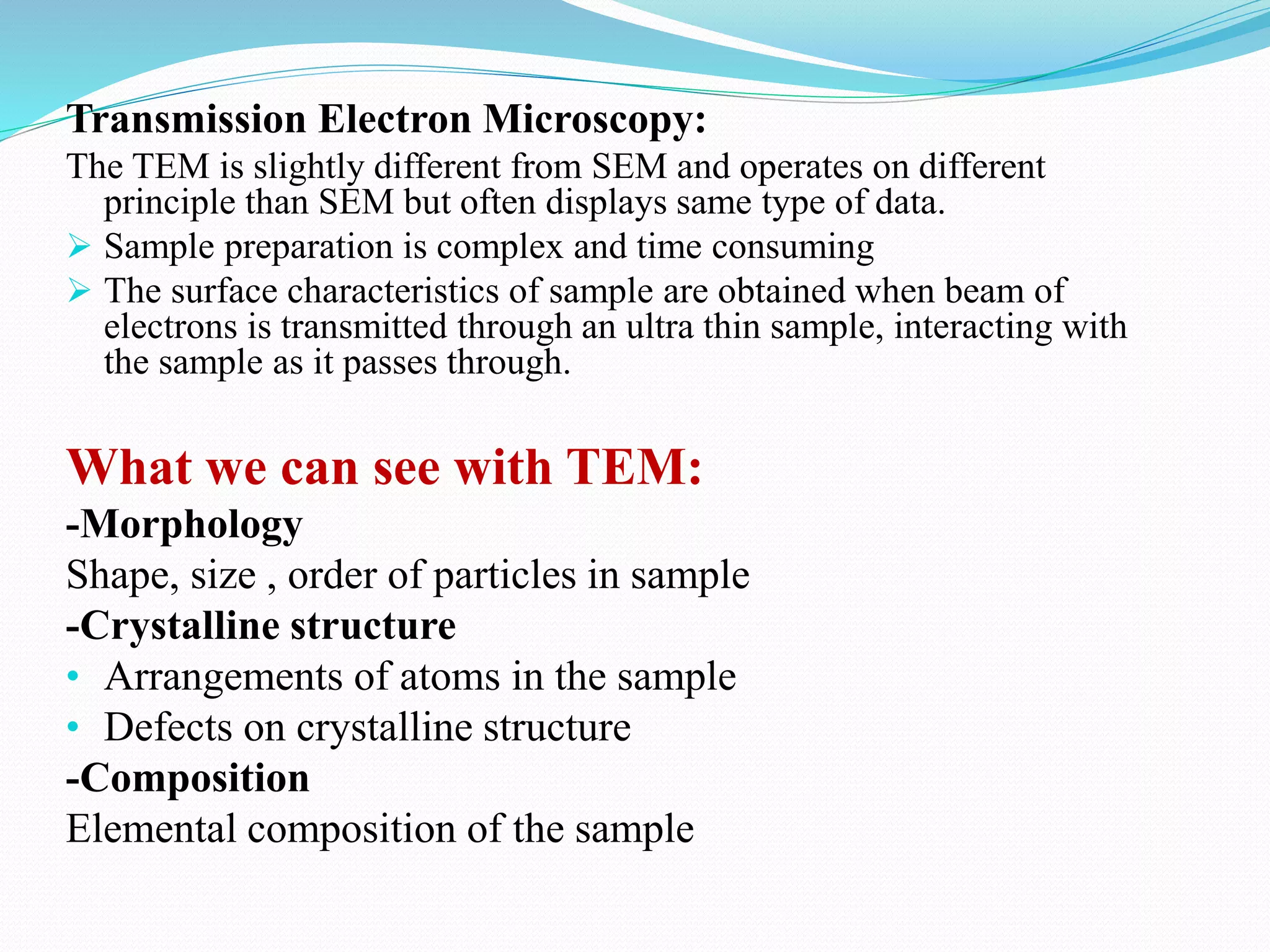
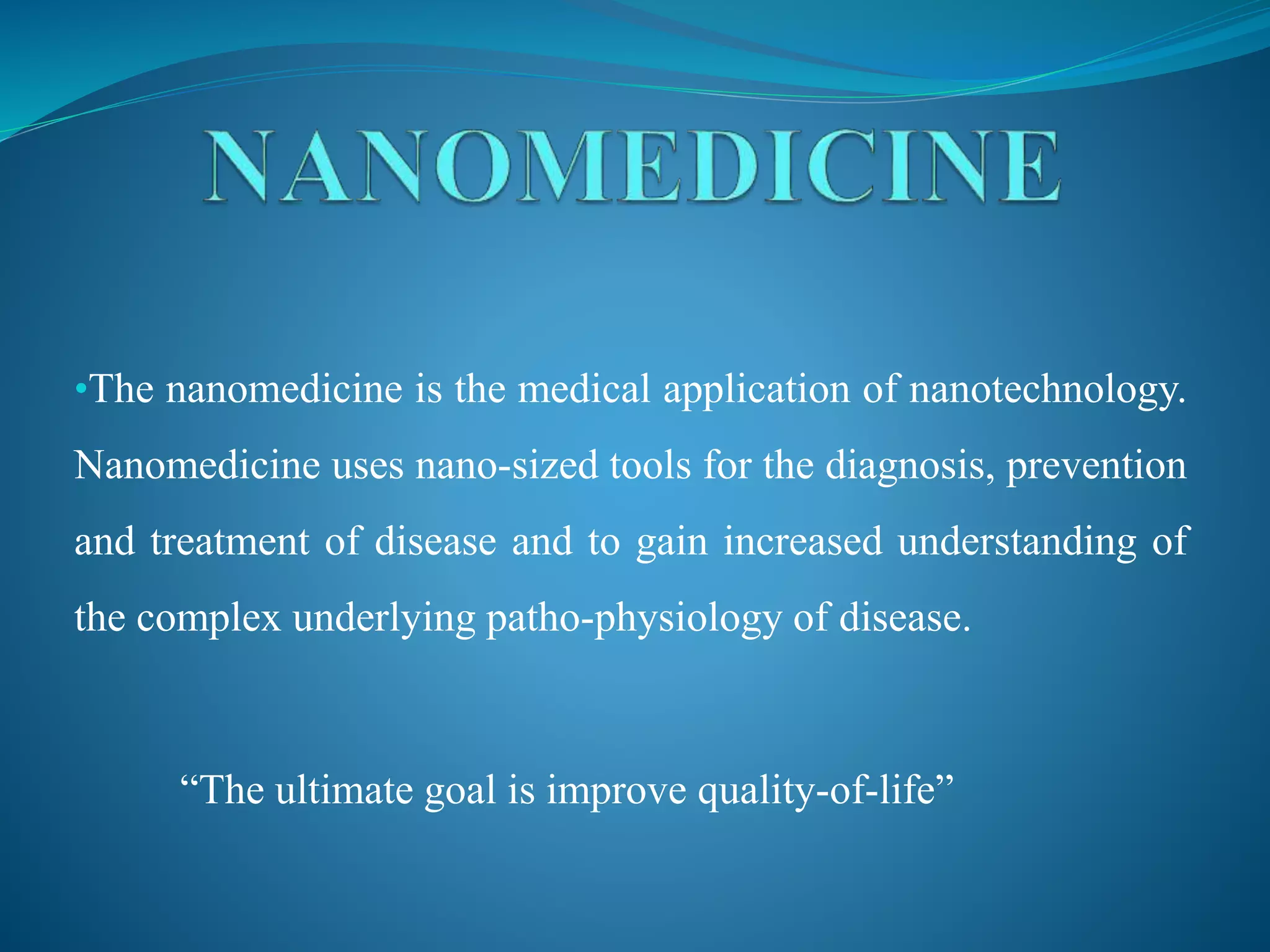
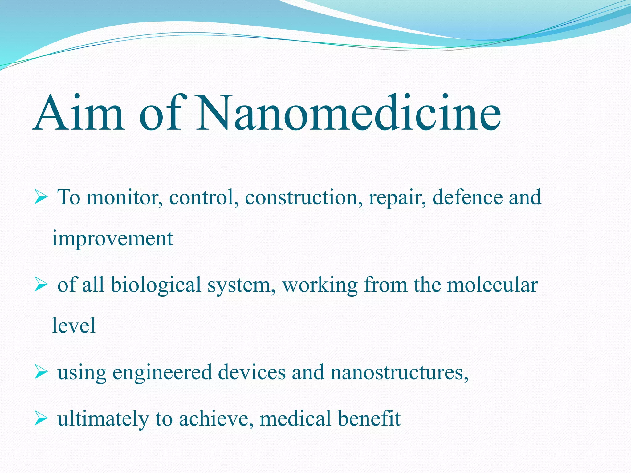
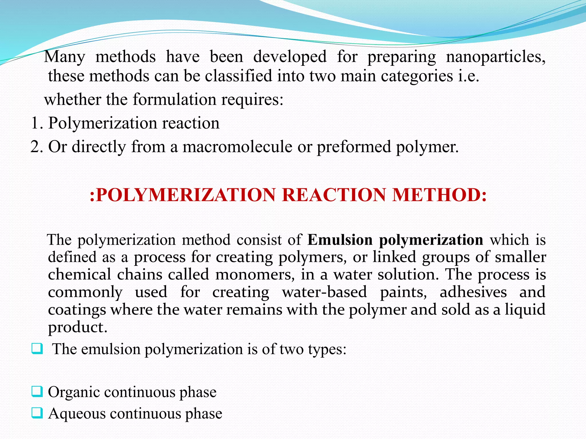
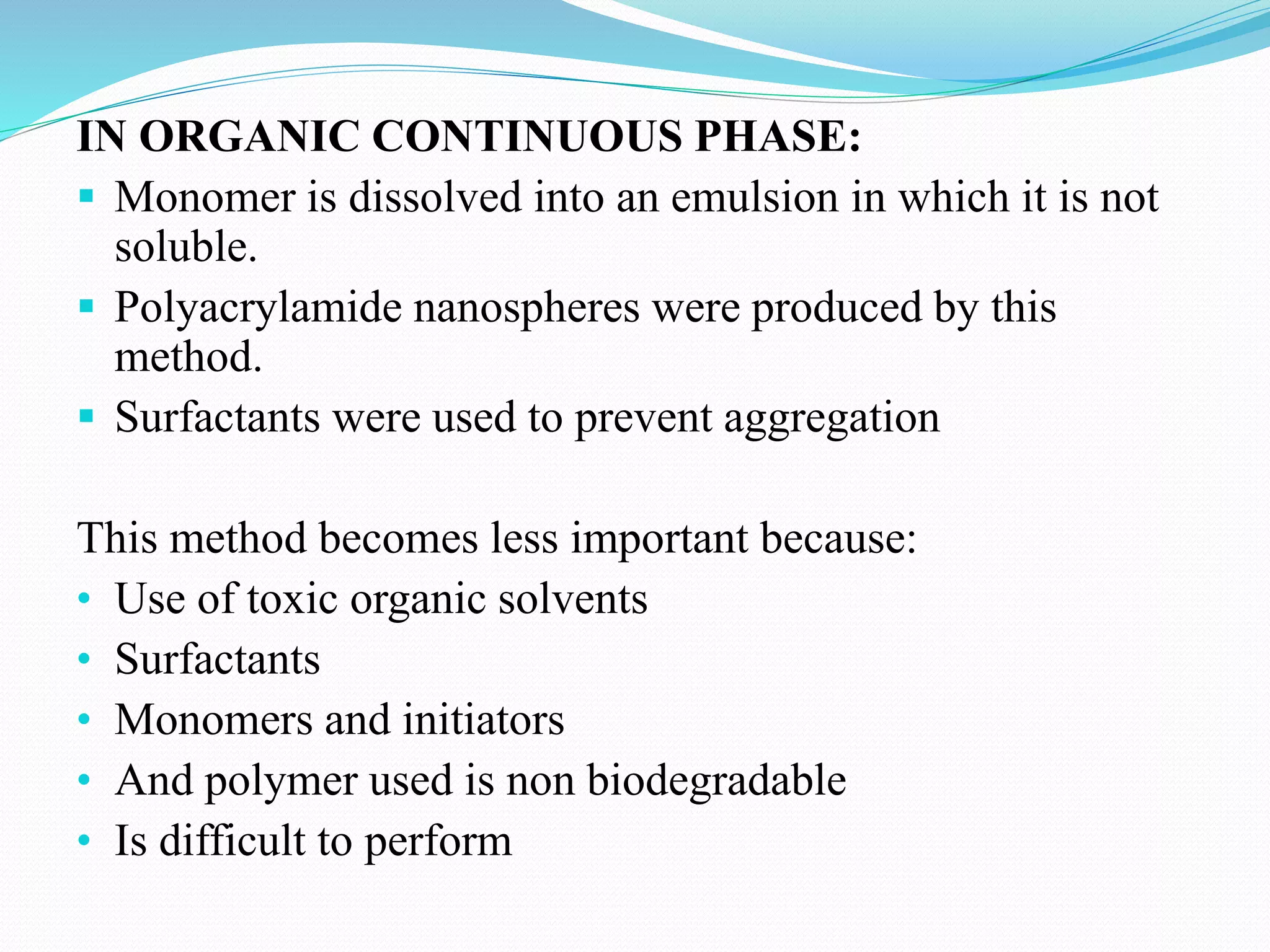
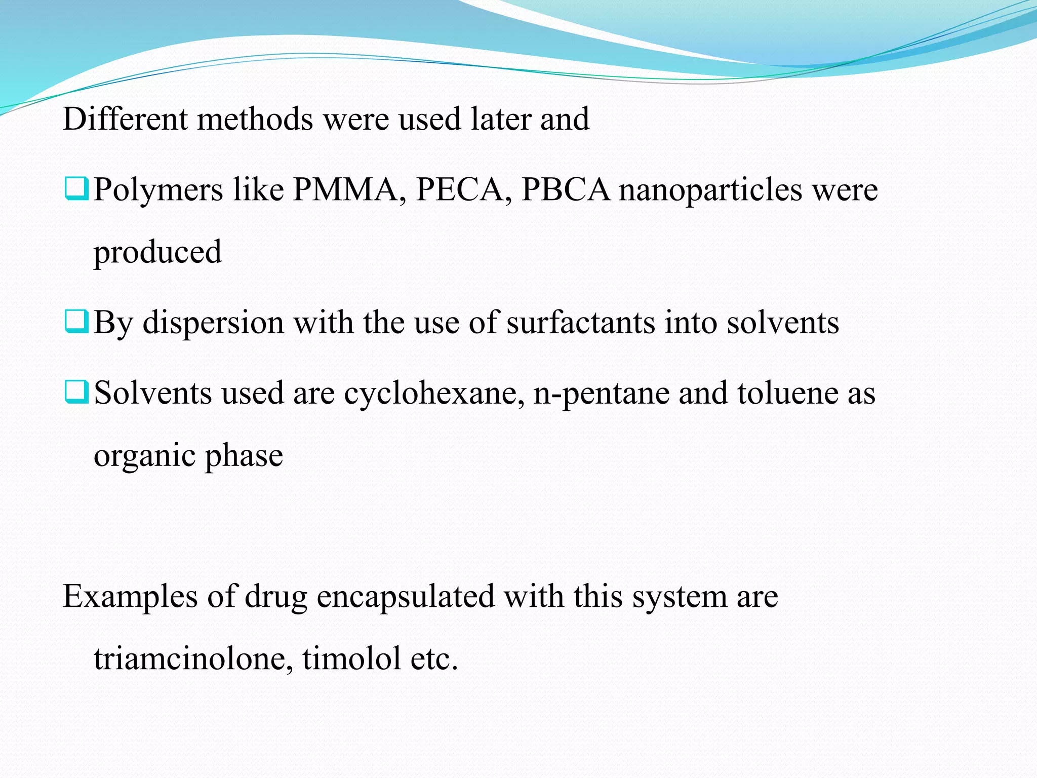
•The nanomedicine is the medical application of nanotechnology.
Nanomedicine uses nano-sized tools for the diagnosis, prevention
and treatment of disease and to gain increased understanding of
the complex underlying patho-physiology of disease.
“The ultimate goal is improve quality-of-life”
Aim of Nanomedicine
 To monitor, control, construction, repair, defence and
improvement
 of all biological system, working from the molecular
level
 using engineered devices and nanostructures,
 ultimately to achieve, medical benefit
Uses of Nanomedicines
It is used in the drug delivery system.
Diagnosis and Imaging
Regenerative medicines
Improved Imaging of Tumors
Cancer treatment
Advantages
 Drug delivery to exact location
Lesser side effects
Molecular targeting by nano engineered devices
Detection is relatively easy
No surgery required
Diseases can be easily cured
Disadvantages
 Not practical yet.
Very costly.
Implementation difficulties.
Many methods have been developed for preparing nanoparticles,
these methods can be classified into two main categories i.e.
whether the formulation requires:
1. Polymerization reaction
2. Or directly from a macromolecule or preformed polymer.
:POLYMERIZATION REACTION METHOD:
The polymerization method consist of Emulsion polymerization which is
defined as a process for creating polymers, or linked groups of smaller
chemical chains called monomers, in a water solution. The process is
commonly used for creating water-based paints, adhesives and
coatings where the water remains with the polymer and sold as a liquid
product.
 The emulsion polymerization is of two types:
 Organic continuous phase
 Aqueous continuous phase
IN ORGANIC CONTINUOUS PHASE:
 Monomer is dissolved into an emulsion in which it is not
soluble.
 Polyacrylamide nanospheres were produced by this
method.
 Surfactants were used to prevent aggregation
This method becomes less important because:
• Use of toxic organic solvents
• Surfactants
• Monomers and initiators
• And polymer used is non biodegradable
• Is difficult to perform
Different methods were used later and
Polymers like PMMA, PECA, PBCA nanoparticles were
produced
By dispersion with the use of surfactants into solvents
Solvents used are cyclohexane, n-pentane and toluene as
organic phase
Examples of drug encapsulated with this system are
triamcinolone, timolol etc.
IN AQUEOUS CONTINUOUS PHASE:
 Monomer is dissolved in a continuous phase which is a aqueous solution
 Surfactants and emulsifiers are not used
 Initiation occurs when monomer is dissolved in continuous phase and
collides with an initiator molecule
 It can be an ion or a free radical
 Chain growth starts when the initiated monomer ions or radicals collide
with other monomer
 PMMA nanoparticles are produced by this mechanism
without the use of emulsifiers.
 Single monomer MMA was used to produce PMMA nanospheres.
NANOPARTICLES OBTAINED FROM
PREFORMED POLYMERS:
It is done with the help of synthetic preformed polymers and
is done by many methods that are listed below.
 emulsification/solvent evaporation
 emulsification/solvent diffusion
 salting out with synthetic polymers
Emulsification/solvent evaporation:
This process involves two steps:
1. Emulsification of polymer into an aqueous phase
2. During 2nd step polymer solvent is evaporated inducing polymer
precipitation as nanospheres
Steps:
A drug is dissolved in a polymer organic solution which is dispersed
into nanodroplets using a dispersing agent & high energy
homogenization
Non-solvent and suspension medium such as chloroform is used
Emulsification of a polymer phase into an aqueous phase containing
surfactant to obtain an O/W emulsion.
Organic solvent is removed from dispersed phase by evaporation
And nanoparticles are formed.
Example- PLA [poly (lactic acid) ]
EMULSIFICATION/SOLVENT DIFFUSION:
Encapsulating polymer is dissolved in a partially water soluble
solvent such as propylene carbonate and saturated with water
The polymer-water saturated solvent phase is emulsified in an
aqueous phase containing stabilizer
Which leads to solvent diffusion and the nanospheres and
nanocapsules are formed
And at last the solvent is eliminated by evaporation or filtration
according to its boiling point.
Examples- Doxorubicin loaded PLGA Nanoparticles.
Salting Out With Synthetic polymers:
It is based on the separation of a water miscible solvent from
aqueous solution via a salting out effect.
Steps:
The polymer and drug are dissolved in a solvent such as acetone
Which is emulsified into an aqueous gel containing the salting out
agent(electrolytes such as MgCl, CaCl) and a colloidal stabilizer
such as hydroxyl ethyl cellulose.
This O/W emulsion is diluted with some water to enhance the
diffusion of acetone into water.
Results in the formation of nanospheres.
Both solvent and salting out agent then eliminated by cross flow
filtration
Nanoparticles are characterized by their size, morphology and by the surface
charge using some microscopic techniques such as scanning electron
microscopy(SEM), transmission electron microscopy(TEM), and atomic
force microscopy(AFM) and characterized by many more techniques.
Scanning Electron Microscopy (SEM):
Basic Principle- It is a type of electron microscope that produces images
of a sample by scanning it with a focused beam of electrons. The
electrons interact with atoms in the sample, producing various signals
that can be detected and that contain information about the sample’s
surface topography, morphology, and composition etc.
Steps:
 Nanoparticles solution first converted into dry powder an then mounted on a
sample holder with a coating of a conductive metal such as gold using
sputter coater.
 then scanned with a focused beam of electrons
 The surface characteristic of sample are obtained from secondary electrons.
 This technique is time consuming, costly and gives high resolution images.
What we can see with a SEM:
-Topography
Texture/surface of a sample
-Morphology
Size, shape, order of particles
-Composition
Elemental composition of a sample
-Crystalline Structure
Arrangement present within sample
Transmission Electron Microscopy:
The TEM is slightly different from SEM and operates on different
principle than SEM but often displays same type of data.
 Sample preparation is complex and time consuming
 The surface characteristics of sample are obtained when beam of
electrons is transmitted through an ultra thin sample, interacting with
the sample as it passes through.
What we can see with TEM:
-Morphology
Shape, size , order of particles in sample
-Crystalline structure
• Arrangements of atoms in the sample
• Defects on crystalline structure
-Composition
Elemental composition of the sample
Atomic Force Microscopy:
Offers ultra high resolution in particle size measurement
Based on physical scanning of samples at submicron level
Provides topographical map of sample
Provides most accurate description of size and size
distribution
And the particle size obtained by AFM gives real picture
which helps to understand the effect of various biological
conditions.

Nanomedicines

  • 1.
    •The nanomedicine isthe medical application of nanotechnology. Nanomedicine uses nano-sized tools for the diagnosis, prevention and treatment of disease and to gain increased understanding of the complex underlying patho-physiology of disease. “The ultimate goal is improve quality-of-life”
  • 2.
    Aim of Nanomedicine To monitor, control, construction, repair, defence and improvement  of all biological system, working from the molecular level  using engineered devices and nanostructures,  ultimately to achieve, medical benefit
  • 3.
    Uses of Nanomedicines Itis used in the drug delivery system. Diagnosis and Imaging Regenerative medicines Improved Imaging of Tumors Cancer treatment
  • 4.
    Advantages  Drug deliveryto exact location Lesser side effects Molecular targeting by nano engineered devices Detection is relatively easy No surgery required Diseases can be easily cured
  • 5.
    Disadvantages  Not practicalyet. Very costly. Implementation difficulties.
  • 7.
    Many methods havebeen developed for preparing nanoparticles, these methods can be classified into two main categories i.e. whether the formulation requires: 1. Polymerization reaction 2. Or directly from a macromolecule or preformed polymer. :POLYMERIZATION REACTION METHOD: The polymerization method consist of Emulsion polymerization which is defined as a process for creating polymers, or linked groups of smaller chemical chains called monomers, in a water solution. The process is commonly used for creating water-based paints, adhesives and coatings where the water remains with the polymer and sold as a liquid product.  The emulsion polymerization is of two types:  Organic continuous phase  Aqueous continuous phase
  • 8.
    IN ORGANIC CONTINUOUSPHASE:  Monomer is dissolved into an emulsion in which it is not soluble.  Polyacrylamide nanospheres were produced by this method.  Surfactants were used to prevent aggregation This method becomes less important because: • Use of toxic organic solvents • Surfactants • Monomers and initiators • And polymer used is non biodegradable • Is difficult to perform
  • 9.
    Different methods wereused later and Polymers like PMMA, PECA, PBCA nanoparticles were produced By dispersion with the use of surfactants into solvents Solvents used are cyclohexane, n-pentane and toluene as organic phase Examples of drug encapsulated with this system are triamcinolone, timolol etc.
  • 10.
    IN AQUEOUS CONTINUOUSPHASE:  Monomer is dissolved in a continuous phase which is a aqueous solution  Surfactants and emulsifiers are not used  Initiation occurs when monomer is dissolved in continuous phase and collides with an initiator molecule  It can be an ion or a free radical  Chain growth starts when the initiated monomer ions or radicals collide with other monomer  PMMA nanoparticles are produced by this mechanism without the use of emulsifiers.  Single monomer MMA was used to produce PMMA nanospheres.
  • 11.
    NANOPARTICLES OBTAINED FROM PREFORMEDPOLYMERS: It is done with the help of synthetic preformed polymers and is done by many methods that are listed below.  emulsification/solvent evaporation  emulsification/solvent diffusion  salting out with synthetic polymers
  • 12.
    Emulsification/solvent evaporation: This processinvolves two steps: 1. Emulsification of polymer into an aqueous phase 2. During 2nd step polymer solvent is evaporated inducing polymer precipitation as nanospheres Steps: A drug is dissolved in a polymer organic solution which is dispersed into nanodroplets using a dispersing agent & high energy homogenization Non-solvent and suspension medium such as chloroform is used Emulsification of a polymer phase into an aqueous phase containing surfactant to obtain an O/W emulsion. Organic solvent is removed from dispersed phase by evaporation And nanoparticles are formed. Example- PLA [poly (lactic acid) ]
  • 13.
    EMULSIFICATION/SOLVENT DIFFUSION: Encapsulating polymeris dissolved in a partially water soluble solvent such as propylene carbonate and saturated with water The polymer-water saturated solvent phase is emulsified in an aqueous phase containing stabilizer Which leads to solvent diffusion and the nanospheres and nanocapsules are formed And at last the solvent is eliminated by evaporation or filtration according to its boiling point. Examples- Doxorubicin loaded PLGA Nanoparticles.
  • 14.
    Salting Out WithSynthetic polymers: It is based on the separation of a water miscible solvent from aqueous solution via a salting out effect. Steps: The polymer and drug are dissolved in a solvent such as acetone Which is emulsified into an aqueous gel containing the salting out agent(electrolytes such as MgCl, CaCl) and a colloidal stabilizer such as hydroxyl ethyl cellulose. This O/W emulsion is diluted with some water to enhance the diffusion of acetone into water. Results in the formation of nanospheres. Both solvent and salting out agent then eliminated by cross flow filtration
  • 16.
    Nanoparticles are characterizedby their size, morphology and by the surface charge using some microscopic techniques such as scanning electron microscopy(SEM), transmission electron microscopy(TEM), and atomic force microscopy(AFM) and characterized by many more techniques. Scanning Electron Microscopy (SEM): Basic Principle- It is a type of electron microscope that produces images of a sample by scanning it with a focused beam of electrons. The electrons interact with atoms in the sample, producing various signals that can be detected and that contain information about the sample’s surface topography, morphology, and composition etc. Steps:  Nanoparticles solution first converted into dry powder an then mounted on a sample holder with a coating of a conductive metal such as gold using sputter coater.  then scanned with a focused beam of electrons  The surface characteristic of sample are obtained from secondary electrons.  This technique is time consuming, costly and gives high resolution images.
  • 17.
    What we cansee with a SEM: -Topography Texture/surface of a sample -Morphology Size, shape, order of particles -Composition Elemental composition of a sample -Crystalline Structure Arrangement present within sample
  • 18.
    Transmission Electron Microscopy: TheTEM is slightly different from SEM and operates on different principle than SEM but often displays same type of data.  Sample preparation is complex and time consuming  The surface characteristics of sample are obtained when beam of electrons is transmitted through an ultra thin sample, interacting with the sample as it passes through. What we can see with TEM: -Morphology Shape, size , order of particles in sample -Crystalline structure • Arrangements of atoms in the sample • Defects on crystalline structure -Composition Elemental composition of the sample
  • 19.
    Atomic Force Microscopy: Offersultra high resolution in particle size measurement Based on physical scanning of samples at submicron level Provides topographical map of sample Provides most accurate description of size and size distribution And the particle size obtained by AFM gives real picture which helps to understand the effect of various biological conditions.