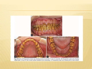
ENAMEL DEFECTS
Dr. Miraat Anser
PGR-Peadiatric Dentistry
WHAT IS ENAMEL?
 An epithilium derived hard substance
covering and protecting the dentine of the
crown of the tooth.
 96% inorganic which is hydroxyapatite,
calcium carbonate, calcium flouride and
magnesium carbonate.
 4% organic substance
DEVELOPMENT
 Development of the enamel is known as
Amelogenesis.
3 stages of enamel development:
1. Inductive/ Formative stage; This is the
morphodifferentiation phase of the crown where the Inner enamel
Epithelium elongate and become preameloblasts following a change in
polarity forming ameloblasts.
2. Secretory/ Calcification stage; Enamel proteins are
released into the surrounding area and contribute to what is
known as the enamel matrix, which is then partially mineralized
by the enzyme alkaline phosphatase.
3. Maturation stage; The ameloblasts transport substances
used in the formation of enamel. As enamel proteins are secreted,
some mineralization occurs by Ca2+ deposition.
ENAMEL FORMING PROTEINS
ENAMEL DEFECTS
Congenital
Aquired
CONGENITAL ENAMEL DEFECTS
 Amelogenesis Imperfecta
 Molar Incisor Hypoplasia
AMELOGENESIS IMPERFECTA/HERIDITARY
BROWN ENAMEL/HERIDITARY ENAMEL
DYSPLASIA/HERIDITARY BROWN OPLACENT
TEETH
 Disorder that affects the structure
and appearance of the enamel of the teeth.
 Can affect both primary and permanent
dentition
 Mainly a genetic disorder
CLASSIFICATION
 Initially there were 2 classifications
depending upon the genotypic and
phenotypic characteristics
 Genotypic Classification
1. Autosomal Dominant
2. Autosomal Recessive
3. X-Linked
4. Sporadic
 Phenotypic Classification
1. Hypoplastic
2. Hypocalcific
3. Hypomaturation
WITKOP 1989 CLASSIFICATION OF
AMELOGENESIS IMPERFECTA
EPIDEMIOLOGY
 The prevalence varies from 1:700 to
1:14,000, according to the populations Orphanet J Rare
Dis. 2007; 2: 17.
 Hypoplastic AI is the most common of all
representing 60-73% cases followed by
 Hypomaturation AI 20-40% and
Hypocalcification AI approx 7% of all cases.
GENETICS
GENES FOUND TO CAUSE AI(NON SYNDROMIC)
 Other than these some other reported are
 C4orf26,SLC24A4,LAMB3,ITGB6
 Seow WK; Aust Dent J.2014;59(suppl1);143-54
 AMELX and ENAM form extracellular
matrix proteins
 KKL4 and MMP20 protenases that
degrade organic matter of matrix during
maturation stage
 SLC24A4 Calcium transporter
 Lesser is known about other genes
 Mutations in ENAM gene is most common
cause of Autosomal dominant AI
 Autosomal recessive AI is caused by mutations
in MMP20, KKL4, FAM20A, C4orf26,SLC24A4
 AMELX gene mutation is seen in X-linked AI
 Analysis of X-linked AI showed a specific
defective gene at locus DXS85 at Xp22, which
is also the location for gene of Amelogenin
which is principle protein in formation of enamel
AI ASSOCIATED WITH SYNDROMES
 AI associated with taurodontism
 AI with neprocalcinosis
 Tricho- dentoosseous syndrome
 Cone-rod dystrophy with AI
CLINICAL FEATURES
HYPOPLASTIC
AI
HYPOMATURATIO
N AI
HYPOCALCIFIC AI
CLINICAL
FEATURES
Not formed to full
thickness; loss of
contacts; pitting
and grooves;
hard on probing
Enamel pierces by
explorer under firm
pressure; structure
lost by chipping
exposing
underlying dentine;
normal shape but
opaque white
yellow to brown
discolouration;
snowcapped
appearance
Enamel removed with
prophycup; yellowish
brown or orange on
eruption; heavily
stained; heavy plaque
and calculus retention;
excessive tooth
strucure loss
RADIOGRAPHIC
FEATURES
Enamel more
radiodense than
dentine
Radiodensity equal
to of dentine
Radiodensity equal to
or less than of dentine
HISTOLOGICAL FEATURES
 Hypoplastic AI: disturbance in differentiation
or viability of ameloblast, defects in matrix
formation and even absence of it
 Hypocalcific AI: defects in matrix structure
and mineral deposition
 Hypomaturation AI: alteration in enamel rod
and rod sheath structure
Problem
List
Sensitivity
Orthodontic
(OVD,
crossbite,
reverse
overjet etc)
Psychosocial
Attrition and
abrasion
Esthetics
Function
 Parekh et al. investigated impact of AI on
patients life(61 patients with response rate of
61%)
 90% patients complained about
discolouration
 77% wanted improvement in their smile
 74% wanted reduction in sensitivity
 60% wanted improvement in size of teeth
 Parkesh S, Almehateb M, Cunningham SJ. How do children with amelogenesis imperfecta feel about their teeth? Intl J. of Pead
Dent. 2014;24:326-35
TREATMENT/MANAGEMENT
 Genetic in nature thus no prevention is
possible yet.
 Management is complex thus management
requires an interdisciplinary approach.
TREATMENT OPTIONS
 Preventive fissure sealants
 Desensitizing agents
 composite fillings/ buildups/veneering
 SS crowns
 PFM
 All ceramic etc
 Orthodontics
 Orthognathic surgery
 Implants
 Dentures
 ????? Are they all affective????
HYPOPLASTIC VS HYPOCALCIFIC VS
HYPOMATURATION
INTERDISCLIPLINATY APPROACH
Restorative
Prosthetic
SurgicalOrthodontic
Psychological
LITERATURE REVIEW
 This child was initially referred by her Dental Therapist was in the early mixed dentition at 7 years of
age.
 She had a familial history of AI on her paternal side.
 O/E, enamel on her teeth appeared rough and the primary teeth were very worn. The permanent first
molar teeth were very sensitive. Radiographic findings confirmed missing enamel and no evidence of
dental caries.
 A clinical diagnosis of hypoplastic amelogenesis imperfecta was made.
 At that time the child was unable to cope with treatment in the dental chair because of extreme
sensitivity of the molars, so initial management involved composite coverage of the permanent molars
under general anaesthetic to stabilise the dentition.
 Composite resin was placed over the defective areas on the molars providing full coverage of the
erupted tissue to gain as much enamel bonding as possible.
 Following this interim treatment an orthodontic opinion was obtained regarding the Class II anterior
open bite and dental crowding.
 The patient was advised to brush with warm water and to use a normal fluoride toothpaste.
AT AGE 12 RX
 At age 12 years girl had a Class II skeletal pattern
with a long face height, incompetent lips and a high
upper lip line.
 She had an Angle Class II Division 1 malocclusion
with an anterior open bite, mild crowding in the lower
arch and moderate crowding in the upper arch.
 The patient was reluctant to undergo comprehensive
orthodontic treatment, but agreed to the placement of
fixed appliances in the upper arch.
13 YEARS
 Further restorative treatment was provided under a second general anaesthetic at 13 years to
restore the newly erupted and highly sensitive second permanent molars prior to commencing
orthodontic treatment.
 Composite resin overlays were placed on the permanent second molars and the restorations on
the first permanent molars were repaired.
 Teeth 14, 24, 53, 63, 65 and 74 were extracted as part of the orthodontic plan.
 A 0.5 percent daily fluoride mouthwash was prescribed to optimise protection of the teeth during
orthodontic treatment.
 An upper fixed appliance was placed to realign the arch and headgear was used to maintain a
Class II molar relationship.
 The following year, composite restorations were placed on all lower premolars.
 The patient continued to decline lower arch orthodontic treatment or to attend a consultation
regarding orthognathic surgery.
 She felt that she had no aesthetic or functional problems. At the completion of orthodontic
treatment for the upper arch, an upper removable retainer was provided.
AT 17 YEARS
 At age 17, the patient considered definitive restorative care. second phase of
orthodontic treatment (fixed appliances to align the lower arch), followed by
maxillofacial surgery (osteotomy and genioplasty)was started.
 Extractions (teeth 38, 35, 45, 48) performed under general anaesthetic.
 At the completion of the second phase of fixed orthodontics, the young woman
was referred to a specialist in restorative dentistry for the placement of definitive
cast crown restorations.
 Full gold crowns were placed on teeth 17 and 27, while porcelain fused to metal
crowns were placed on teeth 16, 15, 25, 26, 37, 36, 46, and 47.
 This treatment was performed over several visits using oral sedation. No
problems with patient management were experienced.
 The young woman chose not to proceed with cast restorations for the anterior
teeth at that stage as she was not experiencing any functional or aesthetic
problems.
AT 18
 A Le Fort I level differential impaction osteotomy (8mm
posterior and 4mm anterior) and mandibular
advancement genioplasty (5mm) were then performed to
correct the skeletal deformity (vertical maxillary excess
and retrognathic chin) and teeth 18 and 28 were
extracted.
 The osteotomy sites were fixed using 2mm titanium mini-
plates and screws.
 This patient, now an adult aged 18, has an aesthetic and
functional dentition and much improved facial proportions.
She reports great satisfaction with her outcome.
CASE REPORT #2
 A 27-year-old Chinese male was referred to the Prosthodontic
Department, Prince Philip Dental Hospital at the University of Hong
Kong.
 He requested to improve his dental appearance and chewing
efficiency.
 He reported having increasing difficulty to grind the food into small
enough pieces for swallowing.
 He also complained of food trapping in between approximal surface
where the enamel had cleaved off which was likely responsible for some
interproximal dental decay.
 The appearance and structural integrity of the patient’s dentition were
significantly compromised.
 The teeth appeared yellow-brown in color and had a rough and soft
surface texture with a marked tooth surface loss of the enamel,
especially on the axial surfaces, which resulted in a change of contour of
the teeth including loss of interproximal enamel marginal ridges which
facilitated food packing. He had no complaints of dentin hypersensitivity.
FAMILY HISTORY
 According to the patient, his father had
experienced similar problems and finally
became edentulous and is now wearing
complete dentures………
Diagnosis
The patient was diagnosed with a hypoplastic
type AI, generalized gingivitis, and caries on
teeth #17M #16D, M #26D #27M. A mild
malocclusion with teeth #12 #13 in reverse
overjet, teeth #13 and #23 mesiobuccally
rotated and a bilateral posterior cross-bite.
TX PLAN
 Upper arch expansion and proclination of the upper
incisors to create posterior interproximal space.
 Correction of the posterior, #12 and #13 cross-bites,
derotation of the teeth #13 and #23, and an increase of
the anterior overjet were planned. The correction of the
malaligned canines would help establish canine guidance.
 Increased overjet will help minimize the amount of labial
reduction required for the lower incisor restorations in
addition; an increase in the prosthetic crown height will
help establish the necessary palatal length for incisor
disclusion.
 A rapid palatal expander and Crozat appliance was
planned to correct the cross-bite and arch wires to
derotate teeth #13 and #23 as well as procline the
upper incisors.
 The occlusal scheme was designed and planned on a
pair of study models, which were mounted on a semi-
adjustable articulator. No change in the occlusal
vertical dimension was planned. A diagnostic wax-up
was performed to plan the proposed occlusal scheme
of incisor and canine disclusion.
 The posterior teeth were prepared first. All second molars were
prepared to receive full gold crowns. All first molars and all premolars
were prepared to receive metal–ceramic crowns. These were
temporized with selfcured temporary restorative material according to
the diagnostic wax-up. After this, the upper anterior teeth were
prepared to receive six individual PROCERA zirconia crowns, and
the lower anterior teeth were prepared to receive six individual IPS
Empress veneers.
 The prepared teeth were temporized with selfcured temporary direct
restorations according to the diagnostic wax-up. After clinical evaluation
of the planned occlusal scheme, an impression was taken and from this,
a study model was used to create a customized incisal guide table by
performing “protrusive” movements of the articulator with cold cure
acrylic on the incisal table.
 This would allow the planned occlusal scheme trailed in the mouth to be
copied to the definitive restoration.
CONCLUSION
 Treatment for AI patients is challenging and complicated due to the
orofacial abnormalities involved and the altered nature of the tooth
tissue.
 As both primary and permanent teeth are affected, diagnosis should be
made during childhood and treatment should be started to avoid
progressive damage and reduce the psychological impact to the patient.
 If the treatment is delayed, the clinical outcome may become more
complicated and a multidisciplinary approach will be indicated.
 In the case presented, the orthodontic interventions can greatly facilitate
the prosthetic planning so as to preserve the remaining tooth structure
and optimize the space for the physical performance of the dental
materials.
MIH(MOLAR INCISOR HYPOPLASIA)
 Type of enamel defect affecting, as the name suggests,
the first molars and incisors in the permanent dentition,
caused by the lack of mineralisation of enamel during its
maturation phase, due to interruption to the function
of ameloblasts.
 MIH often presents as discolouration on one to four
affected permanent molars and the associated incisors.
The enamel of the affected teeth appears yellow, brown,
cream or white.
 Sometimes reffered to as cheese molars.
 Garg, Nishita; Jain, Abhay Kumar; Saha, Sonali; Singh, Jaspal (2012). "Essentiality of Early Diagnosis of Molar Incisor Hypomineralization in
Children and Review of its Clinical Presentation, Etiology and Management". International Journal of Clinical Pediatric Dentistry. 5 (3): 190–
196. doi:10.5005/jp-journals-10005-1164. ISSN 0974-7052. PMC 4155885. PMID 25206166.
AETIOLOGY
 The aetiology of MIH is thought to be multifactorial.
 Pre-natally: risks, such as infection, maternal psychological stress and frequent
exposure to ultrasonic scans were all correlated with increased risks of MIH.
 During the perinatal stage, cesarean section and complications during vaginal
delivery could contribute to an increased chance of MIH.Children born preterm
and those with poor general health or systemic conditions in their first 3 years of
development also run a higher risk of developing MIH. It has also been proposed
that developmental dental defects were associated with long-term breastfeeding
due to exposure to dioxin.
 More recent evidence has suggested a relationship between respiratory
diseases and oxygen shortage of the ameloblasts and MIH. Lastly oxygen
shortage combined with low birth weight is suspected to be a contributing factor.
 It is suggested that there could be pre genetic predisposition in combination with
environmental insult.
CLINICAL FEATURES
 White to brownish lesions
 Post eruptive breakdowns
 Sharp demarcation between normal and
affected enamel
 Higher caries rate
 Sensitivity
 Asymmetric pattern is seen
PREVELANCE
 Many prevalence studies have now been published
from findings across the globe. However, there is a
large amount of variation across the results of
these studies. It is reported that the prevalence of
MIH ranges from 3.6% to 40.2%
 Jälevik, B. (2010-04-01). "Prevalence and Diagnosis of Molar-Incisor-Hypomineralisation (MIH): A systematic
review". European Archives of Paediatric Dentistry. 11 (2): 59–64.
TREATMENT
• Multiple options can be considered depending upon
the location and severity of the problem.
• Primary goal is to prevent caries and early
breakdown of tooth structure.
• Pits and fissure sealing and flouride therapy is
advised along with maintaining a good oral hygiene.
• Hard tooth brush and aggressive brushing is not
advised.
LEAST INVASIVE TO MOST INVASIVE
1. Resin infiltration
2. Etch- rinse- bleach technique
3. Micro abrasion with composite
4. Air abrasion with composite
5. Macro abrasion
6. Direct composite veneering
7. Indirect ceramic veneering
8. Onlay/overlay/vonlay/occlusal veneer
9. Full coverage crown pfm vs ceramic
10. Extraction and implant
Enamel defects
Enamel defects

Enamel defects

  • 1.
    ENAMEL DEFECTS Dr. MiraatAnser PGR-Peadiatric Dentistry
  • 2.
    WHAT IS ENAMEL? An epithilium derived hard substance covering and protecting the dentine of the crown of the tooth.  96% inorganic which is hydroxyapatite, calcium carbonate, calcium flouride and magnesium carbonate.  4% organic substance
  • 3.
    DEVELOPMENT  Development ofthe enamel is known as Amelogenesis. 3 stages of enamel development: 1. Inductive/ Formative stage; This is the morphodifferentiation phase of the crown where the Inner enamel Epithelium elongate and become preameloblasts following a change in polarity forming ameloblasts. 2. Secretory/ Calcification stage; Enamel proteins are released into the surrounding area and contribute to what is known as the enamel matrix, which is then partially mineralized by the enzyme alkaline phosphatase. 3. Maturation stage; The ameloblasts transport substances used in the formation of enamel. As enamel proteins are secreted, some mineralization occurs by Ca2+ deposition.
  • 4.
  • 6.
  • 7.
    CONGENITAL ENAMEL DEFECTS Amelogenesis Imperfecta  Molar Incisor Hypoplasia
  • 9.
    AMELOGENESIS IMPERFECTA/HERIDITARY BROWN ENAMEL/HERIDITARYENAMEL DYSPLASIA/HERIDITARY BROWN OPLACENT TEETH  Disorder that affects the structure and appearance of the enamel of the teeth.  Can affect both primary and permanent dentition  Mainly a genetic disorder
  • 10.
    CLASSIFICATION  Initially therewere 2 classifications depending upon the genotypic and phenotypic characteristics  Genotypic Classification 1. Autosomal Dominant 2. Autosomal Recessive 3. X-Linked 4. Sporadic
  • 11.
     Phenotypic Classification 1.Hypoplastic 2. Hypocalcific 3. Hypomaturation
  • 12.
    WITKOP 1989 CLASSIFICATIONOF AMELOGENESIS IMPERFECTA
  • 13.
    EPIDEMIOLOGY  The prevalencevaries from 1:700 to 1:14,000, according to the populations Orphanet J Rare Dis. 2007; 2: 17.  Hypoplastic AI is the most common of all representing 60-73% cases followed by  Hypomaturation AI 20-40% and Hypocalcification AI approx 7% of all cases.
  • 14.
    GENETICS GENES FOUND TOCAUSE AI(NON SYNDROMIC)
  • 15.
     Other thanthese some other reported are  C4orf26,SLC24A4,LAMB3,ITGB6  Seow WK; Aust Dent J.2014;59(suppl1);143-54
  • 16.
     AMELX andENAM form extracellular matrix proteins  KKL4 and MMP20 protenases that degrade organic matter of matrix during maturation stage  SLC24A4 Calcium transporter  Lesser is known about other genes
  • 17.
     Mutations inENAM gene is most common cause of Autosomal dominant AI  Autosomal recessive AI is caused by mutations in MMP20, KKL4, FAM20A, C4orf26,SLC24A4  AMELX gene mutation is seen in X-linked AI  Analysis of X-linked AI showed a specific defective gene at locus DXS85 at Xp22, which is also the location for gene of Amelogenin which is principle protein in formation of enamel
  • 18.
    AI ASSOCIATED WITHSYNDROMES  AI associated with taurodontism  AI with neprocalcinosis  Tricho- dentoosseous syndrome  Cone-rod dystrophy with AI
  • 19.
    CLINICAL FEATURES HYPOPLASTIC AI HYPOMATURATIO N AI HYPOCALCIFICAI CLINICAL FEATURES Not formed to full thickness; loss of contacts; pitting and grooves; hard on probing Enamel pierces by explorer under firm pressure; structure lost by chipping exposing underlying dentine; normal shape but opaque white yellow to brown discolouration; snowcapped appearance Enamel removed with prophycup; yellowish brown or orange on eruption; heavily stained; heavy plaque and calculus retention; excessive tooth strucure loss RADIOGRAPHIC FEATURES Enamel more radiodense than dentine Radiodensity equal to of dentine Radiodensity equal to or less than of dentine
  • 24.
    HISTOLOGICAL FEATURES  HypoplasticAI: disturbance in differentiation or viability of ameloblast, defects in matrix formation and even absence of it  Hypocalcific AI: defects in matrix structure and mineral deposition  Hypomaturation AI: alteration in enamel rod and rod sheath structure
  • 26.
  • 27.
     Parekh etal. investigated impact of AI on patients life(61 patients with response rate of 61%)  90% patients complained about discolouration  77% wanted improvement in their smile  74% wanted reduction in sensitivity  60% wanted improvement in size of teeth  Parkesh S, Almehateb M, Cunningham SJ. How do children with amelogenesis imperfecta feel about their teeth? Intl J. of Pead Dent. 2014;24:326-35
  • 28.
    TREATMENT/MANAGEMENT  Genetic innature thus no prevention is possible yet.  Management is complex thus management requires an interdisciplinary approach.
  • 29.
    TREATMENT OPTIONS  Preventivefissure sealants  Desensitizing agents  composite fillings/ buildups/veneering  SS crowns  PFM  All ceramic etc  Orthodontics  Orthognathic surgery  Implants  Dentures  ????? Are they all affective????
  • 30.
    HYPOPLASTIC VS HYPOCALCIFICVS HYPOMATURATION
  • 31.
  • 32.
  • 33.
     This childwas initially referred by her Dental Therapist was in the early mixed dentition at 7 years of age.  She had a familial history of AI on her paternal side.  O/E, enamel on her teeth appeared rough and the primary teeth were very worn. The permanent first molar teeth were very sensitive. Radiographic findings confirmed missing enamel and no evidence of dental caries.  A clinical diagnosis of hypoplastic amelogenesis imperfecta was made.  At that time the child was unable to cope with treatment in the dental chair because of extreme sensitivity of the molars, so initial management involved composite coverage of the permanent molars under general anaesthetic to stabilise the dentition.  Composite resin was placed over the defective areas on the molars providing full coverage of the erupted tissue to gain as much enamel bonding as possible.  Following this interim treatment an orthodontic opinion was obtained regarding the Class II anterior open bite and dental crowding.  The patient was advised to brush with warm water and to use a normal fluoride toothpaste.
  • 35.
    AT AGE 12RX  At age 12 years girl had a Class II skeletal pattern with a long face height, incompetent lips and a high upper lip line.  She had an Angle Class II Division 1 malocclusion with an anterior open bite, mild crowding in the lower arch and moderate crowding in the upper arch.  The patient was reluctant to undergo comprehensive orthodontic treatment, but agreed to the placement of fixed appliances in the upper arch.
  • 37.
    13 YEARS  Furtherrestorative treatment was provided under a second general anaesthetic at 13 years to restore the newly erupted and highly sensitive second permanent molars prior to commencing orthodontic treatment.  Composite resin overlays were placed on the permanent second molars and the restorations on the first permanent molars were repaired.  Teeth 14, 24, 53, 63, 65 and 74 were extracted as part of the orthodontic plan.  A 0.5 percent daily fluoride mouthwash was prescribed to optimise protection of the teeth during orthodontic treatment.  An upper fixed appliance was placed to realign the arch and headgear was used to maintain a Class II molar relationship.  The following year, composite restorations were placed on all lower premolars.  The patient continued to decline lower arch orthodontic treatment or to attend a consultation regarding orthognathic surgery.  She felt that she had no aesthetic or functional problems. At the completion of orthodontic treatment for the upper arch, an upper removable retainer was provided.
  • 38.
    AT 17 YEARS At age 17, the patient considered definitive restorative care. second phase of orthodontic treatment (fixed appliances to align the lower arch), followed by maxillofacial surgery (osteotomy and genioplasty)was started.  Extractions (teeth 38, 35, 45, 48) performed under general anaesthetic.  At the completion of the second phase of fixed orthodontics, the young woman was referred to a specialist in restorative dentistry for the placement of definitive cast crown restorations.  Full gold crowns were placed on teeth 17 and 27, while porcelain fused to metal crowns were placed on teeth 16, 15, 25, 26, 37, 36, 46, and 47.  This treatment was performed over several visits using oral sedation. No problems with patient management were experienced.  The young woman chose not to proceed with cast restorations for the anterior teeth at that stage as she was not experiencing any functional or aesthetic problems.
  • 40.
    AT 18  ALe Fort I level differential impaction osteotomy (8mm posterior and 4mm anterior) and mandibular advancement genioplasty (5mm) were then performed to correct the skeletal deformity (vertical maxillary excess and retrognathic chin) and teeth 18 and 28 were extracted.  The osteotomy sites were fixed using 2mm titanium mini- plates and screws.  This patient, now an adult aged 18, has an aesthetic and functional dentition and much improved facial proportions. She reports great satisfaction with her outcome.
  • 41.
  • 42.
     A 27-year-oldChinese male was referred to the Prosthodontic Department, Prince Philip Dental Hospital at the University of Hong Kong.  He requested to improve his dental appearance and chewing efficiency.  He reported having increasing difficulty to grind the food into small enough pieces for swallowing.  He also complained of food trapping in between approximal surface where the enamel had cleaved off which was likely responsible for some interproximal dental decay.  The appearance and structural integrity of the patient’s dentition were significantly compromised.  The teeth appeared yellow-brown in color and had a rough and soft surface texture with a marked tooth surface loss of the enamel, especially on the axial surfaces, which resulted in a change of contour of the teeth including loss of interproximal enamel marginal ridges which facilitated food packing. He had no complaints of dentin hypersensitivity.
  • 43.
    FAMILY HISTORY  Accordingto the patient, his father had experienced similar problems and finally became edentulous and is now wearing complete dentures………
  • 44.
    Diagnosis The patient wasdiagnosed with a hypoplastic type AI, generalized gingivitis, and caries on teeth #17M #16D, M #26D #27M. A mild malocclusion with teeth #12 #13 in reverse overjet, teeth #13 and #23 mesiobuccally rotated and a bilateral posterior cross-bite.
  • 47.
    TX PLAN  Upperarch expansion and proclination of the upper incisors to create posterior interproximal space.  Correction of the posterior, #12 and #13 cross-bites, derotation of the teeth #13 and #23, and an increase of the anterior overjet were planned. The correction of the malaligned canines would help establish canine guidance.  Increased overjet will help minimize the amount of labial reduction required for the lower incisor restorations in addition; an increase in the prosthetic crown height will help establish the necessary palatal length for incisor disclusion.
  • 50.
     A rapidpalatal expander and Crozat appliance was planned to correct the cross-bite and arch wires to derotate teeth #13 and #23 as well as procline the upper incisors.  The occlusal scheme was designed and planned on a pair of study models, which were mounted on a semi- adjustable articulator. No change in the occlusal vertical dimension was planned. A diagnostic wax-up was performed to plan the proposed occlusal scheme of incisor and canine disclusion.
  • 51.
     The posteriorteeth were prepared first. All second molars were prepared to receive full gold crowns. All first molars and all premolars were prepared to receive metal–ceramic crowns. These were temporized with selfcured temporary restorative material according to the diagnostic wax-up. After this, the upper anterior teeth were prepared to receive six individual PROCERA zirconia crowns, and the lower anterior teeth were prepared to receive six individual IPS Empress veneers.  The prepared teeth were temporized with selfcured temporary direct restorations according to the diagnostic wax-up. After clinical evaluation of the planned occlusal scheme, an impression was taken and from this, a study model was used to create a customized incisal guide table by performing “protrusive” movements of the articulator with cold cure acrylic on the incisal table.  This would allow the planned occlusal scheme trailed in the mouth to be copied to the definitive restoration.
  • 53.
    CONCLUSION  Treatment forAI patients is challenging and complicated due to the orofacial abnormalities involved and the altered nature of the tooth tissue.  As both primary and permanent teeth are affected, diagnosis should be made during childhood and treatment should be started to avoid progressive damage and reduce the psychological impact to the patient.  If the treatment is delayed, the clinical outcome may become more complicated and a multidisciplinary approach will be indicated.  In the case presented, the orthodontic interventions can greatly facilitate the prosthetic planning so as to preserve the remaining tooth structure and optimize the space for the physical performance of the dental materials.
  • 54.
    MIH(MOLAR INCISOR HYPOPLASIA) Type of enamel defect affecting, as the name suggests, the first molars and incisors in the permanent dentition, caused by the lack of mineralisation of enamel during its maturation phase, due to interruption to the function of ameloblasts.  MIH often presents as discolouration on one to four affected permanent molars and the associated incisors. The enamel of the affected teeth appears yellow, brown, cream or white.  Sometimes reffered to as cheese molars.  Garg, Nishita; Jain, Abhay Kumar; Saha, Sonali; Singh, Jaspal (2012). "Essentiality of Early Diagnosis of Molar Incisor Hypomineralization in Children and Review of its Clinical Presentation, Etiology and Management". International Journal of Clinical Pediatric Dentistry. 5 (3): 190– 196. doi:10.5005/jp-journals-10005-1164. ISSN 0974-7052. PMC 4155885. PMID 25206166.
  • 58.
    AETIOLOGY  The aetiologyof MIH is thought to be multifactorial.  Pre-natally: risks, such as infection, maternal psychological stress and frequent exposure to ultrasonic scans were all correlated with increased risks of MIH.  During the perinatal stage, cesarean section and complications during vaginal delivery could contribute to an increased chance of MIH.Children born preterm and those with poor general health or systemic conditions in their first 3 years of development also run a higher risk of developing MIH. It has also been proposed that developmental dental defects were associated with long-term breastfeeding due to exposure to dioxin.  More recent evidence has suggested a relationship between respiratory diseases and oxygen shortage of the ameloblasts and MIH. Lastly oxygen shortage combined with low birth weight is suspected to be a contributing factor.  It is suggested that there could be pre genetic predisposition in combination with environmental insult.
  • 59.
    CLINICAL FEATURES  Whiteto brownish lesions  Post eruptive breakdowns  Sharp demarcation between normal and affected enamel  Higher caries rate  Sensitivity  Asymmetric pattern is seen
  • 60.
    PREVELANCE  Many prevalencestudies have now been published from findings across the globe. However, there is a large amount of variation across the results of these studies. It is reported that the prevalence of MIH ranges from 3.6% to 40.2%  Jälevik, B. (2010-04-01). "Prevalence and Diagnosis of Molar-Incisor-Hypomineralisation (MIH): A systematic review". European Archives of Paediatric Dentistry. 11 (2): 59–64.
  • 61.
    TREATMENT • Multiple optionscan be considered depending upon the location and severity of the problem. • Primary goal is to prevent caries and early breakdown of tooth structure. • Pits and fissure sealing and flouride therapy is advised along with maintaining a good oral hygiene. • Hard tooth brush and aggressive brushing is not advised.
  • 62.
    LEAST INVASIVE TOMOST INVASIVE 1. Resin infiltration 2. Etch- rinse- bleach technique 3. Micro abrasion with composite 4. Air abrasion with composite 5. Macro abrasion 6. Direct composite veneering 7. Indirect ceramic veneering 8. Onlay/overlay/vonlay/occlusal veneer 9. Full coverage crown pfm vs ceramic 10. Extraction and implant