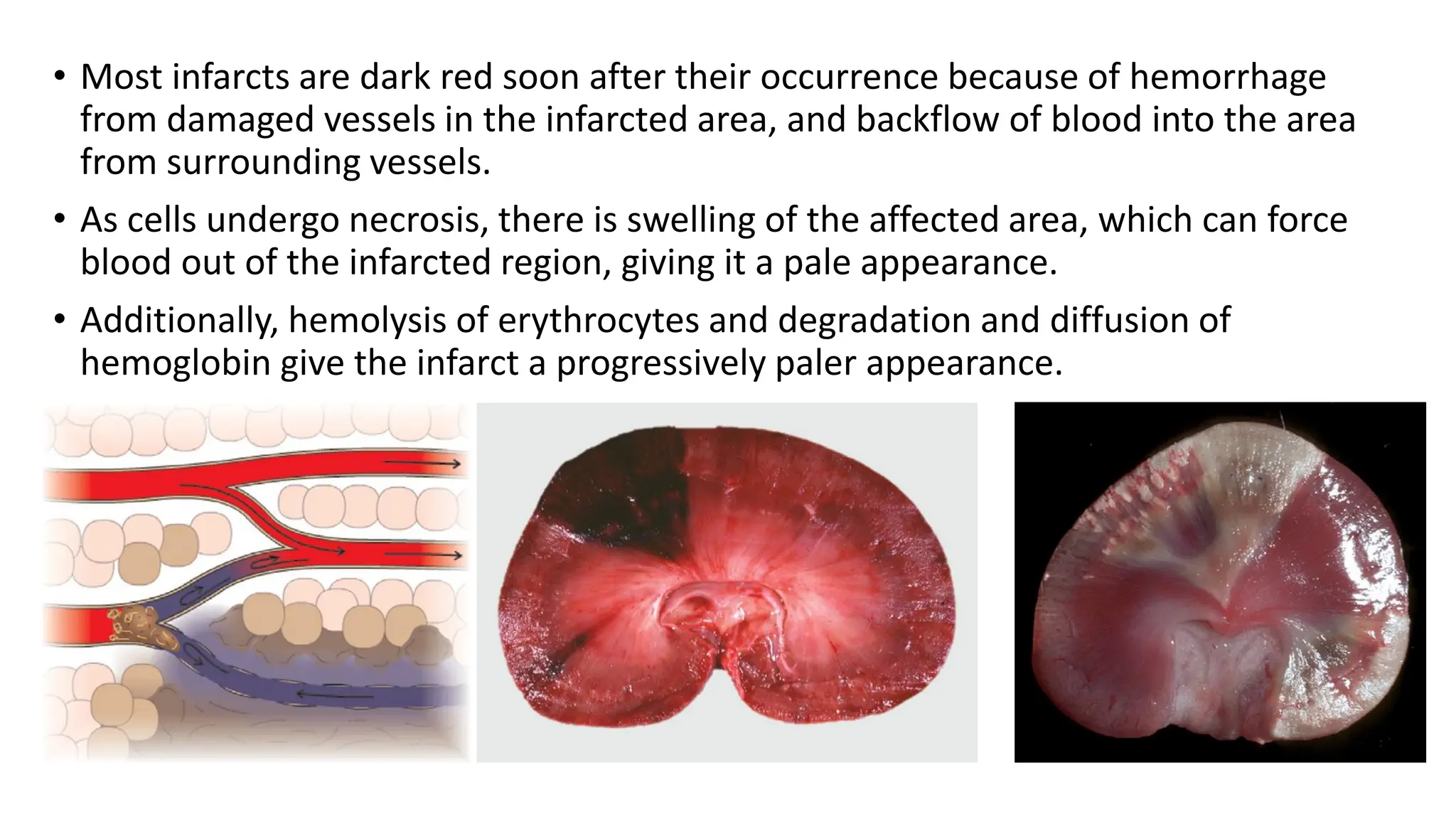
Lecture # 15
• Ischemia occurs in reduced blood flow to a tissue when the perfusion of tissue in the
affected area becomes inadequate to meet the metabolic needs of the tissue.
• Ischemia caused by arterial disease is most commonly due to incomplete luminal
blockage by a thrombus or embolus. The result is a decreased flow of oxygenated
blood into the area. Arteriolar vasoconstriction, if prolonged, can also result in
ischemia.
• Ischemia due to venous lesions can be caused by intraluminal obstruction, such as a
venous thrombus. However, external pressure that occludes the vein, such as
inflammatory or neoplastic masses, is a common cause.
• Venous obstruction leads to congestion characterized by slowing and stagnation of
blood flow, with loss of tissue oxygenation, local increased hydrostatic pressure, and
leakage of fluid into the interstitium (edema).
• Increased interstitial pressure may partially inhibit arterial inflow into the area to
compound the problem.
Ischemia & Infarction
• The severity of ischemia is determined by
1. The local vascular anatomy
2. The degree of anastomoses and collateral circulation
3. The number of microcirculatory vessels
4. The degree of resistance of the arteriole supplying the capillaries,
5. The extent of the decreased perfusion
6. The rate at which the occlusion occurred
7. The metabolic needs of the tissue.
• The brain and heart are most susceptible because of a high need for O2 and nutrients
combined with poor collateral circulation.
• In contrast, organs that recondition blood (e.g., lungs, gastrointestinal tract, kidneys, and
skin) can tolerate substantial reductions in flow because they already receive more
blood than necessary for their metabolic needs.
• Other tissues receive blood based on their immediate needs (e.g., skeletal muscle during
physical activity).
• An infarct is a local area of per-acute ischemia that undergoes coagulative
necrosis.
• Infarction is caused by the same events that result in ischemia and is most
commonly secondary to thrombosis or thromboembolism.
• The characteristics of an infarct are variable and based on
1. The type and size of vessel that was occluded (artery or vein),
2. The duration of the occlusion
3. The tissue in which it occurs
4. The prior perfusion and vitality of the tissue.
• Complete arterial blockage usually results in immediate infarction.
• In contrast, when venous obstruction occurs, such as due to torsions or
displacements of the bowel, there is extensive congestion and edema of
the affected bowel that precedes and promotes infarction.
• Concurrent disease, decreased cardiovascular function, anemia, or
decreased tissue vitality will increase the likelihood of localized areas of
ischemia progressing to infarction.
• In tissue with a single blood supply and minimal anastomoses (e.g., brain, heart,
kidney, and spleen), occlusion of nearly any sized vessel typically results in
infarction of the dependent tissue.
• Nearly 99% of all infarcts result from thrombotic or embolic events. But it can be
due to
• Local vasospasm
• External compression of the vessels. e.g trauma
• Entrapment of vessels at hernial sacks etc.
The development & the size of an infarct are determined by the following factors:
A. The nature of the vascular supply
B. The rate of development of occlusion
C. Suceptibility of the tissue for hypoxia
D. Oxygen content of the blood
E. The severity & duration of ischemia
A. The nature of vascular supply
• Some organs like lungs and liver have dual blood supply so the effect of such a dual
blood supply is that if there is obstruction of one of the arterial supplies, the other
one may offset the rapid occurrence of infarction in these organs
B. Rate of development occlusion
• Slowly developing occlusions are less likely to cause infraction since they provide
time for the development of collaterals.
C. Tissue susceptibility to hypoxia
• The susceptibility of a tissue to hypoxia influences the likelihood of infarction.
Neurons undergo irreversible damage when deprived of their blood supply for only 3
to 4 minutes. Myocardial cells die after 20-30 minutes of ischemia.
D. Oxygen content of blood
• Partial obstruction of the flow of blood in an anemic or cyanotic patient may lead to
tissue infarction.
• Most infarcts are dark red soon after their occurrence because of hemorrhage
from damaged vessels in the infarcted area, and backflow of blood into the area
from surrounding vessels.
• As cells undergo necrosis, there is swelling of the affected area, which can force
blood out of the infarcted region, giving it a pale appearance.
• Additionally, hemolysis of erythrocytes and degradation and diffusion of
hemoglobin give the infarct a progressively paler appearance.
T
Infarcts are classified depending on:
A. the basis of their color (reflecting the amount of hemorrhage) into:
1. Hemorrhagic (Red) infarcts
2. Anemic (White) infarcts
B. the presence or absence of microbial infection into:
1. Septic infarcts
2. Bland infarcts
Red infarcts
• Red infarcts are red due to accumulation of blood in the infarcted tissue. Characteristics
of red infarcts include:
• Occlusion of a vein
• Loose tissues that allow blood to collect in the infarcted zone
• Tissues with a dual circulatory system (lung, small intestines)
Types of infarcts
• White infarcts refer to pale infarcts pale infarcts. This pallor is a result of dead tissue
that is deprived of blood. White infarcts are always arterial infarcts. Arterial
occlusion in organs with a single arterial blood supply.
• Certain types of tissue that have a loose (spongy) consistency, such as the lungs and
storage-type spleens (e.g., dogs and pigs), usually remain red because the interstitial
areas are expandable and necrosis-induced pressure does not build up to force
blood out of the infarcted region.
• Parenchymal tissues with a less expansible interstitium (e.g., kidney) generally
become pale over time because of the pressure that forces blood from the necrotic
area.
White infarcts
• Inflammation occurs at the periphery of the dead tissue so that
leukocytes, then macrophages, enter the area to clear the necrotic debris,
and subsequently neovascularization and granulation occur to replace the
necrotic region with fibrous tissue.
• In contrast to the coagulative necrosis caused by infarction in most tissue,
infarction in the brain and nervous tissue is characterized by liquefactive
necrosis.
Thrombosis → embolism → ischemia → infarction

Lecture # 15, Ischemia and Infarction.pdf

  • 1.
  • 2.
    • Ischemia occursin reduced blood flow to a tissue when the perfusion of tissue in the affected area becomes inadequate to meet the metabolic needs of the tissue. • Ischemia caused by arterial disease is most commonly due to incomplete luminal blockage by a thrombus or embolus. The result is a decreased flow of oxygenated blood into the area. Arteriolar vasoconstriction, if prolonged, can also result in ischemia. • Ischemia due to venous lesions can be caused by intraluminal obstruction, such as a venous thrombus. However, external pressure that occludes the vein, such as inflammatory or neoplastic masses, is a common cause. • Venous obstruction leads to congestion characterized by slowing and stagnation of blood flow, with loss of tissue oxygenation, local increased hydrostatic pressure, and leakage of fluid into the interstitium (edema). • Increased interstitial pressure may partially inhibit arterial inflow into the area to compound the problem. Ischemia & Infarction
  • 3.
    • The severityof ischemia is determined by 1. The local vascular anatomy 2. The degree of anastomoses and collateral circulation 3. The number of microcirculatory vessels 4. The degree of resistance of the arteriole supplying the capillaries, 5. The extent of the decreased perfusion 6. The rate at which the occlusion occurred 7. The metabolic needs of the tissue. • The brain and heart are most susceptible because of a high need for O2 and nutrients combined with poor collateral circulation. • In contrast, organs that recondition blood (e.g., lungs, gastrointestinal tract, kidneys, and skin) can tolerate substantial reductions in flow because they already receive more blood than necessary for their metabolic needs. • Other tissues receive blood based on their immediate needs (e.g., skeletal muscle during physical activity).
  • 4.
    • An infarctis a local area of per-acute ischemia that undergoes coagulative necrosis. • Infarction is caused by the same events that result in ischemia and is most commonly secondary to thrombosis or thromboembolism. • The characteristics of an infarct are variable and based on 1. The type and size of vessel that was occluded (artery or vein), 2. The duration of the occlusion 3. The tissue in which it occurs 4. The prior perfusion and vitality of the tissue. • Complete arterial blockage usually results in immediate infarction. • In contrast, when venous obstruction occurs, such as due to torsions or displacements of the bowel, there is extensive congestion and edema of the affected bowel that precedes and promotes infarction. • Concurrent disease, decreased cardiovascular function, anemia, or decreased tissue vitality will increase the likelihood of localized areas of ischemia progressing to infarction.
  • 5.
    • In tissuewith a single blood supply and minimal anastomoses (e.g., brain, heart, kidney, and spleen), occlusion of nearly any sized vessel typically results in infarction of the dependent tissue. • Nearly 99% of all infarcts result from thrombotic or embolic events. But it can be due to • Local vasospasm • External compression of the vessels. e.g trauma • Entrapment of vessels at hernial sacks etc. The development & the size of an infarct are determined by the following factors: A. The nature of the vascular supply B. The rate of development of occlusion C. Suceptibility of the tissue for hypoxia D. Oxygen content of the blood E. The severity & duration of ischemia
  • 6.
    A. The natureof vascular supply • Some organs like lungs and liver have dual blood supply so the effect of such a dual blood supply is that if there is obstruction of one of the arterial supplies, the other one may offset the rapid occurrence of infarction in these organs B. Rate of development occlusion • Slowly developing occlusions are less likely to cause infraction since they provide time for the development of collaterals. C. Tissue susceptibility to hypoxia • The susceptibility of a tissue to hypoxia influences the likelihood of infarction. Neurons undergo irreversible damage when deprived of their blood supply for only 3 to 4 minutes. Myocardial cells die after 20-30 minutes of ischemia. D. Oxygen content of blood • Partial obstruction of the flow of blood in an anemic or cyanotic patient may lead to tissue infarction.
  • 7.
    • Most infarctsare dark red soon after their occurrence because of hemorrhage from damaged vessels in the infarcted area, and backflow of blood into the area from surrounding vessels. • As cells undergo necrosis, there is swelling of the affected area, which can force blood out of the infarcted region, giving it a pale appearance. • Additionally, hemolysis of erythrocytes and degradation and diffusion of hemoglobin give the infarct a progressively paler appearance.
  • 8.
    • Most infarctsare dark red soon after their occurrence because of hemorrhage from damaged vessels in the infarcted area, and backflow of blood into the area from surrounding vessels. • As cells undergo necrosis, there is swelling of the affected area, which can force blood out of the infarcted region, giving it a pale appearance. • Additionally, hemolysis of erythrocytes and degradation and diffusion of hemoglobin give the infarct a progressively paler appearance.
  • 9.
    T Infarcts are classifieddepending on: A. the basis of their color (reflecting the amount of hemorrhage) into: 1. Hemorrhagic (Red) infarcts 2. Anemic (White) infarcts B. the presence or absence of microbial infection into: 1. Septic infarcts 2. Bland infarcts Red infarcts • Red infarcts are red due to accumulation of blood in the infarcted tissue. Characteristics of red infarcts include: • Occlusion of a vein • Loose tissues that allow blood to collect in the infarcted zone • Tissues with a dual circulatory system (lung, small intestines) Types of infarcts
  • 10.
    • White infarctsrefer to pale infarcts pale infarcts. This pallor is a result of dead tissue that is deprived of blood. White infarcts are always arterial infarcts. Arterial occlusion in organs with a single arterial blood supply. • Certain types of tissue that have a loose (spongy) consistency, such as the lungs and storage-type spleens (e.g., dogs and pigs), usually remain red because the interstitial areas are expandable and necrosis-induced pressure does not build up to force blood out of the infarcted region. • Parenchymal tissues with a less expansible interstitium (e.g., kidney) generally become pale over time because of the pressure that forces blood from the necrotic area. White infarcts
  • 11.
    • Inflammation occursat the periphery of the dead tissue so that leukocytes, then macrophages, enter the area to clear the necrotic debris, and subsequently neovascularization and granulation occur to replace the necrotic region with fibrous tissue. • In contrast to the coagulative necrosis caused by infarction in most tissue, infarction in the brain and nervous tissue is characterized by liquefactive necrosis. Thrombosis → embolism → ischemia → infarction