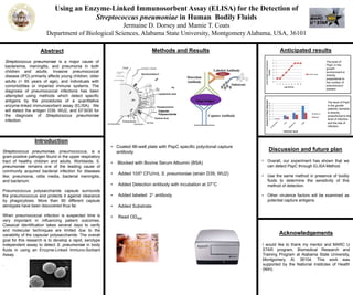
Glycomics, the study of glycans, is applied to biology and chemistry that focuses on the structure and function of carbohydrates, and on glycoform distributions at the cellular, tissue, organ and organism levels. Mass spectrometry plays an important role in glycomics analysis. If you want to know more, please visit https://www.creative-proteomics.com/services/glycomics-service.htm
Image J for analysis of biological image and nanoparticle size distribution Thoria Donia
1- Analysis of gel and western blot images
2- Analysis of fluorescence and colocalization signal of immunofluorescence images
3-Analysis of the nanoparticle size distribution
4- Analysis of stained liver tissue
5-Counting the cells
1. Immunochemical Techniques
The Technique which are used for identification, Characterization, Analysis, Optimization of
Protein, Peptide, Antigen and Antibody Reactions is known as Immunochemical Technique.
It can mainly include,
1. ELISA
2. RADIOIMMUNOASSAY
3. IMMUNOPRECIPITATION
4. IMMUNOELECTROPHOROSIS
Circular Dichroism Spectroscopy
Introduction
Amino Acid Structure & Polarity
Protein Structures
Polarization of Light
Circular Dichroism
Measurement of Circular Dichroism
Instrumentation For CD Spectropolarimeter
CD Spectrum
CD Spectra of Protein Secondary Structures
Other - CD Based Techniques
Conclusion
References
Role of soluble urokinase plasminogen activator receptor (suPAR) as prognosis...IOSR Journals
Biological marker suPAR was used in many pathological conditions, including infection. suPAR
was correlated with the severity of sepsis. The purpose of this study to determine levels of suPAR infants with
risk of infection as a prognostic indicator for sepsis. Groups of infants with the risk of infection (n = 43) were
followed prospectively on days 0, 3rd and 7th and observed for the incidence of sepsis compared to the control
group (n = 10). suPAR was measured by ELISA and the course of infection measured by clinical criteria.
Results suPAR day 0, 3 and 7, displayed in the form of bloxpot and AUC as prognostic power. suPAR control
levels 9.32 ng / mL, sepsis cutoff 15, 41 ng / mL and AUC of 80.3% [95% CI 65.7%, 94.9%, p = 0.00]. Graph
shows ROC AUC sepsis suPAR day 0, the 3rd and 7th respectively 61.9%, 66.6% and 94.4%. Sepsis with
improved output 16.53 ng / mL and worsening 22.19 ng / mL and AUC of 80.8% [95% CI (0.62 to 0.99), p =
0.02]. suPAR levels was increased in neonatal sepsis patients. suPAR could be used as a prognostic factor for
neonatal sepsis.
Background: It is often difficult to predict which newborn with HIE will develop neurological sequlae so there is an urgent need for predictors for adverse neurological outcomes in these infants. Aim of Study: To evaluate the serum levels of serum amyloid A (SAA) protein in newborns with HIE during the first week of life and after 3 and 6 months of follow up to assess its correlation with degree of HIE neurological sequlee. Patients and Methods; This case-control study was conducted on 72 infants; group (1) included 36 full term neonates diagnosed as HIE and group (2)included 36 age and sex matched, infants as a control group, Serum amyloid A by ELIZA technique was measured at post natal age of 1 and 7 days, CT scan was done in justified cases .with follow up at age of 3 and 6 months for neurological sequlee. Results: SAA protein level was elevated in the asphyxiated group in comparison to the control group at day 1 and day 7, SAA level was significantly correlated to the Sarnat scoring system of HIE. SAA level significantly differ on follow up of developmental milestone at age of 3 and 6 months. ROC curve for validity of SAA for severity of HIE at cut off point > 25μg/ml at day 1 and at cut off point > 20 μg/ml at day 7 of HIE diagnosis reported sensitivity 100% and specificity 100% .Conclusion: SAA correlates with the severity of HIE and higher SAA expression is a prognostic marker for morbidity in these infants.
1. Using an Enzyme-Linked Immunosorbent Assay (ELISA) for the Detection of
Streptococcus pneumoniae in Human Bodily Fluids
Jermaine D. Dorsey and Mamie T. Coats
Department of Biological Sciences, Alabama State University, Montgomery Alabama, USA, 36101
Abstract
Streptococcus pneumoniae is a major cause of
bacteremia, meningitis, and pneumonia in both
children and adults. Invasive pneumococcal
disease (IPD) primarily affects young children, older
adults (> 65 years of age), and individuals with
comorbidities or impaired immune systems. The
diagnosis of pneumococcal infections has been
attempted using methods which detect specific
antigens by the procedures of a quantitative
enzyme-linked immunosorbent assay (ELISA). We
will detect the antigen D39, WU2, and EF3030 for
the diagnosis of Streptococcus pneumoniae
infection.
Introduction
Streptococcus pneumoniae, pneumococcus, is a
gram-positive pathogen found in the upper respiratory
tract of healthy children and adults. Worldwide, S.
pneumoniae remains one of the leading cause of
community acquired bacterial infection for diseases
like; pneumonia, otitis media, bacterial meningitis,
and bacterium.
Pneumococcus polysaccharide capsule surrounds
the pneumococcus and protects it against clearance
by phagocytosis. More than 90 different capsule
serotypes have been discovered thus far.
When pneumococcal infection is suspected time is
very important in influencing patient outcomes.
Classical identification takes several days to verify
and molecular techniques are limited due to the
variability of the capsular polysaccharide. The overall
goal for this research is to develop a rapid, serotype
independent assay to detect S. pneumoniae in body
fluids in using an Enzyme-Linked Immuno-Sorbant
Assay.
.
• Coated 96-well plate with PspC specific polyclonal capture
antibody
• Blocked with Bovine Serum Albumin (BSA)
• Added 10X5 CFU/mL S. pneumoniae (strain D39, WU2)
• Added Detection antibody with incubation at 37°C
• Added labeled 2 antibody
• Added Substrate
• Read OD405
Discussion and future plan
• Overall, our experiment has shown that we
can detect PspC through ELISA Method.
• Use the same method in presence of bodily
fluids to determine the sensitivity of this
method of detection.
• Other virulence factors will be examined as
potential capture antigens.
Acknowledgements
I would like to thank my mentor and MARC U
STAR program, Biomedical Research and
Training Program at Alabama State University,
Montgomery, Al. 36104. This work was
supported by the National Institutes of Health
(NIH).
Methods and Results
Capture Antibody
Target Antigen
Detection
Antibody
Labeled Antibody
Biotin
Substrate
Anticipated results
0
1
2
3
4
5
6
7
1 2 3 4 5 6 7 8 9
LevelofPspCinEnvironment
(media)
Log CFU/mL
Free PspC
The level of
PspC in the
growth
environment is
directly
proportional to
the number of
pneumococci
present
The level of PspC
in the growth
patients’ samples
is directly
proportional to the
level of infection
and the site of
infection
pathmicro.med.sc.edu
0
2
4
6
8
10
12
Sputum
Blood
Infection level
AmountofPspCdetected