SlideShare a Scribd company logo
Ehlers Danlos Syndrome<br />( a genetic defect in Collagen Synthesis and structure )<br />King Abdulaziz University<br />Medicine Collage <br />-602742103378Department of Anatomy<br />Waleed Faisal Bokhari<br />Introduction<br />Background <br />Ehlers-Danlos syndrome (EDS) is the name given to a group of more than 10 different inherited disorders; all involve a genetic defect in collagen and connective-tissue synthesis and structure.<br />Ehlers-Danlos syndrome can affect the skin, joints, and blood vessels.This syndrome is clinically heterogeneous; the underlying collagen abnormality is different for each type.Clinical recognition of the types of Ehlers-Danlos syndrome is important. One type, type IV, is associated with arterial rupture and visceral perforation, with possible life-threatening consequences.<br />Pathophysiology<br />Ehlers-Danlos syndrome is a heterogeneous group of inherited connective-tissue disorders characterized by joint hypermobility, cutaneous fragility, and hyperextensibility. The collagen defect has been identified in only 6 of the 11 types of Ehlers-Danlos syndrome. Type IV is characterized by a decreased amount of type III collagen. Types V and VI are characterized by deficiencies in hydroxylase and lysyl oxidase, an important posttranslational modifying enzyme in collagen biosynthesis. Type VII has an amino-terminal procollagen peptidase deficiency. Type IX has abnormal copper metabolism. Type X has nonfunctioning plasma fibronectin. <br />In Ehlers-Danlos syndrome types I and II, the classic variety, identifying the molecular structure in most individuals who are affected is difficult. Causative mutations may involve the COL5A1, COL5A2, and tenascin-X genes and are implied to be in the COL1A2 gene. Nonetheless, in most families with autosomal dominant Ehlers-Danlos syndrome, the disease appears to be linked to loci that contain the COL5A1 or COL5A2 genes. Although half of the mutations that cause Ehlers-Danlos syndrome types I and II are likely to affect the COL5A1 gene, a significant portion of the mutations result in low levels of mRNA from the mutant allele as a consequence of nonsense-mediated mRNA decay.<br />Bouma et al evaluated 3 generations in a family with Ehlers-Danlos syndrome type II. The genomic defect was an A(-2) -> G substitution at the exon 14 splice-acceptor site. Transmission electron micrographs of type I collagen fibrils in a proband dermal biopsy specimen demonstrated heterogeneity in fibril diameter that was greater than that of a matched control sample. The proband was found to have a greater proportion of both larger and smaller fibrils, and occasional fibrils with a cauliflower configuration were observed.<br />Wenstrup and associates identified haploinsufficiency of the COL5A1 gene that encodes the proalpha1(V) chain of type V collagen in the classic form of Ehlers-Danlos syndrome. Eight of 28 probands with classic Ehlers-Danlos syndrome who were heterozygous for expressed polymorphisms in COL5A1 had complete or nearly complete loss of expression of one COL5A1 allele. One third of individuals with classic Ehlers-Danlos syndrome were estimated to have mutations of COL5A1 that result in haploinsufficiency. These findings suggest that the normal formation of the heterotypic collagen fibrils that contain types I, III, and V collagen requires the expression of both COL5A1 alleles. <br />Autosomal recessive–type VI Ehlers-Danlos syndrome, also known as the kyphoscoliotic type, is characterized by neonatal kyphoscoliosis, generalized joint laxity, skin fragility, and severe muscle hypotonia at birth. Biochemically, this type is attributed to a deficiency in lysyl hydroxylase (LH), the enzyme that hydroxylates specific lysine residues in the collagen molecule to form hydroxylysines with 2 important functions. The residues are attachment sites for galactose and glucosylgalactose, and they act as precursors of the cross-linking process that gives collagen its tensile strength.<br />More than 20 mutations are identified in the LH1 gene that contributes to LH deficiency and clinical Ehlers-Danlos syndrome type VI. Yeowell and Walker identified 2 of these mutations in 5 or more unrelated patients: (1) a large duplication of exons 10-16, which arise from a homologous recombination of intronic Alu sequences, and (2) a nonsense mutation, Y511X, in exon 14 of the LH1 gene. Both mutations seem to originate from a single ancestral gene.<br />Tenascin-X is a large extracellular matrix protein, a deficiency of which causes a clinically distinct recessive form of this syndrome. Thus, factors other than collagens or collagen-processing enzymes may cause this syndrome. This newly described form may be associated with additional anomalies.<br />A case with colonic perforation in a young girl, with a fatal outcome, was related to a novel mutation of the COL3A1 gene. Crystal structure of human type III, in the structure G991-G1032 cystine knot, shows both 7/2 and 10/3 symmetries.A novel point mutation has been found in the vascular type of Ehlers-Danlos syndrome. The mutation took place in the second position of exon 24 of COL3A1.Impaired wound healing is a typical feature of Ehlers-Danlos syndrome, probably for a fibroblast defect. Wound repair can be achieved using exogenous type V collagen.Ehlers-Danlos syndrome pediatric patients have been shown to have deficiencies in 3 genes of the glutathione S-transferase family (GSTM1, GSTT1, GSTP1). This leads to the generation of reactive oxygen species.Reduced activity of beta4-galactosyltransferase 7 (beta4GalT-7) is associated with the progeriform Ehlers-Danlos syndrome.<br />Frequency<br />The prevalence of Ehlers-Danlos syndrome is reported to be 1 case in approximately 400,000 people, but mild or incomplete forms appear to be under diagnosed and more common than other forms. It is usually diagnosed in young adults.<br />Mortality/Morbidity<br />20450741046966Type IV Ehlers-Danlos syndrome is a severe form. Patients often have a shortened lifespan because of the spontaneous rupture of a large artery (eg, splenic artery, aorta) or the perforation of internal organs. Surgery can pose life-threatening risks in these patients. The other types are usually not as dangerous, and affected individuals can live a healthy if somewhat restricted life. Type VI is also somewhat dangerous, although it is rare.<br />Clinical<br />History<br />The biochemical collagen defect is present at birth, but clinical manifestations become evident later.<br />Muscle weakness is often present, and patients report a tendency to fall down easily and have poor body control. <br />Sometimes, patients have difficulty walking. <br />Mental development is normal. <br />The newly described tenascin-X–deficient form was described in 8 patients with hyperelastic skin and hypermobile joints. <br />Each patient bruised easily, and most had velvety skin. <br />A few patients also had joint pain and multiple subluxations. <br />None had delayed wound healing or atrophic scars. <br />Dental pathology is common in these patients. Findings include hypodontia of permanent teeth, delayed eruption, and dentin dysplasia.<br />In one patient, splenic rupture due to peliosis led to the diagnosis of vascular Ehlers-Danlos syndrome.<br />Two Ehlers-Danlos syndrome patients with cutaneous metaplastic synovial cysts are described in the literature. <br />Multiple sclerosis can be associated with Ehlers-Danlos syndrome.<br />Absence of the inferior labial or lingual frenula in Ehlers-Danlos syndrome patients has been suggested as a new diagnostic criterion.<br />Subependymal periventricular heterotopia is not a rarity in Ehlers-Danlos syndrome patients.<br />Ehlers-Danlos syndrome and anorexia nervosa have been described in the same patient.<br />Several articles review pregnancy in Ehlers-Danlos syndrome patients. <br />Causes<br />Collagens are a family of proteins that are widely distributed in all organs of the body. Thirteen different subtypes are known, and the number increases constantly. <br />The joints, blood vessels, and skin have different kinds of collagen in their structure; in all these locations, collagen is organized into bundles. <br />Collagen organization is not easily visible by means of light microscopy, and abnormalities are better detected with electron microscopy. <br />Collagen disorganization and/or abnormal bundle size is correlated with clinical evidence in the joints, skin, and blood vessels. <br />In Ehlers-Danlos syndrome, skin collagen alteration can be seen in the reticular dermis. <br />Two abnormalities are evident: irregularities in the diameter of the fibrils and irregular collagen shapes. <br />Fibrils can be large and irregular in some types (types I-III), but they can also be small or varied in others. <br />The most severe form of Ehlers-Danlos syndrome, type IV, is the best studied; biochemically, this type has decreased or absent type III collagen synthesis. Pathologic findings in other skin layers are visible but nonspecific.<br />Physical<br />To date, 11 variants of Ehlers-Danlos syndrome are identified; all have genetic, biochemical, and clinical differences. More than one third of persons with Ehlers-Danlos syndrome do not fit exactly into a single type; overlap is common.<br />Common to almost all groups is a unique appearance of the skin (see Media Files 1-5). <br />The skin is usually white and soft, and underlying vessels are sometimes visible. <br />The skin has a doughy feel. <br />The skin is easily hyperextensible. It is easy to pull, and, once released, it immediately returns to its original state.<br />Molluscoid pseudotumors are small, spongy tumors found over scars and pressure points. <br />Molluscoid pseudotumors consist of fat surrounded by a fibrous capsule. <br />They are commonly seen in patients with type I.<br />Smaller, deep, palpable, and movable nodules are often present in the subcutaneous tissue. <br />These nodules can be found in the arms and over the tibias. <br />Calcification leads to opacity on radiographs.<br />Fragility of dermal skin is common, with frequent bruises and lacerations. <br />Poor wound healing is not rare. <br />The use of sutures is usually a problem in patients, in whom easy dehiscence and cigarette-paper–like scars may be observed. Frequently, these scars are found on the knees.<br />The joints are hyperextensible, sometimes dramatically, but the degree of involvement is variable. <br />The digit joints are most commonly affected, but all the joints can show alterations. <br />Dislocations can occur, but patients are usually able to quickly reduce them with no pain.<br />Type I, the gravis form, affects 43% of patients and is inherited in an autosomal dominant pattern. <br />In this type, the clinical features are usually severe. <br />Patients have marked skin extensibility with frequent lacerations and subsequent scarring in different body locations. <br />Surgical sutures heal poorly, with easy dehiscence. <br />Joint hypermobility is severe and affects all parts of the body. <br />Spontaneous dislocations can occur, but immediate reduction is easy. <br />Varicosities and molluscoid pseudotumors are common. <br />Musculoskeletal features are easily found. These features include kyphoscoliosis, hallus valgus, pes planus (ie, flat feet), and genu recurvatum; bruises are less common in this type than in other forms. <br />Cardiac defects, especially mitral valvular prolapse, are sometimes present. <br />Prematurity with rupture of the fetal membranes is specific to this type.<br />Type II, the mitis form, affects 35% of patients and is inherited in an autosomal dominant pattern. <br />This group is characterized by a mild appearance of the same features of type I. <br />Wide scars are common, but the skin has somewhat less fragility and bruisability. <br />The joints are moderately hyperextensible, and the digits are usually the only body sites affected.<br />Type III, the benign familiar hypermobile form, affects 10% of patients and is inherited in an autosomal dominant pattern. <br />Patients with this variant have minimal or no skin changes, but they do have a striking hyperextensibility in many joints. <br />This hyperextensibility usually causes orthopedic consequences (eg, severe osteoarthritis) in the long term.<br />Type IV, the ecchymotic or arterial forms, affects 6% of patients and is inherited in an autosomal recessive or sometimes autosomal dominant pattern. <br />This variant is relatively rare. <br />Clinically, patients have unique, white, translucent skin, and the underlying vessels are easy to see. <br />The skin is also fragile but not extensible. <br />Scars and molluscoid pseudotumors are numerous, as are bruises and purpuric lesions. <br />Keloids and hyperpigmentation of the scars are common. <br />Joint hyperextensibility is rare or absent. <br />Arterial aneurysms, valvular prolapse, and spontaneous pneumothorax are common complications. <br />Patients also have low weight and short stature. <br />The prognosis for this type is poor, and the patient's life span is shortened. <br />Sudden death can occur after visceral perforation or after the rupture of a large vessel, most commonly an abdominal or splenic vessel. <br />A prenatal diagnosis by means of polymorphic restriction genetic studies is possible.<br />Type V, the X-linked form, affects 5% of patients and is inherited in an X-linked recessive pattern. <br />The skin of patients with this form of Ehlers-Danlos syndrome is highly extensible, and orthopedic abnormalities are common. <br />Bruising and hyperextensibility are rare.<br />Type VI, the ocular form, affects 2% of patients and is inherited in an autosomal recessive pattern. <br />Patients with this type are clinically and severely affected by the disease. <br />The skin is extensible, bruises are common, and wound healing is poor. <br />Patients may have several scars, some of which can be hyperpigmented. <br />The joints are hyperextensible. <br />This subgroup has unique ocular clinical signs. The ocular fragility can cause retinal hemorrhage and detachment, glaucoma, and coloration of the sclera. Rupture of the globe is rare but possible. <br />Measurements of LH in the amniotic fluid can be used to predict the outcome of pregnancy.<br />Type VII, or arthrochalasis multiplex congenita, affects 3% of patients and is inherited in an autosomal recessive or autosomal dominant pattern. <br />Patients with this type have noticeable joint hyperextensibility, but skin changes are less severe than those of other types. <br />Patients have spontaneous joint dislocation, usually with rapid reduction. <br />Patients with this type are usually short in stature.<br />Type VIII, the periodontal form, is rare and inherited in an autosomal dominant pattern. <br />Patients with this type have dental involvement with gingival periodontal inflammation. <br />Skin laxity, joint hyperextensibility, and bruisability are variable. <br />Gingival resorption and permanent loss of the teeth are common by the time the patient is aged 30 years.<br />Type IX, or X-linked cutis laxa, is rare and inherited in an X-linked recessive pattern. <br />Patients with type IX have characteristic bilateral bony prominences on the occiput. <br />Rarely, the skin and joints are dramatically affected. <br />Chronic diarrhea and orthostatic hypotension are unique findings in this group. <br />Scars are usually evident because healing is poor. <br />Patients with this type have a defect in intracellular copper-dependent enzymes, similar to that of patients with Menkes syndrome.<br />Type X (fibronectin deficiency) and type XI (benign hypermobile joint syndrome) are rare forms of Ehlers-Danlos syndrome. Some suggest that these types are so similar that they are better classified as one type rather than 2. <br />In 1997, a new, simpler classification was proposed in an attempt to eliminate the confusion associated with the former classification. Although many dermatology books continue to include both classifications, the Ehlers-Danlos syndrome clinical forms can now be classified as follows: <br />Classic type was formerly types I and II. <br />Hypermobility type was formerly type III. <br />Vascular type was formerly type IV. <br />Kyphoscoliosis type was formerly type VI. <br />Arthrochalasis type was formerly type VII, characterized by deficiency of proA1 or proA2 chains of collagen type I. <br />Dermatosparaxis type was formerly type VII, characterized by deficiency of procollagen N -terminal peptidase. <br />Other was formerly types V, VIII, IX, X and XI.<br />A review of Ehlers-Danlos syndrome type VIII showed distinctive clinical features. The precise underlying molecular defect is unknown, but patients with this type are similar clinically.<br />Treatment <br />Medical Care<br />Treatment is unsatisfactory. <br />One isolated report showed that patients with type VI disease benefited from oral vitamin C at 4 g/d. Scars and bleeding time seemed to improve with this treatment.<br />Surgical Care<br />Extreme caution is mandatory in any surgical maneuver. <br />Plastic re-excision of scars sometimes provides acceptable cosmetic results. <br />Anesthetic implications, although rare, are very important in Ehlers-Danlos syndrome, especially in the type IV (or vascular) patients. Risk of complications is higher, spontaneous vascular rupture can occur, and cervical spine and airway trauma must be kept in mind. Bleeding is also reported.<br />Recombinant factor VIIa has been successful in the treatment of intractable bleeding in vascular-type Ehlers-Danlos syndrome.<br />Activity<br />Patients with Ehlers-Danlos syndrome types IV or VI should avoid participating in dangerous contact sports. <br />Some authors mention risks with activities that can increase intracranial pressure as a result of the Valsalva effect. An example of one such activity is playing the trumpet.<br />Medication<br />The goals of pharmacotherapy are to prevent complications and reduce morbidity.<br />Vitamins<br />Vitamin C may improve morbidity. It is a critical cofactor for collagen fibril synthesis.<br />307314634925Ascorbic acid (Cecon, Cevalin, Cevi-Bid )<br />For collagen synthesis and tissue repair.<br />Further Inpatient Care<br />Type IV Ehlers-Danlos syndrome patients should be carefully monitored because they are at high risk for spontaneous rupture of a large artery (eg, splenic artery, aorta) or perforation of internal organs. These patients should be educated that surgery can pose life-threatening risks. <br />Fernandez-Alcantud reviewed management of anesthesia in vascular type IV Ehlers-Danlos syndrome patients.<br />Jones reports the use of local anesthesia for elective cesarian delivery in a type III Ehlers-Danlos syndrome woman. <br />Deterrence/Prevention<br />Patients should avoid trauma and participation in contact sports. <br />Pregnancy is dangerous for some patients. <br />Bleeding risk should be considered in surgical operations.<br />Prognosis<br />Ehlers-Danlos syndrome type IV is a severe form, and patients with this disease often have a shortened lifespan. <br />Arterial aneurysms, valvular prolapse, and spontaneous pneumothorax are common complications. <br />The prognosis with this type is poor. <br />Sudden death can occur after visceral perforation or after the rupture of a large vessel, most commonly an abdominal and splenic vessel.<br />The other types usually are not as dangerous, and affected individuals can live healthy if somewhat restricted lives.<br />Patient Education<br />For excellent patient education resources, visit eMedicine's Skin, Hair, and Nails Center. Additionally, see eMedicine's patient education article Bruises.<br />Multimedia<br />Patient with Ehlers-Danlos syndrome. Note the abnormal ability to elevate the right toe. <br />Girl with Ehlers-Danlos syndrome. Dorsiflexion of all the fingers is easy and absolutely painless. <br />Patient with Ehlers-Danlos syndrome mitis. Joint hypermobility is less intense than with other conditions.<br />Dorsal view of a patient with Ehlers-Danlos syndrome. Note the S-curved spinal column. <br />Cigarette-paper–like scars over the knees of a patient with Ehlers-Danlos syndrome. Note also the deformity of the left knee.<br />
Ehlers Danlos Syndrome

More Related Content

What's hot

Osteogenesis imperfecta
Osteogenesis imperfectaOsteogenesis imperfecta
Osteogenesis imperfecta
BipulBorthakur
 
Marfan syndrome
Marfan syndromeMarfan syndrome
Marfan syndrome
zakariy al-nuaimi
 
Osteogenesis imperfecta (OI)
Osteogenesis imperfecta (OI) Osteogenesis imperfecta (OI)
Osteogenesis imperfecta (OI)
Carmela Domocmat
 
Bone mineral density (bmd) test
Bone mineral density (bmd) testBone mineral density (bmd) test
Bone mineral density (bmd) test
apoorvaerukulla
 
Epidermolysis bullosa complete
Epidermolysis bullosa  completeEpidermolysis bullosa  complete
Epidermolysis bullosa complete
Dr.vivek ranjan
 
Genetic disorders of human by Jeba Akhtar
Genetic disorders of human by Jeba AkhtarGenetic disorders of human by Jeba Akhtar
Genetic disorders of human by Jeba Akhtar
ZebaAkhtar8
 
Osteogenesis imperfecta
Osteogenesis imperfectaOsteogenesis imperfecta
Osteogenesis imperfectaorthoprince
 
Achondroplasia
AchondroplasiaAchondroplasia
Achondroplasia
Dr. Saad Saleh Al Ani
 
Infantile cortical hyperostosis dr. rohit kumar singh
Infantile cortical hyperostosis  dr. rohit kumar singhInfantile cortical hyperostosis  dr. rohit kumar singh
Infantile cortical hyperostosis dr. rohit kumar singh
rohitkumarsingh104
 
Osteopetrosis(albergs schoberg disease
Osteopetrosis(albergs schoberg diseaseOsteopetrosis(albergs schoberg disease
Osteopetrosis(albergs schoberg disease
Jamil Kifayatullah
 
Hypermobility syndromes
Hypermobility syndromesHypermobility syndromes
Hypermobility syndromes
Mohamed Hefny
 
Osteoarthritis
OsteoarthritisOsteoarthritis
Osteoarthritis
drkmliau
 
Presentation1.pptx, radiological imaging of osteoarthritis.
Presentation1.pptx, radiological imaging of osteoarthritis.Presentation1.pptx, radiological imaging of osteoarthritis.
Presentation1.pptx, radiological imaging of osteoarthritis.
Abdellah Nazeer
 
Rickets
RicketsRickets
Lupus Erythematosus for dermatologists
 Lupus Erythematosus for dermatologists Lupus Erythematosus for dermatologists
Lupus Erythematosus for dermatologists
Mostafa Sanad
 
4.marfansyndrome
4.marfansyndrome4.marfansyndrome
4.marfansyndrome
AhmedAdamMohamud
 
Presentation1.pptx, imaging of genetic diseases. (3)
Presentation1.pptx, imaging of genetic diseases. (3)Presentation1.pptx, imaging of genetic diseases. (3)
Presentation1.pptx, imaging of genetic diseases. (3)Abdellah Nazeer
 
scleroderma
sclerodermascleroderma
scleroderma
aadenitan1
 

What's hot (20)

Osteogenesis imperfecta
Osteogenesis imperfectaOsteogenesis imperfecta
Osteogenesis imperfecta
 
Marfan syndrome
Marfan syndromeMarfan syndrome
Marfan syndrome
 
Osteogenesis imperfecta (OI)
Osteogenesis imperfecta (OI) Osteogenesis imperfecta (OI)
Osteogenesis imperfecta (OI)
 
Bone mineral density (bmd) test
Bone mineral density (bmd) testBone mineral density (bmd) test
Bone mineral density (bmd) test
 
Epidermolysis bullosa complete
Epidermolysis bullosa  completeEpidermolysis bullosa  complete
Epidermolysis bullosa complete
 
Genetic disorders of human by Jeba Akhtar
Genetic disorders of human by Jeba AkhtarGenetic disorders of human by Jeba Akhtar
Genetic disorders of human by Jeba Akhtar
 
Osteogenesis imperfecta
Osteogenesis imperfectaOsteogenesis imperfecta
Osteogenesis imperfecta
 
Achondroplasia
AchondroplasiaAchondroplasia
Achondroplasia
 
Infantile cortical hyperostosis dr. rohit kumar singh
Infantile cortical hyperostosis  dr. rohit kumar singhInfantile cortical hyperostosis  dr. rohit kumar singh
Infantile cortical hyperostosis dr. rohit kumar singh
 
Osteopetrosis(albergs schoberg disease
Osteopetrosis(albergs schoberg diseaseOsteopetrosis(albergs schoberg disease
Osteopetrosis(albergs schoberg disease
 
Hypermobility syndromes
Hypermobility syndromesHypermobility syndromes
Hypermobility syndromes
 
Osteoarthritis
OsteoarthritisOsteoarthritis
Osteoarthritis
 
Presentation1.pptx, radiological imaging of osteoarthritis.
Presentation1.pptx, radiological imaging of osteoarthritis.Presentation1.pptx, radiological imaging of osteoarthritis.
Presentation1.pptx, radiological imaging of osteoarthritis.
 
Rickets
RicketsRickets
Rickets
 
Dwarfism 2
Dwarfism 2Dwarfism 2
Dwarfism 2
 
Lupus Erythematosus for dermatologists
 Lupus Erythematosus for dermatologists Lupus Erythematosus for dermatologists
Lupus Erythematosus for dermatologists
 
4.marfansyndrome
4.marfansyndrome4.marfansyndrome
4.marfansyndrome
 
Presentation1.pptx, imaging of genetic diseases. (3)
Presentation1.pptx, imaging of genetic diseases. (3)Presentation1.pptx, imaging of genetic diseases. (3)
Presentation1.pptx, imaging of genetic diseases. (3)
 
Rheumatoid arthritis
Rheumatoid arthritisRheumatoid arthritis
Rheumatoid arthritis
 
scleroderma
sclerodermascleroderma
scleroderma
 

Similar to Ehlers Danlos Syndrome

Review Article on Ehler Danlos Syndrome Type IV
Review Article on Ehler Danlos Syndrome Type IVReview Article on Ehler Danlos Syndrome Type IV
Review Article on Ehler Danlos Syndrome Type IV
iosrjce
 
Connective tissue disorders
Connective tissue disordersConnective tissue disorders
Connective tissue disorders
LalisaMerga
 
Genetics
GeneticsGenetics
Genetics
drsapnaharsha
 
Genetics for undergraduates
Genetics for undergraduatesGenetics for undergraduates
Genetics for undergraduates
Drsapna Harsha
 
Etiology and pathogenesis of heredity and congenital diseases presentation
Etiology and pathogenesis of heredity and congenital diseases presentationEtiology and pathogenesis of heredity and congenital diseases presentation
Etiology and pathogenesis of heredity and congenital diseases presentation
Muhammad Faisal Mukhtar
 
Genetic disorders pdf
Genetic disorders pdfGenetic disorders pdf
Genetic disorders pdf
jrseema
 
TRADITIONAL INHERITANCE
TRADITIONAL INHERITANCETRADITIONAL INHERITANCE
TRADITIONAL INHERITANCE
ShamilaM3
 
Genetic disorder
Genetic disorderGenetic disorder
Genetic disorder
Chhavi Agarwal
 
GENETIC DISORDERS
GENETIC DISORDERSGENETIC DISORDERS
GENETIC DISORDERS
Rabia Khan Baber
 
human variation marfan syndromeAZ2
human variation marfan syndromeAZ2human variation marfan syndromeAZ2
human variation marfan syndromeAZ2Lee Ann Wayland
 
Arthrogryposis multiplex congenita
Arthrogryposis multiplex congenitaArthrogryposis multiplex congenita
Arthrogryposis multiplex congenita
Naveed Jumani
 
Epidermolysis bullosa
Epidermolysis bullosaEpidermolysis bullosa
Epidermolysis bullosa
Noel Christian
 
Genetics
GeneticsGenetics
ETAS_MCQ_03 a genodermatoses
ETAS_MCQ_03 a genodermatosesETAS_MCQ_03 a genodermatoses
ETAS_MCQ_03 a genodermatosesDerma202
 
Other diseases, called collagen vascular diseases, affect the protein.pdf
Other diseases, called collagen vascular diseases, affect the protein.pdfOther diseases, called collagen vascular diseases, affect the protein.pdf
Other diseases, called collagen vascular diseases, affect the protein.pdf
arkurkuri
 
Progeria
ProgeriaProgeria
Progeria
Monem Alshok
 
Genetic Diseases
Genetic DiseasesGenetic Diseases
Genetic Diseases
Jessabeth Aluba
 

Similar to Ehlers Danlos Syndrome (20)

Review Article on Ehler Danlos Syndrome Type IV
Review Article on Ehler Danlos Syndrome Type IVReview Article on Ehler Danlos Syndrome Type IV
Review Article on Ehler Danlos Syndrome Type IV
 
Connective tissue disorders
Connective tissue disordersConnective tissue disorders
Connective tissue disorders
 
Genetics
GeneticsGenetics
Genetics
 
Genetics for undergraduates
Genetics for undergraduatesGenetics for undergraduates
Genetics for undergraduates
 
Etiology and pathogenesis of heredity and congenital diseases presentation
Etiology and pathogenesis of heredity and congenital diseases presentationEtiology and pathogenesis of heredity and congenital diseases presentation
Etiology and pathogenesis of heredity and congenital diseases presentation
 
Genetic disorders pdf
Genetic disorders pdfGenetic disorders pdf
Genetic disorders pdf
 
Chromosomal aberration syndrome biology
Chromosomal aberration syndrome biologyChromosomal aberration syndrome biology
Chromosomal aberration syndrome biology
 
TRADITIONAL INHERITANCE
TRADITIONAL INHERITANCETRADITIONAL INHERITANCE
TRADITIONAL INHERITANCE
 
Genetic disorder
Genetic disorderGenetic disorder
Genetic disorder
 
GENETIC DISORDERS
GENETIC DISORDERSGENETIC DISORDERS
GENETIC DISORDERS
 
Mutatio
MutatioMutatio
Mutatio
 
human variation marfan syndromeAZ2
human variation marfan syndromeAZ2human variation marfan syndromeAZ2
human variation marfan syndromeAZ2
 
Arthrogryposis multiplex congenita
Arthrogryposis multiplex congenitaArthrogryposis multiplex congenita
Arthrogryposis multiplex congenita
 
Genetic
GeneticGenetic
Genetic
 
Epidermolysis bullosa
Epidermolysis bullosaEpidermolysis bullosa
Epidermolysis bullosa
 
Genetics
GeneticsGenetics
Genetics
 
ETAS_MCQ_03 a genodermatoses
ETAS_MCQ_03 a genodermatosesETAS_MCQ_03 a genodermatoses
ETAS_MCQ_03 a genodermatoses
 
Other diseases, called collagen vascular diseases, affect the protein.pdf
Other diseases, called collagen vascular diseases, affect the protein.pdfOther diseases, called collagen vascular diseases, affect the protein.pdf
Other diseases, called collagen vascular diseases, affect the protein.pdf
 
Progeria
ProgeriaProgeria
Progeria
 
Genetic Diseases
Genetic DiseasesGenetic Diseases
Genetic Diseases
 

Recently uploaded

Triangles of Neck and Clinical Correlation by Dr. RIG.pptx
Triangles of Neck and Clinical Correlation by Dr. RIG.pptxTriangles of Neck and Clinical Correlation by Dr. RIG.pptx
Triangles of Neck and Clinical Correlation by Dr. RIG.pptx
Dr. Rabia Inam Gandapore
 
For Better Surat #ℂall #Girl Service ❤85270-49040❤ Surat #ℂall #Girls
For Better Surat #ℂall #Girl Service ❤85270-49040❤ Surat #ℂall #GirlsFor Better Surat #ℂall #Girl Service ❤85270-49040❤ Surat #ℂall #Girls
For Better Surat #ℂall #Girl Service ❤85270-49040❤ Surat #ℂall #Girls
Savita Shen $i11
 
Ocular injury ppt Upendra pal optometrist upums saifai etawah
Ocular injury  ppt  Upendra pal  optometrist upums saifai etawahOcular injury  ppt  Upendra pal  optometrist upums saifai etawah
Ocular injury ppt Upendra pal optometrist upums saifai etawah
pal078100
 
heat stroke and heat exhaustion in children
heat stroke and heat exhaustion in childrenheat stroke and heat exhaustion in children
heat stroke and heat exhaustion in children
SumeraAhmad5
 
Charaka Samhita Sutra sthana Chapter 15 Upakalpaniyaadhyaya
Charaka Samhita Sutra sthana Chapter 15 UpakalpaniyaadhyayaCharaka Samhita Sutra sthana Chapter 15 Upakalpaniyaadhyaya
Charaka Samhita Sutra sthana Chapter 15 Upakalpaniyaadhyaya
Dr KHALID B.M
 
ARTHROLOGY PPT NCISM SYLLABUS AYURVEDA STUDENTS
ARTHROLOGY PPT NCISM SYLLABUS AYURVEDA STUDENTSARTHROLOGY PPT NCISM SYLLABUS AYURVEDA STUDENTS
ARTHROLOGY PPT NCISM SYLLABUS AYURVEDA STUDENTS
Dr. Vinay Pareek
 
ARTIFICIAL INTELLIGENCE IN HEALTHCARE.pdf
ARTIFICIAL INTELLIGENCE IN  HEALTHCARE.pdfARTIFICIAL INTELLIGENCE IN  HEALTHCARE.pdf
ARTIFICIAL INTELLIGENCE IN HEALTHCARE.pdf
Anujkumaranit
 
Antiulcer drugs Advance Pharmacology .pptx
Antiulcer drugs Advance Pharmacology .pptxAntiulcer drugs Advance Pharmacology .pptx
Antiulcer drugs Advance Pharmacology .pptx
Rohit chaurpagar
 
Prix Galien International 2024 Forum Program
Prix Galien International 2024 Forum ProgramPrix Galien International 2024 Forum Program
Prix Galien International 2024 Forum Program
Levi Shapiro
 
Surgical Site Infections, pathophysiology, and prevention.pptx
Surgical Site Infections, pathophysiology, and prevention.pptxSurgical Site Infections, pathophysiology, and prevention.pptx
Surgical Site Infections, pathophysiology, and prevention.pptx
jval Landero
 
The POPPY STUDY (Preconception to post-partum cardiovascular function in prim...
The POPPY STUDY (Preconception to post-partum cardiovascular function in prim...The POPPY STUDY (Preconception to post-partum cardiovascular function in prim...
The POPPY STUDY (Preconception to post-partum cardiovascular function in prim...
Catherine Liao
 
MANAGEMENT OF ATRIOVENTRICULAR CONDUCTION BLOCK.pdf
MANAGEMENT OF ATRIOVENTRICULAR CONDUCTION BLOCK.pdfMANAGEMENT OF ATRIOVENTRICULAR CONDUCTION BLOCK.pdf
MANAGEMENT OF ATRIOVENTRICULAR CONDUCTION BLOCK.pdf
Jim Jacob Roy
 
Cervical & Brachial Plexus By Dr. RIG.pptx
Cervical & Brachial Plexus By Dr. RIG.pptxCervical & Brachial Plexus By Dr. RIG.pptx
Cervical & Brachial Plexus By Dr. RIG.pptx
Dr. Rabia Inam Gandapore
 
Lung Cancer: Artificial Intelligence, Synergetics, Complex System Analysis, S...
Lung Cancer: Artificial Intelligence, Synergetics, Complex System Analysis, S...Lung Cancer: Artificial Intelligence, Synergetics, Complex System Analysis, S...
Lung Cancer: Artificial Intelligence, Synergetics, Complex System Analysis, S...
Oleg Kshivets
 
Factory Supply Best Quality Pmk Oil CAS 28578–16–7 PMK Powder in Stock
Factory Supply Best Quality Pmk Oil CAS 28578–16–7 PMK Powder in StockFactory Supply Best Quality Pmk Oil CAS 28578–16–7 PMK Powder in Stock
Factory Supply Best Quality Pmk Oil CAS 28578–16–7 PMK Powder in Stock
rebeccabio
 
ANATOMY AND PHYSIOLOGY OF URINARY SYSTEM.pptx
ANATOMY AND PHYSIOLOGY OF URINARY SYSTEM.pptxANATOMY AND PHYSIOLOGY OF URINARY SYSTEM.pptx
ANATOMY AND PHYSIOLOGY OF URINARY SYSTEM.pptx
Swetaba Besh
 
Evaluation of antidepressant activity of clitoris ternatea in animals
Evaluation of antidepressant activity of clitoris ternatea in animalsEvaluation of antidepressant activity of clitoris ternatea in animals
Evaluation of antidepressant activity of clitoris ternatea in animals
Shweta
 
Alcohol_Dr. Jeenal Mistry MD Pharmacology.pdf
Alcohol_Dr. Jeenal Mistry MD Pharmacology.pdfAlcohol_Dr. Jeenal Mistry MD Pharmacology.pdf
Alcohol_Dr. Jeenal Mistry MD Pharmacology.pdf
Dr Jeenal Mistry
 
Surat @ℂall @Girls ꧁❤8527049040❤꧂@ℂall @Girls Service Vip Top Model Safe
Surat @ℂall @Girls ꧁❤8527049040❤꧂@ℂall @Girls Service Vip Top Model SafeSurat @ℂall @Girls ꧁❤8527049040❤꧂@ℂall @Girls Service Vip Top Model Safe
Surat @ℂall @Girls ꧁❤8527049040❤꧂@ℂall @Girls Service Vip Top Model Safe
Savita Shen $i11
 
Ozempic: Preoperative Management of Patients on GLP-1 Receptor Agonists
Ozempic: Preoperative Management of Patients on GLP-1 Receptor Agonists  Ozempic: Preoperative Management of Patients on GLP-1 Receptor Agonists
Ozempic: Preoperative Management of Patients on GLP-1 Receptor Agonists
Saeid Safari
 

Recently uploaded (20)

Triangles of Neck and Clinical Correlation by Dr. RIG.pptx
Triangles of Neck and Clinical Correlation by Dr. RIG.pptxTriangles of Neck and Clinical Correlation by Dr. RIG.pptx
Triangles of Neck and Clinical Correlation by Dr. RIG.pptx
 
For Better Surat #ℂall #Girl Service ❤85270-49040❤ Surat #ℂall #Girls
For Better Surat #ℂall #Girl Service ❤85270-49040❤ Surat #ℂall #GirlsFor Better Surat #ℂall #Girl Service ❤85270-49040❤ Surat #ℂall #Girls
For Better Surat #ℂall #Girl Service ❤85270-49040❤ Surat #ℂall #Girls
 
Ocular injury ppt Upendra pal optometrist upums saifai etawah
Ocular injury  ppt  Upendra pal  optometrist upums saifai etawahOcular injury  ppt  Upendra pal  optometrist upums saifai etawah
Ocular injury ppt Upendra pal optometrist upums saifai etawah
 
heat stroke and heat exhaustion in children
heat stroke and heat exhaustion in childrenheat stroke and heat exhaustion in children
heat stroke and heat exhaustion in children
 
Charaka Samhita Sutra sthana Chapter 15 Upakalpaniyaadhyaya
Charaka Samhita Sutra sthana Chapter 15 UpakalpaniyaadhyayaCharaka Samhita Sutra sthana Chapter 15 Upakalpaniyaadhyaya
Charaka Samhita Sutra sthana Chapter 15 Upakalpaniyaadhyaya
 
ARTHROLOGY PPT NCISM SYLLABUS AYURVEDA STUDENTS
ARTHROLOGY PPT NCISM SYLLABUS AYURVEDA STUDENTSARTHROLOGY PPT NCISM SYLLABUS AYURVEDA STUDENTS
ARTHROLOGY PPT NCISM SYLLABUS AYURVEDA STUDENTS
 
ARTIFICIAL INTELLIGENCE IN HEALTHCARE.pdf
ARTIFICIAL INTELLIGENCE IN  HEALTHCARE.pdfARTIFICIAL INTELLIGENCE IN  HEALTHCARE.pdf
ARTIFICIAL INTELLIGENCE IN HEALTHCARE.pdf
 
Antiulcer drugs Advance Pharmacology .pptx
Antiulcer drugs Advance Pharmacology .pptxAntiulcer drugs Advance Pharmacology .pptx
Antiulcer drugs Advance Pharmacology .pptx
 
Prix Galien International 2024 Forum Program
Prix Galien International 2024 Forum ProgramPrix Galien International 2024 Forum Program
Prix Galien International 2024 Forum Program
 
Surgical Site Infections, pathophysiology, and prevention.pptx
Surgical Site Infections, pathophysiology, and prevention.pptxSurgical Site Infections, pathophysiology, and prevention.pptx
Surgical Site Infections, pathophysiology, and prevention.pptx
 
The POPPY STUDY (Preconception to post-partum cardiovascular function in prim...
The POPPY STUDY (Preconception to post-partum cardiovascular function in prim...The POPPY STUDY (Preconception to post-partum cardiovascular function in prim...
The POPPY STUDY (Preconception to post-partum cardiovascular function in prim...
 
MANAGEMENT OF ATRIOVENTRICULAR CONDUCTION BLOCK.pdf
MANAGEMENT OF ATRIOVENTRICULAR CONDUCTION BLOCK.pdfMANAGEMENT OF ATRIOVENTRICULAR CONDUCTION BLOCK.pdf
MANAGEMENT OF ATRIOVENTRICULAR CONDUCTION BLOCK.pdf
 
Cervical & Brachial Plexus By Dr. RIG.pptx
Cervical & Brachial Plexus By Dr. RIG.pptxCervical & Brachial Plexus By Dr. RIG.pptx
Cervical & Brachial Plexus By Dr. RIG.pptx
 
Lung Cancer: Artificial Intelligence, Synergetics, Complex System Analysis, S...
Lung Cancer: Artificial Intelligence, Synergetics, Complex System Analysis, S...Lung Cancer: Artificial Intelligence, Synergetics, Complex System Analysis, S...
Lung Cancer: Artificial Intelligence, Synergetics, Complex System Analysis, S...
 
Factory Supply Best Quality Pmk Oil CAS 28578–16–7 PMK Powder in Stock
Factory Supply Best Quality Pmk Oil CAS 28578–16–7 PMK Powder in StockFactory Supply Best Quality Pmk Oil CAS 28578–16–7 PMK Powder in Stock
Factory Supply Best Quality Pmk Oil CAS 28578–16–7 PMK Powder in Stock
 
ANATOMY AND PHYSIOLOGY OF URINARY SYSTEM.pptx
ANATOMY AND PHYSIOLOGY OF URINARY SYSTEM.pptxANATOMY AND PHYSIOLOGY OF URINARY SYSTEM.pptx
ANATOMY AND PHYSIOLOGY OF URINARY SYSTEM.pptx
 
Evaluation of antidepressant activity of clitoris ternatea in animals
Evaluation of antidepressant activity of clitoris ternatea in animalsEvaluation of antidepressant activity of clitoris ternatea in animals
Evaluation of antidepressant activity of clitoris ternatea in animals
 
Alcohol_Dr. Jeenal Mistry MD Pharmacology.pdf
Alcohol_Dr. Jeenal Mistry MD Pharmacology.pdfAlcohol_Dr. Jeenal Mistry MD Pharmacology.pdf
Alcohol_Dr. Jeenal Mistry MD Pharmacology.pdf
 
Surat @ℂall @Girls ꧁❤8527049040❤꧂@ℂall @Girls Service Vip Top Model Safe
Surat @ℂall @Girls ꧁❤8527049040❤꧂@ℂall @Girls Service Vip Top Model SafeSurat @ℂall @Girls ꧁❤8527049040❤꧂@ℂall @Girls Service Vip Top Model Safe
Surat @ℂall @Girls ꧁❤8527049040❤꧂@ℂall @Girls Service Vip Top Model Safe
 
Ozempic: Preoperative Management of Patients on GLP-1 Receptor Agonists
Ozempic: Preoperative Management of Patients on GLP-1 Receptor Agonists  Ozempic: Preoperative Management of Patients on GLP-1 Receptor Agonists
Ozempic: Preoperative Management of Patients on GLP-1 Receptor Agonists
 

Ehlers Danlos Syndrome

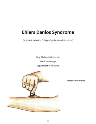
  • 1. Ehlers Danlos Syndrome<br />( a genetic defect in Collagen Synthesis and structure )<br />King Abdulaziz University<br />Medicine Collage <br />-602742103378Department of Anatomy<br />Waleed Faisal Bokhari<br />Introduction<br />Background <br />Ehlers-Danlos syndrome (EDS) is the name given to a group of more than 10 different inherited disorders; all involve a genetic defect in collagen and connective-tissue synthesis and structure.<br />Ehlers-Danlos syndrome can affect the skin, joints, and blood vessels.This syndrome is clinically heterogeneous; the underlying collagen abnormality is different for each type.Clinical recognition of the types of Ehlers-Danlos syndrome is important. One type, type IV, is associated with arterial rupture and visceral perforation, with possible life-threatening consequences.<br />Pathophysiology<br />Ehlers-Danlos syndrome is a heterogeneous group of inherited connective-tissue disorders characterized by joint hypermobility, cutaneous fragility, and hyperextensibility. The collagen defect has been identified in only 6 of the 11 types of Ehlers-Danlos syndrome. Type IV is characterized by a decreased amount of type III collagen. Types V and VI are characterized by deficiencies in hydroxylase and lysyl oxidase, an important posttranslational modifying enzyme in collagen biosynthesis. Type VII has an amino-terminal procollagen peptidase deficiency. Type IX has abnormal copper metabolism. Type X has nonfunctioning plasma fibronectin. <br />In Ehlers-Danlos syndrome types I and II, the classic variety, identifying the molecular structure in most individuals who are affected is difficult. Causative mutations may involve the COL5A1, COL5A2, and tenascin-X genes and are implied to be in the COL1A2 gene. Nonetheless, in most families with autosomal dominant Ehlers-Danlos syndrome, the disease appears to be linked to loci that contain the COL5A1 or COL5A2 genes. Although half of the mutations that cause Ehlers-Danlos syndrome types I and II are likely to affect the COL5A1 gene, a significant portion of the mutations result in low levels of mRNA from the mutant allele as a consequence of nonsense-mediated mRNA decay.<br />Bouma et al evaluated 3 generations in a family with Ehlers-Danlos syndrome type II. The genomic defect was an A(-2) -> G substitution at the exon 14 splice-acceptor site. Transmission electron micrographs of type I collagen fibrils in a proband dermal biopsy specimen demonstrated heterogeneity in fibril diameter that was greater than that of a matched control sample. The proband was found to have a greater proportion of both larger and smaller fibrils, and occasional fibrils with a cauliflower configuration were observed.<br />Wenstrup and associates identified haploinsufficiency of the COL5A1 gene that encodes the proalpha1(V) chain of type V collagen in the classic form of Ehlers-Danlos syndrome. Eight of 28 probands with classic Ehlers-Danlos syndrome who were heterozygous for expressed polymorphisms in COL5A1 had complete or nearly complete loss of expression of one COL5A1 allele. One third of individuals with classic Ehlers-Danlos syndrome were estimated to have mutations of COL5A1 that result in haploinsufficiency. These findings suggest that the normal formation of the heterotypic collagen fibrils that contain types I, III, and V collagen requires the expression of both COL5A1 alleles. <br />Autosomal recessive–type VI Ehlers-Danlos syndrome, also known as the kyphoscoliotic type, is characterized by neonatal kyphoscoliosis, generalized joint laxity, skin fragility, and severe muscle hypotonia at birth. Biochemically, this type is attributed to a deficiency in lysyl hydroxylase (LH), the enzyme that hydroxylates specific lysine residues in the collagen molecule to form hydroxylysines with 2 important functions. The residues are attachment sites for galactose and glucosylgalactose, and they act as precursors of the cross-linking process that gives collagen its tensile strength.<br />More than 20 mutations are identified in the LH1 gene that contributes to LH deficiency and clinical Ehlers-Danlos syndrome type VI. Yeowell and Walker identified 2 of these mutations in 5 or more unrelated patients: (1) a large duplication of exons 10-16, which arise from a homologous recombination of intronic Alu sequences, and (2) a nonsense mutation, Y511X, in exon 14 of the LH1 gene. Both mutations seem to originate from a single ancestral gene.<br />Tenascin-X is a large extracellular matrix protein, a deficiency of which causes a clinically distinct recessive form of this syndrome. Thus, factors other than collagens or collagen-processing enzymes may cause this syndrome. This newly described form may be associated with additional anomalies.<br />A case with colonic perforation in a young girl, with a fatal outcome, was related to a novel mutation of the COL3A1 gene. Crystal structure of human type III, in the structure G991-G1032 cystine knot, shows both 7/2 and 10/3 symmetries.A novel point mutation has been found in the vascular type of Ehlers-Danlos syndrome. The mutation took place in the second position of exon 24 of COL3A1.Impaired wound healing is a typical feature of Ehlers-Danlos syndrome, probably for a fibroblast defect. Wound repair can be achieved using exogenous type V collagen.Ehlers-Danlos syndrome pediatric patients have been shown to have deficiencies in 3 genes of the glutathione S-transferase family (GSTM1, GSTT1, GSTP1). This leads to the generation of reactive oxygen species.Reduced activity of beta4-galactosyltransferase 7 (beta4GalT-7) is associated with the progeriform Ehlers-Danlos syndrome.<br />Frequency<br />The prevalence of Ehlers-Danlos syndrome is reported to be 1 case in approximately 400,000 people, but mild or incomplete forms appear to be under diagnosed and more common than other forms. It is usually diagnosed in young adults.<br />Mortality/Morbidity<br />20450741046966Type IV Ehlers-Danlos syndrome is a severe form. Patients often have a shortened lifespan because of the spontaneous rupture of a large artery (eg, splenic artery, aorta) or the perforation of internal organs. Surgery can pose life-threatening risks in these patients. The other types are usually not as dangerous, and affected individuals can live a healthy if somewhat restricted life. Type VI is also somewhat dangerous, although it is rare.<br />Clinical<br />History<br />The biochemical collagen defect is present at birth, but clinical manifestations become evident later.<br />Muscle weakness is often present, and patients report a tendency to fall down easily and have poor body control. <br />Sometimes, patients have difficulty walking. <br />Mental development is normal. <br />The newly described tenascin-X–deficient form was described in 8 patients with hyperelastic skin and hypermobile joints. <br />Each patient bruised easily, and most had velvety skin. <br />A few patients also had joint pain and multiple subluxations. <br />None had delayed wound healing or atrophic scars. <br />Dental pathology is common in these patients. Findings include hypodontia of permanent teeth, delayed eruption, and dentin dysplasia.<br />In one patient, splenic rupture due to peliosis led to the diagnosis of vascular Ehlers-Danlos syndrome.<br />Two Ehlers-Danlos syndrome patients with cutaneous metaplastic synovial cysts are described in the literature. <br />Multiple sclerosis can be associated with Ehlers-Danlos syndrome.<br />Absence of the inferior labial or lingual frenula in Ehlers-Danlos syndrome patients has been suggested as a new diagnostic criterion.<br />Subependymal periventricular heterotopia is not a rarity in Ehlers-Danlos syndrome patients.<br />Ehlers-Danlos syndrome and anorexia nervosa have been described in the same patient.<br />Several articles review pregnancy in Ehlers-Danlos syndrome patients. <br />Causes<br />Collagens are a family of proteins that are widely distributed in all organs of the body. Thirteen different subtypes are known, and the number increases constantly. <br />The joints, blood vessels, and skin have different kinds of collagen in their structure; in all these locations, collagen is organized into bundles. <br />Collagen organization is not easily visible by means of light microscopy, and abnormalities are better detected with electron microscopy. <br />Collagen disorganization and/or abnormal bundle size is correlated with clinical evidence in the joints, skin, and blood vessels. <br />In Ehlers-Danlos syndrome, skin collagen alteration can be seen in the reticular dermis. <br />Two abnormalities are evident: irregularities in the diameter of the fibrils and irregular collagen shapes. <br />Fibrils can be large and irregular in some types (types I-III), but they can also be small or varied in others. <br />The most severe form of Ehlers-Danlos syndrome, type IV, is the best studied; biochemically, this type has decreased or absent type III collagen synthesis. Pathologic findings in other skin layers are visible but nonspecific.<br />Physical<br />To date, 11 variants of Ehlers-Danlos syndrome are identified; all have genetic, biochemical, and clinical differences. More than one third of persons with Ehlers-Danlos syndrome do not fit exactly into a single type; overlap is common.<br />Common to almost all groups is a unique appearance of the skin (see Media Files 1-5). <br />The skin is usually white and soft, and underlying vessels are sometimes visible. <br />The skin has a doughy feel. <br />The skin is easily hyperextensible. It is easy to pull, and, once released, it immediately returns to its original state.<br />Molluscoid pseudotumors are small, spongy tumors found over scars and pressure points. <br />Molluscoid pseudotumors consist of fat surrounded by a fibrous capsule. <br />They are commonly seen in patients with type I.<br />Smaller, deep, palpable, and movable nodules are often present in the subcutaneous tissue. <br />These nodules can be found in the arms and over the tibias. <br />Calcification leads to opacity on radiographs.<br />Fragility of dermal skin is common, with frequent bruises and lacerations. <br />Poor wound healing is not rare. <br />The use of sutures is usually a problem in patients, in whom easy dehiscence and cigarette-paper–like scars may be observed. Frequently, these scars are found on the knees.<br />The joints are hyperextensible, sometimes dramatically, but the degree of involvement is variable. <br />The digit joints are most commonly affected, but all the joints can show alterations. <br />Dislocations can occur, but patients are usually able to quickly reduce them with no pain.<br />Type I, the gravis form, affects 43% of patients and is inherited in an autosomal dominant pattern. <br />In this type, the clinical features are usually severe. <br />Patients have marked skin extensibility with frequent lacerations and subsequent scarring in different body locations. <br />Surgical sutures heal poorly, with easy dehiscence. <br />Joint hypermobility is severe and affects all parts of the body. <br />Spontaneous dislocations can occur, but immediate reduction is easy. <br />Varicosities and molluscoid pseudotumors are common. <br />Musculoskeletal features are easily found. These features include kyphoscoliosis, hallus valgus, pes planus (ie, flat feet), and genu recurvatum; bruises are less common in this type than in other forms. <br />Cardiac defects, especially mitral valvular prolapse, are sometimes present. <br />Prematurity with rupture of the fetal membranes is specific to this type.<br />Type II, the mitis form, affects 35% of patients and is inherited in an autosomal dominant pattern. <br />This group is characterized by a mild appearance of the same features of type I. <br />Wide scars are common, but the skin has somewhat less fragility and bruisability. <br />The joints are moderately hyperextensible, and the digits are usually the only body sites affected.<br />Type III, the benign familiar hypermobile form, affects 10% of patients and is inherited in an autosomal dominant pattern. <br />Patients with this variant have minimal or no skin changes, but they do have a striking hyperextensibility in many joints. <br />This hyperextensibility usually causes orthopedic consequences (eg, severe osteoarthritis) in the long term.<br />Type IV, the ecchymotic or arterial forms, affects 6% of patients and is inherited in an autosomal recessive or sometimes autosomal dominant pattern. <br />This variant is relatively rare. <br />Clinically, patients have unique, white, translucent skin, and the underlying vessels are easy to see. <br />The skin is also fragile but not extensible. <br />Scars and molluscoid pseudotumors are numerous, as are bruises and purpuric lesions. <br />Keloids and hyperpigmentation of the scars are common. <br />Joint hyperextensibility is rare or absent. <br />Arterial aneurysms, valvular prolapse, and spontaneous pneumothorax are common complications. <br />Patients also have low weight and short stature. <br />The prognosis for this type is poor, and the patient's life span is shortened. <br />Sudden death can occur after visceral perforation or after the rupture of a large vessel, most commonly an abdominal or splenic vessel. <br />A prenatal diagnosis by means of polymorphic restriction genetic studies is possible.<br />Type V, the X-linked form, affects 5% of patients and is inherited in an X-linked recessive pattern. <br />The skin of patients with this form of Ehlers-Danlos syndrome is highly extensible, and orthopedic abnormalities are common. <br />Bruising and hyperextensibility are rare.<br />Type VI, the ocular form, affects 2% of patients and is inherited in an autosomal recessive pattern. <br />Patients with this type are clinically and severely affected by the disease. <br />The skin is extensible, bruises are common, and wound healing is poor. <br />Patients may have several scars, some of which can be hyperpigmented. <br />The joints are hyperextensible. <br />This subgroup has unique ocular clinical signs. The ocular fragility can cause retinal hemorrhage and detachment, glaucoma, and coloration of the sclera. Rupture of the globe is rare but possible. <br />Measurements of LH in the amniotic fluid can be used to predict the outcome of pregnancy.<br />Type VII, or arthrochalasis multiplex congenita, affects 3% of patients and is inherited in an autosomal recessive or autosomal dominant pattern. <br />Patients with this type have noticeable joint hyperextensibility, but skin changes are less severe than those of other types. <br />Patients have spontaneous joint dislocation, usually with rapid reduction. <br />Patients with this type are usually short in stature.<br />Type VIII, the periodontal form, is rare and inherited in an autosomal dominant pattern. <br />Patients with this type have dental involvement with gingival periodontal inflammation. <br />Skin laxity, joint hyperextensibility, and bruisability are variable. <br />Gingival resorption and permanent loss of the teeth are common by the time the patient is aged 30 years.<br />Type IX, or X-linked cutis laxa, is rare and inherited in an X-linked recessive pattern. <br />Patients with type IX have characteristic bilateral bony prominences on the occiput. <br />Rarely, the skin and joints are dramatically affected. <br />Chronic diarrhea and orthostatic hypotension are unique findings in this group. <br />Scars are usually evident because healing is poor. <br />Patients with this type have a defect in intracellular copper-dependent enzymes, similar to that of patients with Menkes syndrome.<br />Type X (fibronectin deficiency) and type XI (benign hypermobile joint syndrome) are rare forms of Ehlers-Danlos syndrome. Some suggest that these types are so similar that they are better classified as one type rather than 2. <br />In 1997, a new, simpler classification was proposed in an attempt to eliminate the confusion associated with the former classification. Although many dermatology books continue to include both classifications, the Ehlers-Danlos syndrome clinical forms can now be classified as follows: <br />Classic type was formerly types I and II. <br />Hypermobility type was formerly type III. <br />Vascular type was formerly type IV. <br />Kyphoscoliosis type was formerly type VI. <br />Arthrochalasis type was formerly type VII, characterized by deficiency of proA1 or proA2 chains of collagen type I. <br />Dermatosparaxis type was formerly type VII, characterized by deficiency of procollagen N -terminal peptidase. <br />Other was formerly types V, VIII, IX, X and XI.<br />A review of Ehlers-Danlos syndrome type VIII showed distinctive clinical features. The precise underlying molecular defect is unknown, but patients with this type are similar clinically.<br />Treatment <br />Medical Care<br />Treatment is unsatisfactory. <br />One isolated report showed that patients with type VI disease benefited from oral vitamin C at 4 g/d. Scars and bleeding time seemed to improve with this treatment.<br />Surgical Care<br />Extreme caution is mandatory in any surgical maneuver. <br />Plastic re-excision of scars sometimes provides acceptable cosmetic results. <br />Anesthetic implications, although rare, are very important in Ehlers-Danlos syndrome, especially in the type IV (or vascular) patients. Risk of complications is higher, spontaneous vascular rupture can occur, and cervical spine and airway trauma must be kept in mind. Bleeding is also reported.<br />Recombinant factor VIIa has been successful in the treatment of intractable bleeding in vascular-type Ehlers-Danlos syndrome.<br />Activity<br />Patients with Ehlers-Danlos syndrome types IV or VI should avoid participating in dangerous contact sports. <br />Some authors mention risks with activities that can increase intracranial pressure as a result of the Valsalva effect. An example of one such activity is playing the trumpet.<br />Medication<br />The goals of pharmacotherapy are to prevent complications and reduce morbidity.<br />Vitamins<br />Vitamin C may improve morbidity. It is a critical cofactor for collagen fibril synthesis.<br />307314634925Ascorbic acid (Cecon, Cevalin, Cevi-Bid )<br />For collagen synthesis and tissue repair.<br />Further Inpatient Care<br />Type IV Ehlers-Danlos syndrome patients should be carefully monitored because they are at high risk for spontaneous rupture of a large artery (eg, splenic artery, aorta) or perforation of internal organs. These patients should be educated that surgery can pose life-threatening risks. <br />Fernandez-Alcantud reviewed management of anesthesia in vascular type IV Ehlers-Danlos syndrome patients.<br />Jones reports the use of local anesthesia for elective cesarian delivery in a type III Ehlers-Danlos syndrome woman. <br />Deterrence/Prevention<br />Patients should avoid trauma and participation in contact sports. <br />Pregnancy is dangerous for some patients. <br />Bleeding risk should be considered in surgical operations.<br />Prognosis<br />Ehlers-Danlos syndrome type IV is a severe form, and patients with this disease often have a shortened lifespan. <br />Arterial aneurysms, valvular prolapse, and spontaneous pneumothorax are common complications. <br />The prognosis with this type is poor. <br />Sudden death can occur after visceral perforation or after the rupture of a large vessel, most commonly an abdominal and splenic vessel.<br />The other types usually are not as dangerous, and affected individuals can live healthy if somewhat restricted lives.<br />Patient Education<br />For excellent patient education resources, visit eMedicine's Skin, Hair, and Nails Center. Additionally, see eMedicine's patient education article Bruises.<br />Multimedia<br />Patient with Ehlers-Danlos syndrome. Note the abnormal ability to elevate the right toe. <br />Girl with Ehlers-Danlos syndrome. Dorsiflexion of all the fingers is easy and absolutely painless. <br />Patient with Ehlers-Danlos syndrome mitis. Joint hypermobility is less intense than with other conditions.<br />Dorsal view of a patient with Ehlers-Danlos syndrome. Note the S-curved spinal column. <br />Cigarette-paper–like scars over the knees of a patient with Ehlers-Danlos syndrome. Note also the deformity of the left knee.<br />