
Recommended
More Related Content
What's hot
What's hot (20)
Similar to Genetic disorders of human by Jeba Akhtar
Similar to Genetic disorders of human by Jeba Akhtar (20)
Recently uploaded
Recently uploaded (20)
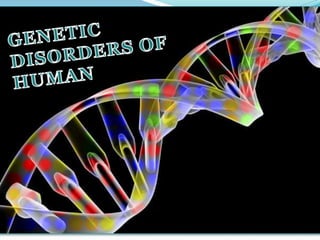
Genetic disorders of human by Jeba Akhtar
- 2. In recent years, a large number of human diseases, deformities and disorders have been found to be hereditary. The various genetic disorders can be grouped under two broad categories- 1. Mendelian disorders and 2.Chromosomal disorders The above disorders can further be classified into the following categories:
- 3. Mendelian disorders Autosomal Dominant gene mutation Autosomal Recessive gene mutation Sex-linked Recessive 1.Polydactyly 1.Alcaptonuria 1.Haemophilia 2.Huntington’s disorder 2.Sickle cell anaemia 2.Duchenne’s muscular dystrophy
- 4. Chromosomal disorders Autosomal disorders Sex-chromosomal disorders 1.Down’s syndrome 1.klinefelter’s syndrome 2.Edward syndrome 2.Turner’s syndrome
- 5. Mendelian disorders A congenital physical anomaly in human. It occurs due Results as an irregularity in the process of formation of hand One single finger splits into two. More than usual number of fingers on hand or foot.
- 6. Picture 1.a: polydactyly on feet Picture 1.b: on hand 1.a 1.b
- 7. Also known as Huntington's chorea, is an inherited disorder. Due to mutation in either of an parent's two copies of a gene called This gene provides the genetic information for a ) triplet repeats in the gene coding for the Huntingtin protein results in an abnormal protein, which gradually jerky, random, and uncontrollable movements called chorea. may be initially exhibited as general restlessness, small unintentionally initiated or uncompleted motions & lack of coordination.
- 8. Picture 2: showing the most affected regions i.e., the basal ganglia region due to Huntington’s disorder
- 9. It is an inborn metabolic disorder produced by the action of a single gene. Due to lack of normal gene that controls the synthesis of the enzyme , which catalyse the breakdown of in liver. Homogentisic acid accumulates and is excreted in urine. Blackening of urine on exposure to air. Darkening of certain cartilages. It also causes mild arthritis.
- 10. Picture 3.a: A-normal urine; B-alcaptonuric urine Picture 3.b: Hands of a severely affected alcaptonuric person. 3.a 3.b
- 11. . Can be transmitted from parents to offspring when both the partners are carrier for the gene. Controlled by a single pair of gene. Due to substitution of at the 6th position of molecule. The oxygen carrying capacity of blood cell decreases. RBC becomes sickle shaped. Surface area decreases. Extremely painful, causing abdominal, chest and bone pain, fatigue, shortness of breath, delayed puberty etc.
- 12. Picture 4: showing RBC of a normal man and a affected man
- 13. First reported by . disease which shows its transmission from Affects only male child. caused by the gene, say h located in the . Lack of blood clotting factor (i.e., factor VIII). Haemophiliacs bleed excessively when injured. A seriously affected person may bleed to death even after minor skin cut.
- 14. Picture 5: HAEMOPHILIA (THE BLEEDING CONDITION)
- 15. Also known as Duchenne Muscular Dystrophy. It is a disorder. a group of Due mutation of gene on , hence fails to produce a protein called . Dystrophin is thought to relay the nerve’s signal to the in the muscle cell, as a result is not released and Progressive muscular wasting. Poor balance. Scoliosis (curvature of the spine and the back). Progressive inability to walk. Limited range of movement. Muscle spasms.
- 16. Picture 6.a: Normal muscle picture 6.b: In affected muscle the tissue has become disorganized and the concentration of dystrophin (green) is greatly reduced. 6.a 6.b 6.c Picture 6.c: showing difference between normal and affected muscles tissue.
- 17. Chromosomal disorders Chromosomal disorders are caused due of one or more chromosomes. Failure of segregation of chromatids during cell division cycle results in the gain or loss of a chromosome(s) is called
- 18. First reported by Langdon Down in 1866. But actual cause was identified by Lejeune and his co-workers. trisomic, extra chromosome no.21 is present i.e., Due to during oogenesis in the mother’s ovary. Rounded face, broad forehead, a Mongolian type of eyelid fold, furrowed tongue & partially open mouth. Palm is broad with characteristics palm crease. Physical, psychomotor and mental retardation.
- 19. Picture 7.a: Showing a boy with Down’s syndrome Picture 7.b: Feet of a boy with the disorder. 7.a 7.b
- 20. Due to the presence of all or a part of a , hence also known as . Most cases of this disease occur due to problems during the formation of the reproductive cells or during early development. Heart defect. Small head. Small or abnormal jaw. Clenched fist with overlapping fingers. Malformed legs.
- 21. Picture 8.a and 8.b showing babies affected with Edward’s syndrome 8.a 8.b
- 22. Due to the presence of an resulting into karyotype of This genotype results from the union of a and . Sterile male with small testes. unusually long legs. Obesity. Sparse body hair. And many female characteristics such as breasts.
- 23. Picture 9.a and 9.b: showing a person with klinefelter’s syndrome 9.a 9.b
- 24. It is an with a single sex ). (i.e., total no. of chromosome is 45) Due to union of an abnormal egg with a normal X sperm or a normal X egg and an abnormal O sperm. Sterile female. Underdeveloped breasts. Rudimentary ovaries. Small uterus.
- 25. Picture 10.a: A girl with Turner’s syndrome picture 10.b: Clinical features of an individual affected with the disorder. 10.a 10.b
- 26. Conclusion: We have learned that there are a number of genetic disorders that can affect the human population which can vary in causes, severity and characteristics.