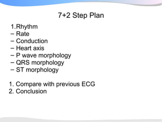
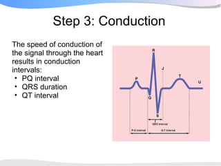
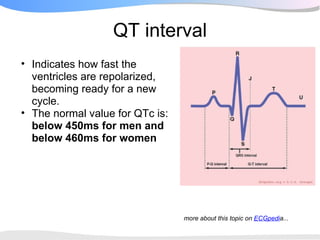
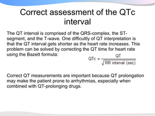
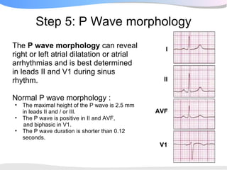
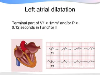
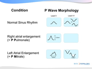
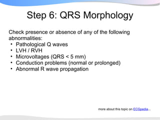
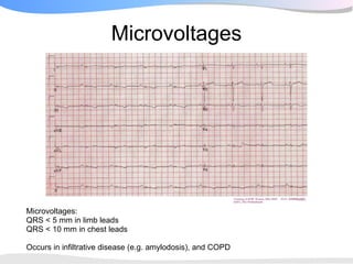
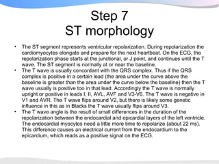
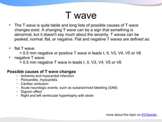
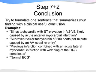
SlideShare a Scribd company logo
1 of 43
When confronted with an ECG, always use a systematic
approach, following all steps to come to the correct
conclusion.
In this course we use the 7+2 step plan
7+2 Step Plan
1.Rhythm
– Rate
– Conduction
– Heart axis
– P wave morphology
– QRS morphology
– ST morphology

1. Compare with previous ECG
2. Conclusion
Step 1: What is the Rhythm?                     P wave


Normal (conducted) sinus rhythm
 • A P wave (atrial contraction) precedes          I
   every QRS complex.
 • A QRS complex follows every P wave.
 • The rhythm is regular, but varies slightly
   during respirations.                           II
 • The rate ranges between 60 and 100
   beats per minute.
 • The P waves maximum height is 2.5 mm
   in II and/or III.                            AVF
 • The P wave is positive in I and II, and
   biphasic in V1.
 • The PQ time is between 0,12 and 0,2
   seconds.
                                                 V1
An example of normal sinus rhythm
Not sinus rhythm?

• If the rhythm is not sinus rhythm you proceed with the
  arrhythmia algorithm (which you will learn with the next
  topics) after determining the heart rate, heart axis and
  conduction intervals.
• (A purist definition of sinus rhythm is that sinus rhythm is
  present in the atria and not by definition in the ventricles,
  e.g. there may be sinus rhythm in the atria, complete AV
  block and a nodal escape rhythm in the ventricles).
Step 2: What is the heart rate?
Knowing the ECG paperspeed
(usually 25 mm/sec) allows us to
calculate the heart rate from the
ECG using any of these methods:
 • Use the "square counting"
    method
 • Use a calculator
 • Use a separate ECG ruler
The square counting method
                          The count method to determine the
    The square counting method is ideal The second QRS
                          heart frequency.
    for regular heart rates. Use the between 75 and 60 beats
                          complex is
    sequence:             per minute. This heartbeat is between
                          that, around 65 beats per minute.
    300-150-100-75-60-50-43-37.

    Count from the first QRS complex
    (preferably occuring on a thick line). If
    the next QRS complex would occur on
    the next thick line, the heart rate would
    be 300, 150 if at the second thick line,
    100 if at the third thick line and so on.
    When the second QRS complex is
    between two lines, take the mean of the
    two numbers from the sequence.
Step 3: Conduction
The speed of conduction of
the signal through the heart
results in conduction
intervals:
 • PQ interval
 • QRS duration
 • QT interval
PQ interval
• Normal between 0.12 and 0.2 seconds.
• Starts at the beginning of the atrial complex and ends at
  the beginning of the ventricular complex.

May be shortened if there is pre-excitation of the ventricles
through abnormal conduction between the atria and
ventricles (accessory pathway).
If the PQ interval is prolonged there is a degree of AV
block (which will be discussed later).
QRS duration
• Normal < 0.10 - 0.12 seconds
• Indicates how fast the ventricles depolarize

• QRS longer than 120 milliseconds may result from:
  o   Left bundle branch block
  o   Right bundle branch block
  o   Electrolyte disorders
  o   Idioventricular rhythm or paced rhythm
QT interval
• Indicates how fast the
  ventricles are repolarized,
  becoming ready for a new
  cycle.
• The normal value for QTc is:
  below 450ms for men and
  below 460ms for women


                                 more about this topic on ECGpedia...
Correct assessment of the QTc
                 interval
The QT interval is comprised of the QRS-complex, the ST-
segment, and the T-wave. One difficultly of QT interpretation is
that the QT interval gets shorter as the heart rate increases. This
problem can be solved by correcting the QT time for heart rate
using the Bazett formula:


Correct QT measurements are important because QT prolongation
may make the patient prone to arrhythmias, especially when
combined with QT-prolonging drugs.
Stepwise approach to correct measurement of the QT
                         interval
1. Use lead II. Use lead V5 alternatively if lead II cannot be read.
– Draw a line through the baseline (PR segment, or TP alternatively)
– Draw a tangent against the steepest part of the end of the T wave. If the T
   wave has two positive deflections, the taller deflection should be chosen. If the
   T wave is biphasic, the end of the taller deflection should be chosen.
– The QT interval starts at the beginning of the QRS interval and ends where
   the tangent and baseline cross.
– If the QRS duration exceeds 120ms, the amount surpassing 120ms should be
   deducted from the QT interval (i.e. QT=QT-(QRS width-120ms) )
– Calculate QTc according to Bazett. You can use the QTc calculator for this.
Causes of QT prolongation
• Congenital long QT syndrome.

But QT prolongation can also occur as a consequence of
(a.o.):
 • Medication (anti-arrhythmics, tricyclic antidepressants,
   phenothiazides). See torsades.org for a full list.
 • Electrolyte imbalances.
 • Ischemia.
Step 4: Heart axis
The heart axis points in the direction of
the average electrical vector of all the
depolarizing heart cells.

A change of the heart axis or an extreme
deviation can be an indication of
pathology.

A positive QRS complex (more above
than below the baseline) in a certain lead
means that the heart axis is going (at
least slightly) in that lead's direction.

The heart axis is normal between -30
and +90 degrees.

Therefore, if QRS is positive in both
leads I and II, the heart axis is normal.
Interpretation
There are four areas where
the QRS vector can point:
• Left axis deviation (between
  -30º and -90º)
• Normal axis between -30º and
  90º
• Right lower quadrant --> right
  axis deviation (between 90º
  and -180º)
• Right upper quadrant -->
  extreme right axis
  deviation(between -90º and
  -180º)


                                   more about this topic on ECGpedia...
Abnormal heart axis


Heart axis deviation to the left in   Heart axis deviation to the right
case of an inferior infarct. Left     can result from right
anterior hemiblock is another         ventricular overload as in COPD
common cause. A left axis is          or pulmonary embolism. A right
present between -30 and -90           axis is between +90 and +180
degrees.                              degrees.
                                      A left - right arm lead exchange is
                                      the most common cause of right
                                      axis deviation!
Step 5: P Wave morphology
The P wave morphology can reveal                    I
right or left atrial dilatation or atrial
arrhythmias and is best determined
in leads II and V1 during sinus
rhythm.                                            II


Normal P wave morphology :
•   The maximal height of the P wave is 2.5 mm
    in leads II and / or III.                    AVF
•   The P wave is positive in II and AVF,
     and biphasic in V1.
•   The P wave duration is shorter than 0.12
    seconds.
                                                  V1
Left atrial dilatation

Terminal part of V1 > 1mm2 and/or P >
0.12 seconds in I and/ or II
Right atrial dilatation
P > 2.5 mm in II and/ or III and/ or aVF
and/ or P > 1.5 mm in V1
Condition                P Wave Morphology


Normal Sinus Rhythm


Right atrial enlargement
(= P Pulmonale)


Left Atrial Enlargement
(= P Mitrale)
Step 6: QRS Morphology
Check presence or absence of any of the following
abnormalities:
 • Pathological Q waves
 • LVH / RVH
 • Microvoltages (QRS < 5 mm)
 • Conduction problems (normal or prolonged)
 • Abnormal R wave propagation


                                    more about this topic on ECGpedia...
Pathological Q wave
• Q waves point at electrically
  silent areas and can be a sign of
  previous myocardial infarction
• Definition of a pathologic Q
  wave:
   o Any Q wave in leads V2–V3
   o Q wave ≥ 0.03 s and > 0.1 mV
     deep in other leads.
   o To be defined as pathologic, Q
     waves need to be present in
     two contiguous leads (e.g. II
     and AVF or I and AVL or V1
     and V2)
Left ventricular hypertrophy
Hypertrophic myocardium
has more electrical activity,
resulting in larger peaks.

Definition of LVH: R in V5 or
V6 + S in VI > 35mm
(Sokolow-Lyon criteria)
Often a "strain pattern" is
seen in V5 and V6.
Right ventricular hypertrophy
is probably present when R is   V1
larger than S in VI
Microvoltages


Microvoltages:
QRS < 5 mm in limb leads
QRS < 10 mm in chest leads

Occurs in infiltrative disease (e.g. amylodosis), and COPD
Conduction disorders
If the QRS duration is more than 0.12 seconds there may
be a block in the conductive tissue. Most often it will be
either right or left bundle branch block.
Rule of thumb: when distinguishing left
and right bundle branch block--look at
V1 only!

Does the signal end negative (below
the baseline) in V1? (away from V1)
 >> the ventricle farther from V1 is
depolarized later>> it must be a left
bundle branch block

Does the signal end positive in V1?
(towards V1) >> the ventricle closer to
V1 is depolarized later >> it must be a
right bundle branch block
Conduction disorders: right bundle branch
                  block
• QRS > 0.12 seconds                   V1
• RSR'-pattern in V1 where
  R' > R
• Slurred S wave in lead I
  and V6
Conduction disorders: left bundle branch
                   block
• QRS > 0.12 seconds
• Broad monomorphic S waves in V1, may have a small
  initial R wave
• Broad monomorphic R waves in I and V6 with no Q
  waves
Normal R wave progression


Normally R waves become larger from V1-V5. At V5 it should be maximal. If
the R wave in V2 is larger than in V3, this could be a sign of a (previous)
posterior myocardial infarction.
Step 7
                       ST morphology
•   The ST segment represents ventricular repolarization. During repolarization the
    cardiomyocytes elongate and prepare for the next heartbeat. On the ECG, the
    repolarization phase starts at the junctional, or J point, and continues until the T
    wave. The ST segment is normally at or near the baseline.
•   The T wave is usually concordant with the QRS complex. Thus if the QRS
    complex is positive in a certain lead (the area under the curve above the
    baseline is greater than the area under the curve below the baseline) then the T
    wave usually is positive too in that lead. Accordingly the T wave is normally
    upright or positive in leads I, II, AVL, AVF and V3-V6. The T wave is negative in
    V1 and AVR. The T wave flips around V2, but there is likely some genetic
    influence in this as in Blacks the T wave usually flips around V3.
•   The T wave angle is the result of small differences in the duration of the
    repolarization between the endocardial and epicardial layers of the left ventricle.
    The endocardial myocytes need a little more time to repolarize (about 22 ms).
    This difference causes an electrical current from the endocardium to the
    epicardium, which reads as a positive signal on the ECG.
Step 7
                           ST morphology
• ST segment elevation
•   Ischemia
•   Pericarditis
•   Aneurysma cordis
•   Normal variant

• ST depression
•   Ischemia
•   LVH
•   Digitalis
•   Low potassium/ low magnesium
•   Neurologic

• T wave changes
•   Ischemia
•   Pericarditis
•   Myocarditis
•   LVH / RVH
•   Electrolyte changes (especially potassium)
Common causes of ST shift
ST elevation
                       normal
90% of healthy (young) men and women to a lesser extent
have ST elevation in precordial leads.

Normal variants of
ST segment elevation are:
• 1: normal
• 2: ‘early repolarization’
• 3: normal 'variant'
Abnormal ST segment elevation


1: LVH
2: LBBB
3: Pericarditis
4: High pottasium
5: Acute AS infarct
6: Acute AS infarct +
RBBB
7: Brugada syndrome
Diffuse ST elevation in pericarditis


ST segment elevation (upper ECG) due to pericarditis. The lower ECG
    shows PTa depression, which is typically seen in pericarditis.
ST depression
The most important cause of ST segment depression is ischemia. Causes of
ST segment depression include:
  Reciprocal ST segment depression during ischemia. If one lead shows
   ST segment elevation then usually the lead "on the other side" shows ST
   segment depression.
  Left ventricular hypertophy with "strain" or depolarization abnormality
  Digoxin effect
  Low potassium/low magnesium
  Heart rate-induced changes (post tachycardia), 'cardiac memory'
  During acute neurologic events


                                                 more about this topic on ECGpedia...
ST segment elevation due to high potassium levels
T wave
•   The T wave is quite labile and long lists of possible causes of T wave
    changes exist. A changing T wave can be a sign that something is
    abnormal, but it doesn't say much about the severity. T waves can be
    peaked, normal, flat, or negative. Flat and negative T waves are defined as:

•   flat T wave:
        < 0.5 mm negative or positive T wave in leads I, II, V3, V4, V5 or V6
•   negative T wave:
        > 0.5 mm negative T wave in leads I, II, V3, V4, V5 or V6

Possible causes of T wave changes
     o   Ischemia and myocardial infarction
     o   Pericarditis, myocarditis
     o   Cardiac contusion
     o   Acute neurologic events, such as subarachnoid bleeding (SAB)
     o   Digoxin effect
     o   Right and left ventricular hypertrophy with strain


                                                             more about this topic on ECGpedia...
Prominent U wave
Sometimes a U wave is present: an extra wave following the T wave. During
hypokalemia (and hypocalcemia) the U wave can become more prominent:
Step 7+1
     Compare with previous ECG
•   New LBBB?
•   Change in heart axis?
•   New pathologic Q?
•   Decreased R wave height?


All of these can exist as chronic abnormalities, but when
the are new it can be a sign of acute ischemia or another
condition.

                                      more about this topic on ECGpedia...
Step 7+2
                   Conclusion
Try to formulate one sentence that summarizes your
finding with a clinical useful conclusion.
Examples:
 "Sinus tachycardia with ST elevation in V2-V5, likely
  caused by acute anterior myocardial infarction"
 "Supraventricular tachycardia of 200 beats per minute
  caused by an AV nodal re-entry"
 "Previous infarction combined with an acute lateral
  myocardial infarction with widening of the QRS
  complexes"
 "Normal ECG"

More Related Content

What's hot (20)

Localization of WPW( accessory Pathway) by surface ECG
Localization of WPW( accessory Pathway) by surface ECGLocalization of WPW( accessory Pathway) by surface ECG
Localization of WPW( accessory Pathway) by surface ECG
 
Ecg changes in mi
Ecg changes in miEcg changes in mi
Ecg changes in mi
 
WIDE QRS TACHYCARDIA
WIDE  QRS TACHYCARDIAWIDE  QRS TACHYCARDIA
WIDE QRS TACHYCARDIA
 
ECG Quiz
ECG QuizECG Quiz
ECG Quiz
 
ECG in Emergency Department - Advances in ACS ECG
ECG in Emergency Department - Advances in ACS ECGECG in Emergency Department - Advances in ACS ECG
ECG in Emergency Department - Advances in ACS ECG
 
Bundle branch blocks
Bundle branch blocksBundle branch blocks
Bundle branch blocks
 
ECG: Quiz
ECG: QuizECG: Quiz
ECG: Quiz
 
Cardiac Arrhythmias
Cardiac ArrhythmiasCardiac Arrhythmias
Cardiac Arrhythmias
 
Ecg quiz @ SEMICON 1018
Ecg quiz @ SEMICON 1018Ecg quiz @ SEMICON 1018
Ecg quiz @ SEMICON 1018
 
ECG: Ventricular Premature Beats
ECG: Ventricular Premature BeatsECG: Ventricular Premature Beats
ECG: Ventricular Premature Beats
 
Basics of ecg interpretation by dr sai
Basics of ecg interpretation by dr saiBasics of ecg interpretation by dr sai
Basics of ecg interpretation by dr sai
 
Ecg test 2
Ecg test 2Ecg test 2
Ecg test 2
 
Wide Complex Tachycardia
Wide Complex TachycardiaWide Complex Tachycardia
Wide Complex Tachycardia
 
ECG in Acute Myocardial Infarction
ECG in Acute Myocardial InfarctionECG in Acute Myocardial Infarction
ECG in Acute Myocardial Infarction
 
Ecg
EcgEcg
Ecg
 
LBBB
LBBBLBBB
LBBB
 
Ecg assessment of ihd
Ecg assessment of ihdEcg assessment of ihd
Ecg assessment of ihd
 
ECG: Myocardial Infarction with CHB
ECG: Myocardial Infarction with CHBECG: Myocardial Infarction with CHB
ECG: Myocardial Infarction with CHB
 
Ecg interpretation
Ecg interpretationEcg interpretation
Ecg interpretation
 
8 ECG WORKSHOP BUNDLE BRANCH BLOCK & FASCICULAR BLOCK.pptx
8 ECG WORKSHOP BUNDLE BRANCH BLOCK & FASCICULAR BLOCK.pptx8 ECG WORKSHOP BUNDLE BRANCH BLOCK & FASCICULAR BLOCK.pptx
8 ECG WORKSHOP BUNDLE BRANCH BLOCK & FASCICULAR BLOCK.pptx
 

Viewers also liked

Ecg interpritation
Ecg interpritationEcg interpritation
Ecg interpritationvijay dihora
 
Basics of ECG.ppt dr.k.subramanyam
Basics of ECG.ppt dr.k.subramanyamBasics of ECG.ppt dr.k.subramanyam
Basics of ECG.ppt dr.k.subramanyamAdarsh
 
ECG BASICS AND ARRTHYMIAS
ECG BASICS AND ARRTHYMIASECG BASICS AND ARRTHYMIAS
ECG BASICS AND ARRTHYMIASPrudhvi Krishna
 
Electrocardiogaram - ECG EKG
Electrocardiogaram - ECG EKGElectrocardiogaram - ECG EKG
Electrocardiogaram - ECG EKGFuad Farooq
 
ECG interpretation: the basics
ECG interpretation: the basicsECG interpretation: the basics
ECG interpretation: the basicsJamie Ranse
 
Understanding rhythm strips
Understanding rhythm stripsUnderstanding rhythm strips
Understanding rhythm stripsAnn Bentley
 
St segment elevations
St  segment elevationsSt  segment elevations
St segment elevationsRamesh Babu
 
Cardiac Electrophysiology
Cardiac ElectrophysiologyCardiac Electrophysiology
Cardiac ElectrophysiologyTeleClinEd
 
Cardiac Ecg Interpretation
Cardiac Ecg InterpretationCardiac Ecg Interpretation
Cardiac Ecg InterpretationNorthTec
 
Mean cardiac axis (cardiac vector)
Mean cardiac axis (cardiac vector)Mean cardiac axis (cardiac vector)
Mean cardiac axis (cardiac vector)Seyed Yahya Moradi
 
Ecg interpretation reading 20031
Ecg interpretation  reading 20031Ecg interpretation  reading 20031
Ecg interpretation reading 20031Joj Ram
 
Electrocardiographi crvmi
Electrocardiographi crvmiElectrocardiographi crvmi
Electrocardiographi crvmiasadsoomro1960
 

Viewers also liked (20)

Ecg lecture
Ecg lectureEcg lecture
Ecg lecture
 
ECG
ECGECG
ECG
 
ECG Basics
ECG BasicsECG Basics
ECG Basics
 
Ecg interpritation
Ecg interpritationEcg interpritation
Ecg interpritation
 
Basics of ECG.ppt dr.k.subramanyam
Basics of ECG.ppt dr.k.subramanyamBasics of ECG.ppt dr.k.subramanyam
Basics of ECG.ppt dr.k.subramanyam
 
ECG BASICS AND ARRTHYMIAS
ECG BASICS AND ARRTHYMIASECG BASICS AND ARRTHYMIAS
ECG BASICS AND ARRTHYMIAS
 
Electrocardiogaram - ECG EKG
Electrocardiogaram - ECG EKGElectrocardiogaram - ECG EKG
Electrocardiogaram - ECG EKG
 
Shortcut to ECG
Shortcut to ECGShortcut to ECG
Shortcut to ECG
 
ECG interpretation: the basics
ECG interpretation: the basicsECG interpretation: the basics
ECG interpretation: the basics
 
Basics of ECG
Basics of ECGBasics of ECG
Basics of ECG
 
Understanding rhythm strips
Understanding rhythm stripsUnderstanding rhythm strips
Understanding rhythm strips
 
Ecg usm
Ecg   usmEcg   usm
Ecg usm
 
St segment elevations
St  segment elevationsSt  segment elevations
St segment elevations
 
ECG from Alpha to Omega
ECG from Alpha to OmegaECG from Alpha to Omega
ECG from Alpha to Omega
 
Cardiac Electrophysiology
Cardiac ElectrophysiologyCardiac Electrophysiology
Cardiac Electrophysiology
 
Cardiac Ecg Interpretation
Cardiac Ecg InterpretationCardiac Ecg Interpretation
Cardiac Ecg Interpretation
 
Mean cardiac axis (cardiac vector)
Mean cardiac axis (cardiac vector)Mean cardiac axis (cardiac vector)
Mean cardiac axis (cardiac vector)
 
Ecg interpretation reading 20031
Ecg interpretation  reading 20031Ecg interpretation  reading 20031
Ecg interpretation reading 20031
 
Electrocardiographi crvmi
Electrocardiographi crvmiElectrocardiographi crvmi
Electrocardiographi crvmi
 
Ekgnperusteet1
Ekgnperusteet1Ekgnperusteet1
Ekgnperusteet1
 

Similar to ECGpedia - ECG Presentation

Similar to ECGpedia - ECG Presentation (20)

Basic ecg
Basic ecgBasic ecg
Basic ecg
 
Ecg easy way
Ecg easy wayEcg easy way
Ecg easy way
 
simple ecg learningMEM.pptx
simple ecg learningMEM.pptxsimple ecg learningMEM.pptx
simple ecg learningMEM.pptx
 
ECG easy way
ECG easy way ECG easy way
ECG easy way
 
13973762 all-about-ecg
13973762 all-about-ecg13973762 all-about-ecg
13973762 all-about-ecg
 
44 E C G
44 E C G44 E C G
44 E C G
 
44 E C G
44 E C G44 E C G
44 E C G
 
All info about ecg
All info about ecgAll info about ecg
All info about ecg
 
A Guide TO ECG Interpretation
A Guide TO ECG InterpretationA Guide TO ECG Interpretation
A Guide TO ECG Interpretation
 
Beginners Guide for ECG Interpretation
Beginners Guide for ECG InterpretationBeginners Guide for ECG Interpretation
Beginners Guide for ECG Interpretation
 
Ecg basics
Ecg basicsEcg basics
Ecg basics
 
ECG-2 RAMA.pptx
ECG-2 RAMA.pptxECG-2 RAMA.pptx
ECG-2 RAMA.pptx
 
Ecg interpretation , Upgraded
Ecg interpretation , UpgradedEcg interpretation , Upgraded
Ecg interpretation , Upgraded
 
Edited_-_ECG_Interpretation_and_Arrhythmia_Recognition_-_Azeren.pptx
Edited_-_ECG_Interpretation_and_Arrhythmia_Recognition_-_Azeren.pptxEdited_-_ECG_Interpretation_and_Arrhythmia_Recognition_-_Azeren.pptx
Edited_-_ECG_Interpretation_and_Arrhythmia_Recognition_-_Azeren.pptx
 
E C G M O H A M E D R A M A D A N
E C G  M O H A M E D  R A M A D A NE C G  M O H A M E D  R A M A D A N
E C G M O H A M E D R A M A D A N
 
Ecg workshop
Ecg workshopEcg workshop
Ecg workshop
 
ecgworkshop-180204063605vvvvvvvvvvvb.pdf
ecgworkshop-180204063605vvvvvvvvvvvb.pdfecgworkshop-180204063605vvvvvvvvvvvb.pdf
ecgworkshop-180204063605vvvvvvvvvvvb.pdf
 
Tutorial in Basic ECG for Medical Students
Tutorial in Basic ECG for Medical StudentsTutorial in Basic ECG for Medical Students
Tutorial in Basic ECG for Medical Students
 
Basics of ECG.ppt
Basics of ECG.pptBasics of ECG.ppt
Basics of ECG.ppt
 
All About ECG
All About ECGAll About ECG
All About ECG
 

More from DR YONG & ASSOCIATES Family Health

More from DR YONG & ASSOCIATES Family Health (7)

Differential Diagnosis in Clinical Examination (2012) [PDF] [UnitedVRG].pdf
Differential Diagnosis in Clinical Examination (2012) [PDF] [UnitedVRG].pdfDifferential Diagnosis in Clinical Examination (2012) [PDF] [UnitedVRG].pdf
Differential Diagnosis in Clinical Examination (2012) [PDF] [UnitedVRG].pdf
 
394 infant-milestone-chart
394 infant-milestone-chart394 infant-milestone-chart
394 infant-milestone-chart
 
Adrenal suppression -a practical guide to the screening and management of thi...
Adrenal suppression -a practical guide to the screening and management of thi...Adrenal suppression -a practical guide to the screening and management of thi...
Adrenal suppression -a practical guide to the screening and management of thi...
 
Common medical diagnosis -an algorithmic approach --3rd ed
Common medical diagnosis -an algorithmic approach --3rd edCommon medical diagnosis -an algorithmic approach --3rd ed
Common medical diagnosis -an algorithmic approach --3rd ed
 
Diagnosis --checklist--diffdx-2
Diagnosis --checklist--diffdx-2Diagnosis --checklist--diffdx-2
Diagnosis --checklist--diffdx-2
 
Diabetes_Flowsheet
Diabetes_FlowsheetDiabetes_Flowsheet
Diabetes_Flowsheet
 
Top10_Differential_Diagnosis_In_Primary_Care
Top10_Differential_Diagnosis_In_Primary_CareTop10_Differential_Diagnosis_In_Primary_Care
Top10_Differential_Diagnosis_In_Primary_Care
 

Recently uploaded

Russian Call Girls in Pune Tanvi 9907093804 Short 1500 Night 6000 Best call g...
Russian Call Girls in Pune Tanvi 9907093804 Short 1500 Night 6000 Best call g...Russian Call Girls in Pune Tanvi 9907093804 Short 1500 Night 6000 Best call g...
Russian Call Girls in Pune Tanvi 9907093804 Short 1500 Night 6000 Best call g...Miss joya
 
Russian Call Girls in Chennai Pallavi 9907093804 Independent Call Girls Servi...
Russian Call Girls in Chennai Pallavi 9907093804 Independent Call Girls Servi...Russian Call Girls in Chennai Pallavi 9907093804 Independent Call Girls Servi...
Russian Call Girls in Chennai Pallavi 9907093804 Independent Call Girls Servi...Nehru place Escorts
 
Call Girls Service in Bommanahalli - 7001305949 with real photos and phone nu...
Call Girls Service in Bommanahalli - 7001305949 with real photos and phone nu...Call Girls Service in Bommanahalli - 7001305949 with real photos and phone nu...
Call Girls Service in Bommanahalli - 7001305949 with real photos and phone nu...narwatsonia7
 
Call Girls Service Surat Samaira ❤️🍑 8250192130 👄 Independent Escort Service ...
Call Girls Service Surat Samaira ❤️🍑 8250192130 👄 Independent Escort Service ...Call Girls Service Surat Samaira ❤️🍑 8250192130 👄 Independent Escort Service ...
Call Girls Service Surat Samaira ❤️🍑 8250192130 👄 Independent Escort Service ...CALL GIRLS
 
Housewife Call Girls Hoskote | 7001305949 At Low Cost Cash Payment Booking
Housewife Call Girls Hoskote | 7001305949 At Low Cost Cash Payment BookingHousewife Call Girls Hoskote | 7001305949 At Low Cost Cash Payment Booking
Housewife Call Girls Hoskote | 7001305949 At Low Cost Cash Payment Bookingnarwatsonia7
 
Russian Call Girls in Pune Riya 9907093804 Short 1500 Night 6000 Best call gi...
Russian Call Girls in Pune Riya 9907093804 Short 1500 Night 6000 Best call gi...Russian Call Girls in Pune Riya 9907093804 Short 1500 Night 6000 Best call gi...
Russian Call Girls in Pune Riya 9907093804 Short 1500 Night 6000 Best call gi...Miss joya
 
Kesar Bagh Call Girl Price 9548273370 , Lucknow Call Girls Service
Kesar Bagh Call Girl Price 9548273370 , Lucknow Call Girls ServiceKesar Bagh Call Girl Price 9548273370 , Lucknow Call Girls Service
Kesar Bagh Call Girl Price 9548273370 , Lucknow Call Girls Servicemakika9823
 
Russian Call Girls Chennai Madhuri 9907093804 Independent Call Girls Service ...
Russian Call Girls Chennai Madhuri 9907093804 Independent Call Girls Service ...Russian Call Girls Chennai Madhuri 9907093804 Independent Call Girls Service ...
Russian Call Girls Chennai Madhuri 9907093804 Independent Call Girls Service ...Nehru place Escorts
 
Call Girls Service Bellary Road Just Call 7001305949 Enjoy College Girls Service
Call Girls Service Bellary Road Just Call 7001305949 Enjoy College Girls ServiceCall Girls Service Bellary Road Just Call 7001305949 Enjoy College Girls Service
Call Girls Service Bellary Road Just Call 7001305949 Enjoy College Girls Servicenarwatsonia7
 
VIP Call Girls Pune Vani 9907093804 Short 1500 Night 6000 Best call girls Ser...
VIP Call Girls Pune Vani 9907093804 Short 1500 Night 6000 Best call girls Ser...VIP Call Girls Pune Vani 9907093804 Short 1500 Night 6000 Best call girls Ser...
VIP Call Girls Pune Vani 9907093804 Short 1500 Night 6000 Best call girls Ser...Miss joya
 
CALL ON ➥9907093804 🔝 Call Girls Hadapsar ( Pune) Girls Service
CALL ON ➥9907093804 🔝 Call Girls Hadapsar ( Pune)  Girls ServiceCALL ON ➥9907093804 🔝 Call Girls Hadapsar ( Pune)  Girls Service
CALL ON ➥9907093804 🔝 Call Girls Hadapsar ( Pune) Girls ServiceMiss joya
 
College Call Girls Pune Mira 9907093804 Short 1500 Night 6000 Best call girls...
College Call Girls Pune Mira 9907093804 Short 1500 Night 6000 Best call girls...College Call Girls Pune Mira 9907093804 Short 1500 Night 6000 Best call girls...
College Call Girls Pune Mira 9907093804 Short 1500 Night 6000 Best call girls...Miss joya
 
Call Girl Chennai Indira 9907093804 Independent Call Girls Service Chennai
Call Girl Chennai Indira 9907093804 Independent Call Girls Service ChennaiCall Girl Chennai Indira 9907093804 Independent Call Girls Service Chennai
Call Girl Chennai Indira 9907093804 Independent Call Girls Service ChennaiNehru place Escorts
 
Call Girls Colaba Mumbai ❤️ 9920874524 👈 Cash on Delivery
Call Girls Colaba Mumbai ❤️ 9920874524 👈 Cash on DeliveryCall Girls Colaba Mumbai ❤️ 9920874524 👈 Cash on Delivery
Call Girls Colaba Mumbai ❤️ 9920874524 👈 Cash on Deliverynehamumbai
 
Call Girls Chennai Megha 9907093804 Independent Call Girls Service Chennai
Call Girls Chennai Megha 9907093804 Independent Call Girls Service ChennaiCall Girls Chennai Megha 9907093804 Independent Call Girls Service Chennai
Call Girls Chennai Megha 9907093804 Independent Call Girls Service ChennaiNehru place Escorts
 
Call Girls Service In Shyam Nagar Whatsapp 8445551418 Independent Escort Service
Call Girls Service In Shyam Nagar Whatsapp 8445551418 Independent Escort ServiceCall Girls Service In Shyam Nagar Whatsapp 8445551418 Independent Escort Service
Call Girls Service In Shyam Nagar Whatsapp 8445551418 Independent Escort Serviceparulsinha
 
Call Girls Service Pune Vaishnavi 9907093804 Short 1500 Night 6000 Best call ...
Call Girls Service Pune Vaishnavi 9907093804 Short 1500 Night 6000 Best call ...Call Girls Service Pune Vaishnavi 9907093804 Short 1500 Night 6000 Best call ...
Call Girls Service Pune Vaishnavi 9907093804 Short 1500 Night 6000 Best call ...Miss joya
 
VIP Mumbai Call Girls Hiranandani Gardens Just Call 9920874524 with A/C Room ...
VIP Mumbai Call Girls Hiranandani Gardens Just Call 9920874524 with A/C Room ...VIP Mumbai Call Girls Hiranandani Gardens Just Call 9920874524 with A/C Room ...
VIP Mumbai Call Girls Hiranandani Gardens Just Call 9920874524 with A/C Room ...Garima Khatri
 

Recently uploaded (20)

Russian Call Girls in Pune Tanvi 9907093804 Short 1500 Night 6000 Best call g...
Russian Call Girls in Pune Tanvi 9907093804 Short 1500 Night 6000 Best call g...Russian Call Girls in Pune Tanvi 9907093804 Short 1500 Night 6000 Best call g...
Russian Call Girls in Pune Tanvi 9907093804 Short 1500 Night 6000 Best call g...
 
Russian Call Girls in Chennai Pallavi 9907093804 Independent Call Girls Servi...
Russian Call Girls in Chennai Pallavi 9907093804 Independent Call Girls Servi...Russian Call Girls in Chennai Pallavi 9907093804 Independent Call Girls Servi...
Russian Call Girls in Chennai Pallavi 9907093804 Independent Call Girls Servi...
 
Call Girls Service in Bommanahalli - 7001305949 with real photos and phone nu...
Call Girls Service in Bommanahalli - 7001305949 with real photos and phone nu...Call Girls Service in Bommanahalli - 7001305949 with real photos and phone nu...
Call Girls Service in Bommanahalli - 7001305949 with real photos and phone nu...
 
Call Girls Service Surat Samaira ❤️🍑 8250192130 👄 Independent Escort Service ...
Call Girls Service Surat Samaira ❤️🍑 8250192130 👄 Independent Escort Service ...Call Girls Service Surat Samaira ❤️🍑 8250192130 👄 Independent Escort Service ...
Call Girls Service Surat Samaira ❤️🍑 8250192130 👄 Independent Escort Service ...
 
Housewife Call Girls Hoskote | 7001305949 At Low Cost Cash Payment Booking
Housewife Call Girls Hoskote | 7001305949 At Low Cost Cash Payment BookingHousewife Call Girls Hoskote | 7001305949 At Low Cost Cash Payment Booking
Housewife Call Girls Hoskote | 7001305949 At Low Cost Cash Payment Booking
 
Russian Call Girls in Pune Riya 9907093804 Short 1500 Night 6000 Best call gi...
Russian Call Girls in Pune Riya 9907093804 Short 1500 Night 6000 Best call gi...Russian Call Girls in Pune Riya 9907093804 Short 1500 Night 6000 Best call gi...
Russian Call Girls in Pune Riya 9907093804 Short 1500 Night 6000 Best call gi...
 
Kesar Bagh Call Girl Price 9548273370 , Lucknow Call Girls Service
Kesar Bagh Call Girl Price 9548273370 , Lucknow Call Girls ServiceKesar Bagh Call Girl Price 9548273370 , Lucknow Call Girls Service
Kesar Bagh Call Girl Price 9548273370 , Lucknow Call Girls Service
 
Russian Call Girls Chennai Madhuri 9907093804 Independent Call Girls Service ...
Russian Call Girls Chennai Madhuri 9907093804 Independent Call Girls Service ...Russian Call Girls Chennai Madhuri 9907093804 Independent Call Girls Service ...
Russian Call Girls Chennai Madhuri 9907093804 Independent Call Girls Service ...
 
Call Girls Service Bellary Road Just Call 7001305949 Enjoy College Girls Service
Call Girls Service Bellary Road Just Call 7001305949 Enjoy College Girls ServiceCall Girls Service Bellary Road Just Call 7001305949 Enjoy College Girls Service
Call Girls Service Bellary Road Just Call 7001305949 Enjoy College Girls Service
 
sauth delhi call girls in Bhajanpura 🔝 9953056974 🔝 escort Service
sauth delhi call girls in Bhajanpura 🔝 9953056974 🔝 escort Servicesauth delhi call girls in Bhajanpura 🔝 9953056974 🔝 escort Service
sauth delhi call girls in Bhajanpura 🔝 9953056974 🔝 escort Service
 
VIP Call Girls Pune Vani 9907093804 Short 1500 Night 6000 Best call girls Ser...
VIP Call Girls Pune Vani 9907093804 Short 1500 Night 6000 Best call girls Ser...VIP Call Girls Pune Vani 9907093804 Short 1500 Night 6000 Best call girls Ser...
VIP Call Girls Pune Vani 9907093804 Short 1500 Night 6000 Best call girls Ser...
 
CALL ON ➥9907093804 🔝 Call Girls Hadapsar ( Pune) Girls Service
CALL ON ➥9907093804 🔝 Call Girls Hadapsar ( Pune)  Girls ServiceCALL ON ➥9907093804 🔝 Call Girls Hadapsar ( Pune)  Girls Service
CALL ON ➥9907093804 🔝 Call Girls Hadapsar ( Pune) Girls Service
 
College Call Girls Pune Mira 9907093804 Short 1500 Night 6000 Best call girls...
College Call Girls Pune Mira 9907093804 Short 1500 Night 6000 Best call girls...College Call Girls Pune Mira 9907093804 Short 1500 Night 6000 Best call girls...
College Call Girls Pune Mira 9907093804 Short 1500 Night 6000 Best call girls...
 
Russian Call Girls in Delhi Tanvi ➡️ 9711199012 💋📞 Independent Escort Service...
Russian Call Girls in Delhi Tanvi ➡️ 9711199012 💋📞 Independent Escort Service...Russian Call Girls in Delhi Tanvi ➡️ 9711199012 💋📞 Independent Escort Service...
Russian Call Girls in Delhi Tanvi ➡️ 9711199012 💋📞 Independent Escort Service...
 
Call Girl Chennai Indira 9907093804 Independent Call Girls Service Chennai
Call Girl Chennai Indira 9907093804 Independent Call Girls Service ChennaiCall Girl Chennai Indira 9907093804 Independent Call Girls Service Chennai
Call Girl Chennai Indira 9907093804 Independent Call Girls Service Chennai
 
Call Girls Colaba Mumbai ❤️ 9920874524 👈 Cash on Delivery
Call Girls Colaba Mumbai ❤️ 9920874524 👈 Cash on DeliveryCall Girls Colaba Mumbai ❤️ 9920874524 👈 Cash on Delivery
Call Girls Colaba Mumbai ❤️ 9920874524 👈 Cash on Delivery
 
Call Girls Chennai Megha 9907093804 Independent Call Girls Service Chennai
Call Girls Chennai Megha 9907093804 Independent Call Girls Service ChennaiCall Girls Chennai Megha 9907093804 Independent Call Girls Service Chennai
Call Girls Chennai Megha 9907093804 Independent Call Girls Service Chennai
 
Call Girls Service In Shyam Nagar Whatsapp 8445551418 Independent Escort Service
Call Girls Service In Shyam Nagar Whatsapp 8445551418 Independent Escort ServiceCall Girls Service In Shyam Nagar Whatsapp 8445551418 Independent Escort Service
Call Girls Service In Shyam Nagar Whatsapp 8445551418 Independent Escort Service
 
Call Girls Service Pune Vaishnavi 9907093804 Short 1500 Night 6000 Best call ...
Call Girls Service Pune Vaishnavi 9907093804 Short 1500 Night 6000 Best call ...Call Girls Service Pune Vaishnavi 9907093804 Short 1500 Night 6000 Best call ...
Call Girls Service Pune Vaishnavi 9907093804 Short 1500 Night 6000 Best call ...
 
VIP Mumbai Call Girls Hiranandani Gardens Just Call 9920874524 with A/C Room ...
VIP Mumbai Call Girls Hiranandani Gardens Just Call 9920874524 with A/C Room ...VIP Mumbai Call Girls Hiranandani Gardens Just Call 9920874524 with A/C Room ...
VIP Mumbai Call Girls Hiranandani Gardens Just Call 9920874524 with A/C Room ...
 

ECGpedia - ECG Presentation

  • 1. When confronted with an ECG, always use a systematic approach, following all steps to come to the correct conclusion. In this course we use the 7+2 step plan
  • 2. 7+2 Step Plan 1.Rhythm – Rate – Conduction – Heart axis – P wave morphology – QRS morphology – ST morphology 1. Compare with previous ECG 2. Conclusion
  • 3. Step 1: What is the Rhythm? P wave Normal (conducted) sinus rhythm • A P wave (atrial contraction) precedes I every QRS complex. • A QRS complex follows every P wave. • The rhythm is regular, but varies slightly during respirations. II • The rate ranges between 60 and 100 beats per minute. • The P waves maximum height is 2.5 mm in II and/or III. AVF • The P wave is positive in I and II, and biphasic in V1. • The PQ time is between 0,12 and 0,2 seconds. V1
  • 4. An example of normal sinus rhythm
  • 5. Not sinus rhythm? • If the rhythm is not sinus rhythm you proceed with the arrhythmia algorithm (which you will learn with the next topics) after determining the heart rate, heart axis and conduction intervals. • (A purist definition of sinus rhythm is that sinus rhythm is present in the atria and not by definition in the ventricles, e.g. there may be sinus rhythm in the atria, complete AV block and a nodal escape rhythm in the ventricles).
  • 6. Step 2: What is the heart rate? Knowing the ECG paperspeed (usually 25 mm/sec) allows us to calculate the heart rate from the ECG using any of these methods: • Use the "square counting" method • Use a calculator • Use a separate ECG ruler
  • 7. The square counting method The count method to determine the The square counting method is ideal The second QRS heart frequency. for regular heart rates. Use the between 75 and 60 beats complex is sequence: per minute. This heartbeat is between that, around 65 beats per minute. 300-150-100-75-60-50-43-37. Count from the first QRS complex (preferably occuring on a thick line). If the next QRS complex would occur on the next thick line, the heart rate would be 300, 150 if at the second thick line, 100 if at the third thick line and so on. When the second QRS complex is between two lines, take the mean of the two numbers from the sequence.
  • 8. Step 3: Conduction The speed of conduction of the signal through the heart results in conduction intervals: • PQ interval • QRS duration • QT interval
  • 9. PQ interval • Normal between 0.12 and 0.2 seconds. • Starts at the beginning of the atrial complex and ends at the beginning of the ventricular complex. May be shortened if there is pre-excitation of the ventricles through abnormal conduction between the atria and ventricles (accessory pathway). If the PQ interval is prolonged there is a degree of AV block (which will be discussed later).
  • 10. QRS duration • Normal < 0.10 - 0.12 seconds • Indicates how fast the ventricles depolarize • QRS longer than 120 milliseconds may result from: o Left bundle branch block o Right bundle branch block o Electrolyte disorders o Idioventricular rhythm or paced rhythm
  • 11. QT interval • Indicates how fast the ventricles are repolarized, becoming ready for a new cycle. • The normal value for QTc is: below 450ms for men and below 460ms for women more about this topic on ECGpedia...
  • 12. Correct assessment of the QTc interval The QT interval is comprised of the QRS-complex, the ST- segment, and the T-wave. One difficultly of QT interpretation is that the QT interval gets shorter as the heart rate increases. This problem can be solved by correcting the QT time for heart rate using the Bazett formula: Correct QT measurements are important because QT prolongation may make the patient prone to arrhythmias, especially when combined with QT-prolonging drugs.
  • 13. Stepwise approach to correct measurement of the QT interval 1. Use lead II. Use lead V5 alternatively if lead II cannot be read. – Draw a line through the baseline (PR segment, or TP alternatively) – Draw a tangent against the steepest part of the end of the T wave. If the T wave has two positive deflections, the taller deflection should be chosen. If the T wave is biphasic, the end of the taller deflection should be chosen. – The QT interval starts at the beginning of the QRS interval and ends where the tangent and baseline cross. – If the QRS duration exceeds 120ms, the amount surpassing 120ms should be deducted from the QT interval (i.e. QT=QT-(QRS width-120ms) ) – Calculate QTc according to Bazett. You can use the QTc calculator for this.
  • 14. Causes of QT prolongation • Congenital long QT syndrome. But QT prolongation can also occur as a consequence of (a.o.): • Medication (anti-arrhythmics, tricyclic antidepressants, phenothiazides). See torsades.org for a full list. • Electrolyte imbalances. • Ischemia.
  • 15. Step 4: Heart axis The heart axis points in the direction of the average electrical vector of all the depolarizing heart cells. A change of the heart axis or an extreme deviation can be an indication of pathology. A positive QRS complex (more above than below the baseline) in a certain lead means that the heart axis is going (at least slightly) in that lead's direction. The heart axis is normal between -30 and +90 degrees. Therefore, if QRS is positive in both leads I and II, the heart axis is normal.
  • 16. Interpretation There are four areas where the QRS vector can point: • Left axis deviation (between -30º and -90º) • Normal axis between -30º and 90º • Right lower quadrant --> right axis deviation (between 90º and -180º) • Right upper quadrant --> extreme right axis deviation(between -90º and -180º) more about this topic on ECGpedia...
  • 17. Abnormal heart axis Heart axis deviation to the left in Heart axis deviation to the right case of an inferior infarct. Left can result from right anterior hemiblock is another ventricular overload as in COPD common cause. A left axis is or pulmonary embolism. A right present between -30 and -90 axis is between +90 and +180 degrees. degrees. A left - right arm lead exchange is the most common cause of right axis deviation!
  • 18. Step 5: P Wave morphology The P wave morphology can reveal I right or left atrial dilatation or atrial arrhythmias and is best determined in leads II and V1 during sinus rhythm. II Normal P wave morphology : • The maximal height of the P wave is 2.5 mm in leads II and / or III. AVF • The P wave is positive in II and AVF, and biphasic in V1. • The P wave duration is shorter than 0.12 seconds. V1
  • 19. Left atrial dilatation Terminal part of V1 > 1mm2 and/or P > 0.12 seconds in I and/ or II
  • 20. Right atrial dilatation P > 2.5 mm in II and/ or III and/ or aVF and/ or P > 1.5 mm in V1
  • 21. Condition P Wave Morphology Normal Sinus Rhythm Right atrial enlargement (= P Pulmonale) Left Atrial Enlargement (= P Mitrale)
  • 22. Step 6: QRS Morphology Check presence or absence of any of the following abnormalities: • Pathological Q waves • LVH / RVH • Microvoltages (QRS < 5 mm) • Conduction problems (normal or prolonged) • Abnormal R wave propagation more about this topic on ECGpedia...
  • 23. Pathological Q wave • Q waves point at electrically silent areas and can be a sign of previous myocardial infarction • Definition of a pathologic Q wave: o Any Q wave in leads V2–V3 o Q wave ≥ 0.03 s and > 0.1 mV deep in other leads. o To be defined as pathologic, Q waves need to be present in two contiguous leads (e.g. II and AVF or I and AVL or V1 and V2)
  • 24. Left ventricular hypertrophy Hypertrophic myocardium has more electrical activity, resulting in larger peaks. Definition of LVH: R in V5 or V6 + S in VI > 35mm (Sokolow-Lyon criteria) Often a "strain pattern" is seen in V5 and V6.
  • 25. Right ventricular hypertrophy Right ventricular hypertrophy is probably present when R is V1 larger than S in VI
  • 26. Microvoltages Microvoltages: QRS < 5 mm in limb leads QRS < 10 mm in chest leads Occurs in infiltrative disease (e.g. amylodosis), and COPD
  • 27. Conduction disorders If the QRS duration is more than 0.12 seconds there may be a block in the conductive tissue. Most often it will be either right or left bundle branch block. Rule of thumb: when distinguishing left and right bundle branch block--look at V1 only! Does the signal end negative (below the baseline) in V1? (away from V1) >> the ventricle farther from V1 is depolarized later>> it must be a left bundle branch block Does the signal end positive in V1? (towards V1) >> the ventricle closer to V1 is depolarized later >> it must be a right bundle branch block
  • 28. Conduction disorders: right bundle branch block • QRS > 0.12 seconds V1 • RSR'-pattern in V1 where R' > R • Slurred S wave in lead I and V6
  • 29. Conduction disorders: left bundle branch block • QRS > 0.12 seconds • Broad monomorphic S waves in V1, may have a small initial R wave • Broad monomorphic R waves in I and V6 with no Q waves
  • 30. Normal R wave progression Normally R waves become larger from V1-V5. At V5 it should be maximal. If the R wave in V2 is larger than in V3, this could be a sign of a (previous) posterior myocardial infarction.
  • 31. Step 7 ST morphology • The ST segment represents ventricular repolarization. During repolarization the cardiomyocytes elongate and prepare for the next heartbeat. On the ECG, the repolarization phase starts at the junctional, or J point, and continues until the T wave. The ST segment is normally at or near the baseline. • The T wave is usually concordant with the QRS complex. Thus if the QRS complex is positive in a certain lead (the area under the curve above the baseline is greater than the area under the curve below the baseline) then the T wave usually is positive too in that lead. Accordingly the T wave is normally upright or positive in leads I, II, AVL, AVF and V3-V6. The T wave is negative in V1 and AVR. The T wave flips around V2, but there is likely some genetic influence in this as in Blacks the T wave usually flips around V3. • The T wave angle is the result of small differences in the duration of the repolarization between the endocardial and epicardial layers of the left ventricle. The endocardial myocytes need a little more time to repolarize (about 22 ms). This difference causes an electrical current from the endocardium to the epicardium, which reads as a positive signal on the ECG.
  • 32. Step 7 ST morphology • ST segment elevation • Ischemia • Pericarditis • Aneurysma cordis • Normal variant • ST depression • Ischemia • LVH • Digitalis • Low potassium/ low magnesium • Neurologic • T wave changes • Ischemia • Pericarditis • Myocarditis • LVH / RVH • Electrolyte changes (especially potassium)
  • 33. Common causes of ST shift
  • 34. ST elevation normal 90% of healthy (young) men and women to a lesser extent have ST elevation in precordial leads. Normal variants of ST segment elevation are: • 1: normal • 2: ‘early repolarization’ • 3: normal 'variant'
  • 35. Abnormal ST segment elevation 1: LVH 2: LBBB 3: Pericarditis 4: High pottasium 5: Acute AS infarct 6: Acute AS infarct + RBBB 7: Brugada syndrome
  • 36. Diffuse ST elevation in pericarditis ST segment elevation (upper ECG) due to pericarditis. The lower ECG shows PTa depression, which is typically seen in pericarditis.
  • 37. ST depression The most important cause of ST segment depression is ischemia. Causes of ST segment depression include:  Reciprocal ST segment depression during ischemia. If one lead shows ST segment elevation then usually the lead "on the other side" shows ST segment depression.  Left ventricular hypertophy with "strain" or depolarization abnormality  Digoxin effect  Low potassium/low magnesium  Heart rate-induced changes (post tachycardia), 'cardiac memory'  During acute neurologic events more about this topic on ECGpedia...
  • 38. ST segment elevation due to high potassium levels
  • 39. T wave • The T wave is quite labile and long lists of possible causes of T wave changes exist. A changing T wave can be a sign that something is abnormal, but it doesn't say much about the severity. T waves can be peaked, normal, flat, or negative. Flat and negative T waves are defined as: • flat T wave: < 0.5 mm negative or positive T wave in leads I, II, V3, V4, V5 or V6 • negative T wave: > 0.5 mm negative T wave in leads I, II, V3, V4, V5 or V6 Possible causes of T wave changes o Ischemia and myocardial infarction o Pericarditis, myocarditis o Cardiac contusion o Acute neurologic events, such as subarachnoid bleeding (SAB) o Digoxin effect o Right and left ventricular hypertrophy with strain more about this topic on ECGpedia...
  • 40.
  • 41. Prominent U wave Sometimes a U wave is present: an extra wave following the T wave. During hypokalemia (and hypocalcemia) the U wave can become more prominent:
  • 42. Step 7+1 Compare with previous ECG • New LBBB? • Change in heart axis? • New pathologic Q? • Decreased R wave height? All of these can exist as chronic abnormalities, but when the are new it can be a sign of acute ischemia or another condition. more about this topic on ECGpedia...
  • 43. Step 7+2 Conclusion Try to formulate one sentence that summarizes your finding with a clinical useful conclusion. Examples:  "Sinus tachycardia with ST elevation in V2-V5, likely caused by acute anterior myocardial infarction"  "Supraventricular tachycardia of 200 beats per minute caused by an AV nodal re-entry"  "Previous infarction combined with an acute lateral myocardial infarction with widening of the QRS complexes"  "Normal ECG"