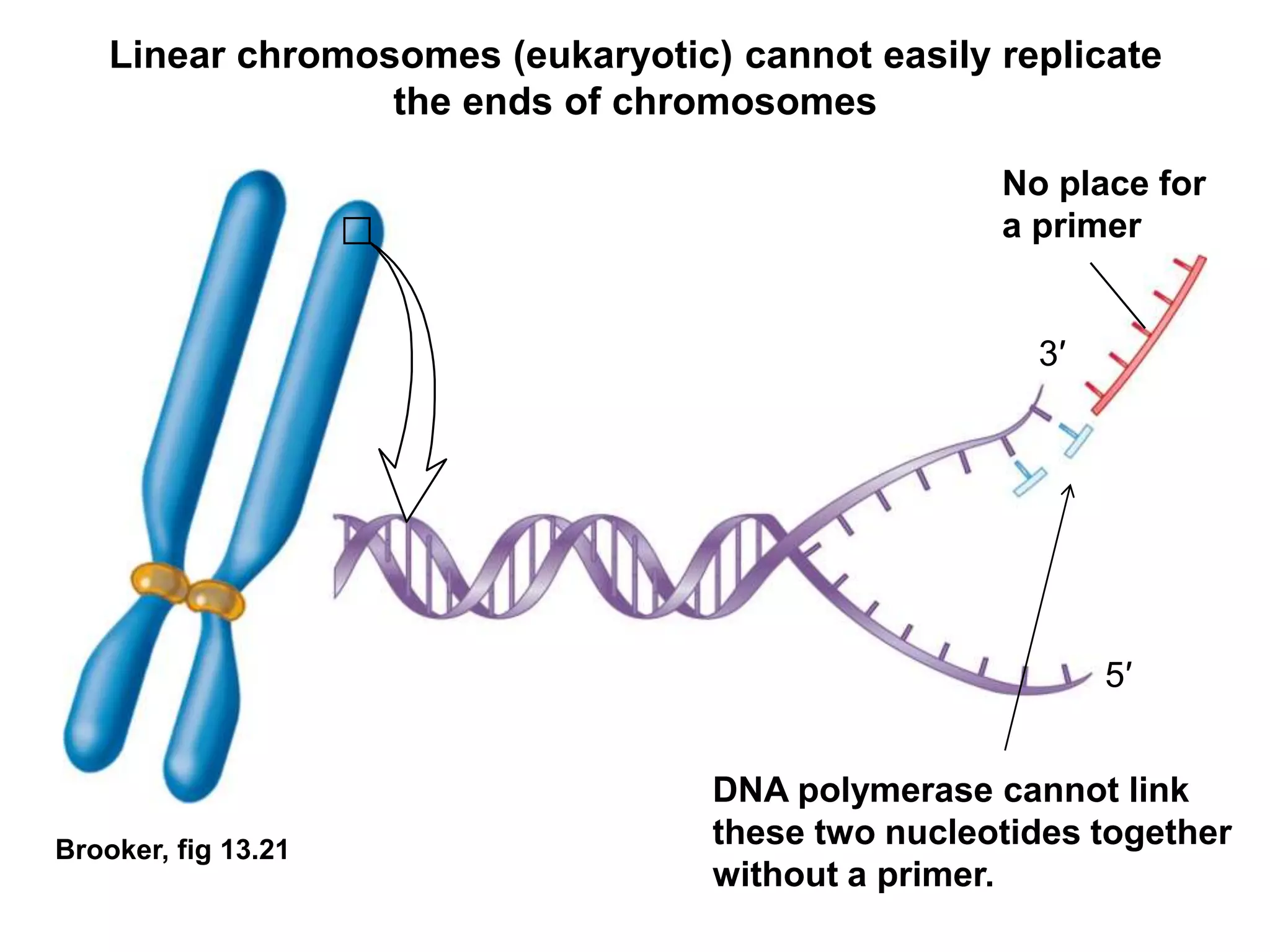
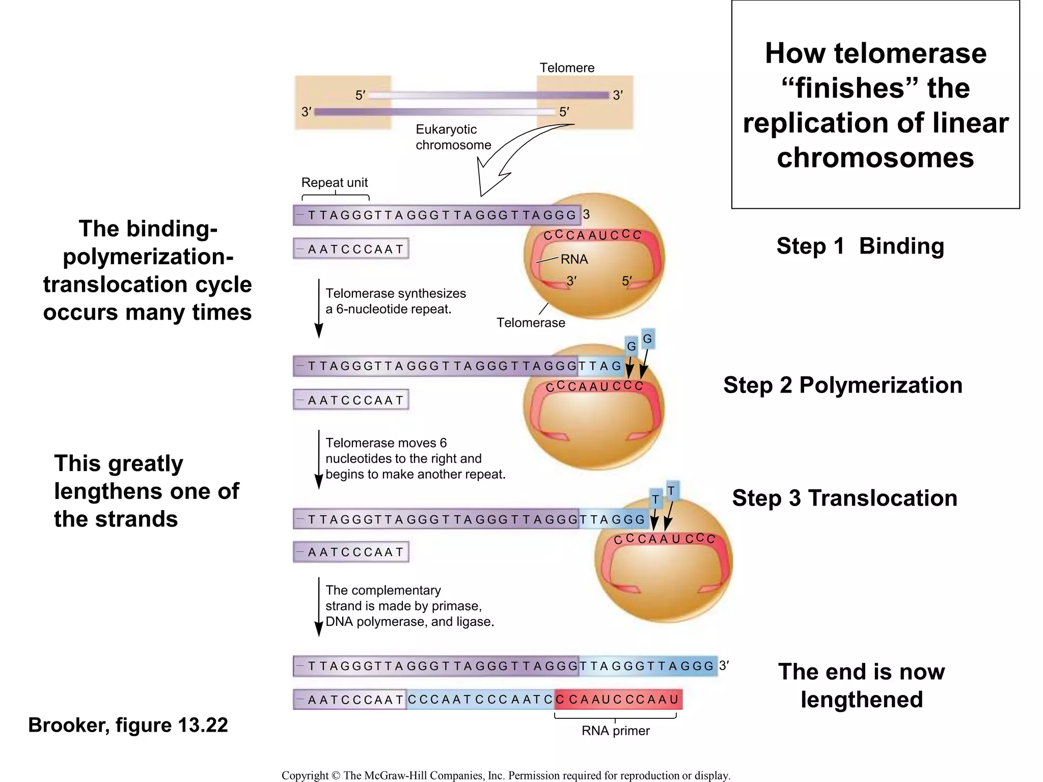
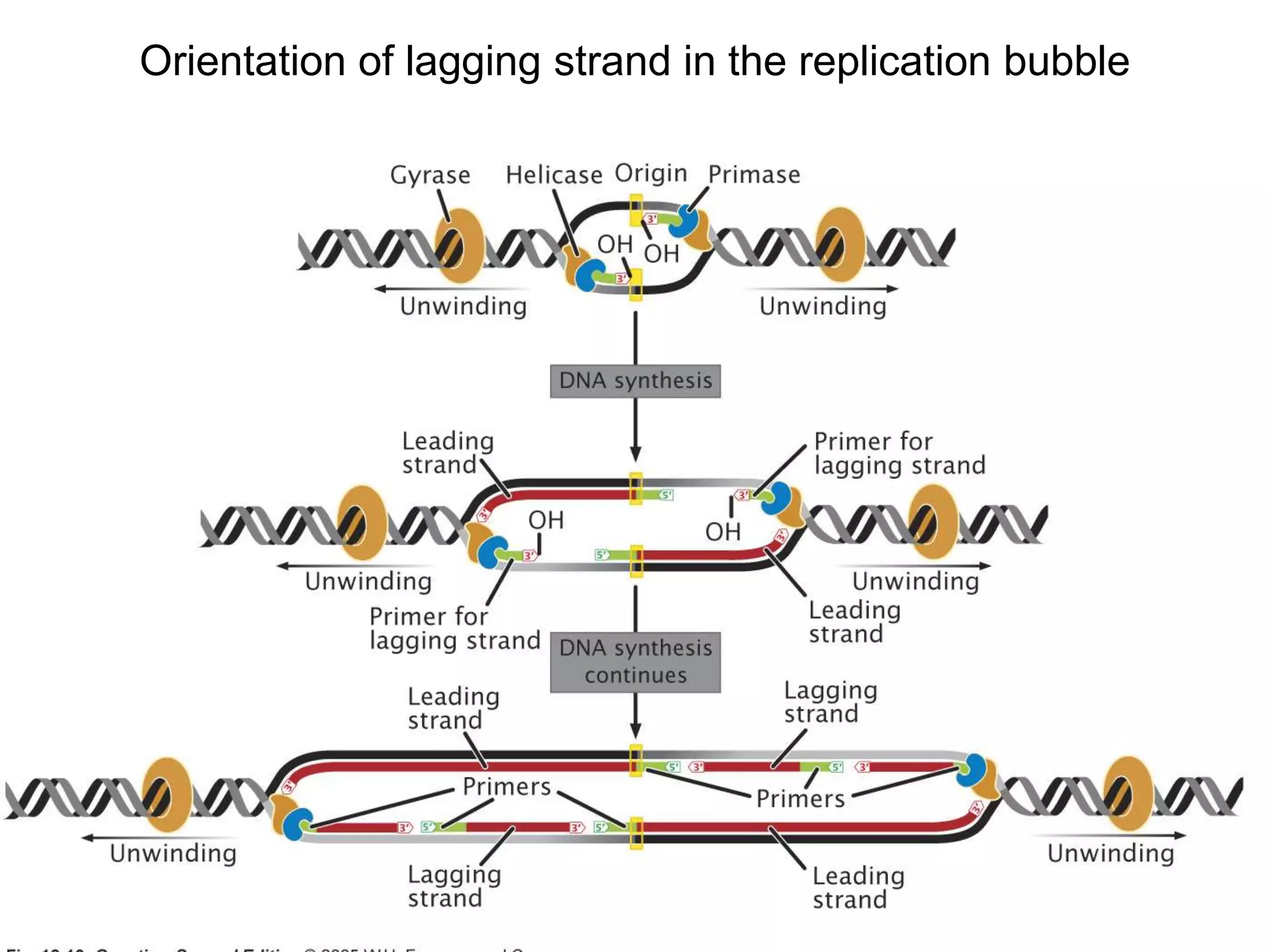
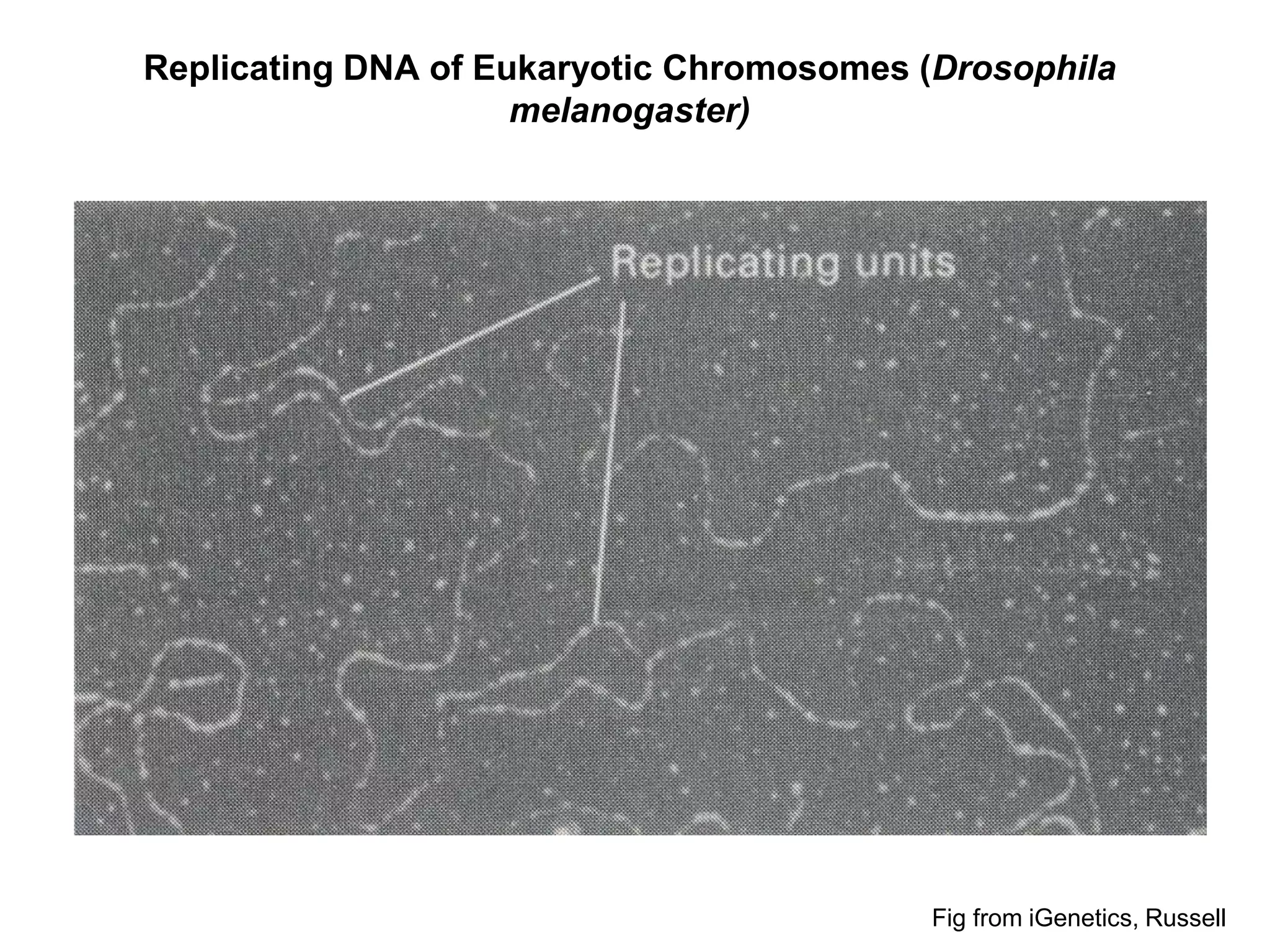
Broad Course Objectives for DNA Replication
Students will be able to:
• describe the historic experiment that demonstrated
DNA replication follows a semi-conservative model.
• describe the process of DNA replication in
prokaryotes at the biochemical level
• explain how proofreading and repair is accomplished
during DNA synthesis
Outline/study guide—DNA Replication
• At what point in the cell cycle does DNA replication occur?
• When two DNA molecules (or chromosomes) are made from one, where
do the parental strands end up, vs. the newly synthesized strands? (i.e.
semiconservative replication)
• Why can DNA only be synthesized in the 5’  3’ direction?
• What are the enzymes and proteins involved in DNA synthesis? What is
the function of each and at what point do they act?
• At what point does RNA function in DNA replication?
• What determines the lagging strand vs. the leading strand? How does
this change on the “other” side of the replication origin?
• How are the Okazaki fragments joined into one continuous DNA strand?
• How does the DNA replication machinery correct errors made during
replication?
• Are human chromosomes linear or circular? Bacteria?
• Why do linear chromosomes (but not circular chromosomes) have a
problem with telomeres becoming shorter and shorter with each round of
replication? How do some cells get around this?
48 year old woman with Werner Syndrome
Progeria
T A
G
C
A
G
A T
T A
T
G
G
A
A
C
C
C
T
T
G
C G
T A
T A
C
G
A T
T A
C G
T
A T
C G
C
C G
A T
C G
A
C
G
Incoming
nucleotides
Original
(template)
strand
Original
(template)
strand
Newly
synthesized
daughter strand
Replication
fork
(a) The mechanism of DNA replication (b) The products of replication
Leading
strand
Lagging
strand
5′ 3′
3′ 5′
A T
A T
T A
T A
T A
C G
C G
G C
G C
G C
G C
C G
A T
5′ 3′
5′ 3′
3′ 5′
A T
A T
T A
T A
T A
C G
C G
G C
G C
G C
G C
C G
A T
3′ 3′
3′ 5′
A T
A T
T A
T A
T A
C G
C G
G C
G C
G C
G C
C G
A T
Copyright © The McGraw-Hill Companies, Inc. Permission required for reproduction or display.
5′
3′
A
A T
Brooker Fig 13.1
Identical
base sequences
Each strand of
the parent DNA
molecule
becomes a
template for the
new
molecule(s)
The width of the
nucleotides
reflect larger
purines and
smaller
pyrimidines
Brooker fig 13.2
Conservative model
First
replication
Second
replication
Original
DNA
Semiconservative model Dispersive model
Copyright © The McGraw-Hill Companies, Inc. Permission required for reproduction or display.
Different models of DNA Replication
Brooker fig 13.2
Conservative model
First
Replication
(N14)
Second
Replication
(N14)
Original
DNA (N15)
Semiconservative model Dispersive model
Copyright © The McGraw-Hill Companies, Inc. Permission required for reproduction or display.
How do you expect the different models to appear in the
centrifuge experiment?
Brooker fig 13.3 Copyright © The McGraw-Hill Companies, Inc. Permission required for reproduction or display.
Experimental level Conceptual level
2. Incubate the cells for various cell generations
3. Lyse cells to release DNA
4. Load sample of lysate onto CsCl gradient.
5. Centrifuge until the DNA molecules reach equilibrium
densities.
6. View DNA within the gradient using a UV light.
DNA
Cell wall
Cell membrane
Light DNA
Half-heavy DNA
Heavy DNA
(after 2 generations.)
CsCl
gradient
Lysate
37°C
14N
solution
Suspension of
bacterial
cells labeled
with 15N
Up to 4 generations
Density centrifugation
Generation
0
1
Add 14N
2
1. Grow bacteria in excess of 15N-containing
compounds. Switch to 14N at Generation 1.
15N-DNA = purple
14N-DNA = blue
Experiment to distinguish between DNA replication models
Light
Half-heavy
Heavy
Generations After 14N Addition
4.1 3.0 2.5 1.9 1.5 1.1 1.0 0.7 0.3
*Data from: Meselson, M. and Stahl, F.W. (1958) The Replication of DNA in Escherichia coli. Proc. Natl. Acad. Sci. USA 44: 671−682
Interpreting the Data
Copyright ©The McGraw-Hill Companies, Inc. Permission required for reproduction or display
After one generation,
DNA is “half-heavy”
(consistent with both semi-
conservative and dispersive
models)
After ~ two generations: DNA
is “light” and “half-heavy”
(Consistent with which model?)
Why does DNA (and RNA) only “grow” in the 3’ direction?
New strand Template strand
5’ end 3’ end
Sugar A T
Base
C
G
G
C
A
C
OH
P P
5’ end 3’ end
5’ end 5’ end
A T
C
G
G
C
A
C
T
3’ end
Nucleoside
triphosphate
Pyrophosphate
2 P
OH
Phosphate
Fig from Cambell and Reece, 7th ed
(Like Brooker, fig 13-15)
Brooker, fig 13.10
Origin of replication
Replication
forks
Direction of
replication fork
1st Okazaki
fragment
First and second Okazaki
fragments have been
connected to each other.
1st Okazaki fragment
of the lagging strand
2nd Okazaki
fragment
3rd
Okazaki
fragment
Primer
Primer
The leading strand elongates,
and a second Okazaki fragment
is made.
The leading strand continues to elongate. A
third Okazaki fragment is made, and the first
and second are connected together.
Primers initiate DNA synthesis.
Synthesis of the leading strand occurs in
the same direction as movement of the
replication fork. 1st Okazaki
fragment of lagging strand is
made in opposite direction.
5′
5′
5′
5′
3′
5′
3′
3′
5′
3′
3′
3′
5′
5′
5′
5′
3′
3′
3′
3′
5′
5′
3′
3′
Copyright © The McGraw-Hill Companies, Inc. Permission required for reproduction or display.
Leading
strand
DNA strands separate at
origin
Overview of
DNA
Replication
Copyright ©The McGraw-Hill Companies, Inc. Permission required for reproduction or display
Brooker, fig 13.7
5′
3′
5′
5′
3′
3′
DNA polymerase III
Origin
Leading
strand
Lagging strand
Linked Okazaki
fragments
Direction of fork movement
DNA polymerase III
RNA
primer
Okazaki fragment
DNA
ligase
RNA primer
Single-strand
binding protein
DNA helicase
Topoisomerase II
Parental DNA
Primase
Replication fork
• DNA helicase breaks the hydrogen
bonds between the DNA strands.
• Topoisomerase alleviates positive
supercoiling.
• Single-strand binding proteins keep
the parental strands apart.
• Primase synthesizes an RNA
primer.
• DNA polymerase III synthesizes a
daughter strand of DNA.
• DNA polymerase I excises the
RNA primers and fills in with
DNA (not shown).
• DNA ligase covalently links the
Okazaki fragments together.
Functions of key proteins involved with DNA
replication
Brooker, fig 13.5
E. coli
chromosome
oriC
G G
G G G
GG
A GAGA
AAAAA G
AA A
A
T
T
T T ATT TT
TA A
TTT
T
T
C T T
C AT
T
C
T T
C
C
C
1
C
C C C
CC
T CTCT
TTTTT C
TT T
T
A A A TAA AA
AT T
AAA
A
A
G A A
G TA
A
G
A A
G
G
T A
G T C
CT
T AACA
AGGAT A
GC C
A
G T T CCT T
T
C
G
DnaA box
DnaA box
DnaA box
DnaA box
DnaA box
T T
GGA
T
C
A T C
G CT
G
G
A G
G
A T
C A G
GA
A TTGT
TCCT A T
CG G
T
C A A GGA AG
CA A
CCT
A
G
T A G
C GA
C
C
T C
C
A
T C
T A
CA
T GAAT
CCTGG G
AA G
C
A A A ATT GG
AA T
CTG
A
A
A A C
T AT
G
T
G T
A
A
G
C C
C C G
GT
T TACA
GCTGG C
T
T
T
A
T
G A A TGA TC
GG A
GTT
A
C
G G A
A AA
A
A
C G
A
A
G G
G G C
CA
A ATGT
CGACC G
T A T
A
C T T ACT AG
CC T
CAA
T
G
C C T
T TT
T
T
G C
T
T
A G
C A T
AC
T GA C
GTTCT G
TG A
G
G G T CTA CT
CC T
GGT
T
C
A T A
A CT
C
T
C A
A
A
T C
G T A
TG
A CT AG
CAAGA ACCT
C
C C A GAT GA
GG A
CCA
A
G
T A T
T GA
G
A
G T
T
T
GA T G
TA
C CAGTA C
A G
C
A T CA
G
G C
A
CT A C
AT
G GTCAT G
TA C
G
T A GT
C
C G
T
A G
A A T
GT
A CTTA
GGACC C
TT C
G
T T T TAA CC
TT A
GAC
T
T
T T G
A TA
C
A
C A
T
C
AT-rich region
5′–
–
50
51 100
101 150
201
251 275
250
151 200
Copyright © The McGraw-Hill Companies, Inc. Permission required for reproduction or display.
3′
Origin of Replication
Brooker fig 13.6
AT-rich region DnaA boxes
DnaA proteins bind to DnaA boxes and to
each other. Additional proteins that cause
the DNA to bend also bind (not shown).
This causes the region to wrap around
the DnaA proteins and separates the
AT-rich region.
DNA helicase
DNA helicase (DnaB protein) binds to the
origin. DnaC protein (not shown) assists
this process.
DNA helicase separates the DNA in both
directions, creating 2 replication forks.
DnaA protein
Copyright © The McGraw-Hill Companies, Inc. Permission required for reproduction or display.
5′ 3′
AT-
rich
region
Fork
Fork
3′ 5′
5′
3′
5′
3′
5′
3′
3′
5′
3′
5′
3′
5′
How the origin
sequence initiates
replication
DNA helicase
DNA helicase separates the DNA in both
directions, creating 2 replication forks.
Fork
Fork
5′
3′
5′
3′
3′
5′
3′
5′
Brooker fig 13.6
Travels along the DNA
in the 5’ to 3’ direction
Bidirectional
replication
Copyright © The McGraw-Hill Companies, Inc. Permission required for reproduction or display.
Replication initiation cont.
Copyright ©The McGraw-Hill Companies, Inc. Permission required for reproduction or display
Brooker, fig 13.8
3′
3′ exonuclease
site
3′
5′ 5′
Fingers
Thumb
DNA polymerase
catalytic site
Template
strand
Palm
Incoming
DNA nucleotides (triphosphates)
(dNTPs)
Schematic side view of DNA polymerase III
(bacterial)
Model for how the leading strand and lagging strand
coordinate at the replication fork
Brooker Fig 13.12
5′
3′
5′
3′
3′
5′ 5′
DNA helicase
Replisome
Primosome
Topoisomerase
Leading strand
DNA
polymerase III
Single-strand
binding proteins
Region
where
next Okazaki
fragment
will be made
Primase
RNA primer
New Okazaki
fragment
Older Okazaki
fragment
Replication
fork
5′
Copyright © The McGraw-Hill Companies, Inc. Permission required for reproduction or display.
Orientation of lagging strand in the replication bubble
Brooker, ch 13
Chromosome Sister chromatids
Before S phase During S phase End of S phase
Origin
Origin
Origin
Origin
Origin
Centromere
(DNA under the
kinetochore)
Copyright © The McGraw-Hill Companies, Inc. Permission required for reproduction or display.
Eukaryotes
have hundreds
of origins of
replication on
their (linear)
chromosome
Replicating DNA of Eukaryotic Chromosomes (Drosophila
melanogaster)
Fig from iGenetics, Russell
Brooker,
fig 13.4a
0.25 μm
(b) Autoradiograph of an E. coli chromosome in the act of replication
(a) Bacterial chromosome replication
Replication
forks
Origin of
replication
Replication
fork
Site where
replication
ends
Copyright
©
The
McGraw-Hill
Companies,
Inc.
Permission
required
for
reproduction
or
display.
From Cold Spring Harbor Symposia of Quantitative Biology, 28, p. 43 (1963).
Copyright holder is Cold Spring Habour Laboratory Press.
Replication
fork
Bacteria only have one origin on their
(circular) chromosome
Replication rate
• Eukaryotic DNA replication
– Typical human chromosome length: 100 million bp
– Time to replicate a chromosome: minutes to hours
– Hundreds of origins per chromosome
– Replicon = ~20,000 to 300,000 bp long
– 500-5000 bp / minute at each replication fork
(slower than bacterial replication; that much harder
to “unwind” the DNA for replication).
• Bacterial (prokaryotic) replication:
– Single circular chromosome (~4.6 million base pairs
[bp])
– Single origin of replication  single replicon
(“Replication Bubble”)
Requirements of DNA Replication in a complex
organism
• Very low error rate:
– One human cell: 6 billion bp of DNA. A
copying error rate of 1 error/million nt
6000 errors with every cell division
• Very fast copy rate
– E. coli –1000 nt per minute  3 days to
replicate (real life: 20 minutes per cell
cycle; 1000 nt per second)
Brooker, fig 13.21
DNA polymerase cannot link
these two nucleotides together
without a primer.
No place for
a primer
3′
5′
Linear chromosomes (eukaryotic) cannot easily replicate
the ends of chromosomes
Copyright ©The McGraw-Hill Companies, Inc. Permission required for reproduction or display
Brooker, fig 13.20
Telomeric repeat sequences
Overhang
C
G
C
G
C
G
T
A
A
T
A
T
C
G
C
G
C
G
T
A
A
T
A
T
C
G
C
G
C
G
T
A
A
T
A
T
C
G
C
G
C
G
T
A
A
T
A
T
C
G
C
G
C
G
T
A
A
T
A
T
C
G
C
G
C
G
T
A
A
T
A
T
C
G
C
G
C
G
T
A
A
T
T
A
T G G G
A
A
T
A
T G G G
A
T A
T T T G
G
G
5′
3′
Chromosome gets
shorter at the
telomeres with each
replication if
overhang is left.
In humans and most complex organisms, telomerase is only used in
continuously dividing stem cells (e.g. spermatogonia stem cells) 
most cells get shorter telomeres over time (age). What happened to
Dolly, the cloned sheep? (she was generated from a skin cell with
shorter telomeres, and she aged early)
Linear chromosomes (eukaryotic) must fill in
gap left by RNA primer
Telomere
Telomerase
Eukaryotic
chromosome
Repeat unit
3′
3
5′
T T A G G G T T A
A A T C C C A A T
C A A U C
G G G A G G G
T T A T T
G G G
T T A G G G T T A
C A A U C
G G G T T A T T G
G
G
T T A
G G G A G G G
C A A U C
T
T
C C C A A T A A A A
T C C C U A A
C U
C C
C C C
T T A G G G T T A G G G T T A T T G
T T A
G G G A G G G G G
T T A G G G T T A G G G T T A T T G
T T A
G G G A G G G G G G
T T A G G
A A T C C C A A T
A A T C C C A A T
A A T C C C A A T
RNA
RNA primer
Telomerase synthesizes
a 6-nucleotide repeat.
Telomerase moves 6
nucleotides to the right and
begins to make another repeat.
The complementary
strand is made by primase,
DNA polymerase, and ligase.
3′ 5′
5′ 3′
3′
Copyright © The McGraw-Hill Companies, Inc. Permission required for reproduction or display.
Brooker, figure 13.22
Step 1 Binding
Step 3 Translocation
The binding-
polymerization-
translocation cycle
occurs many times
This greatly
lengthens one of
the strands
Step 2 Polymerization
The end is now
lengthened
How telomerase
“finishes” the
replication of linear
chromosomes
Go over lecture outline at end of
lecture
Concept Checks
• In the Meselson and Stahl experiment,
how was switching the bacterial media
from N15 to N14 important for supporting
the Semi-conservative model?
Concept check
• What are the functions of the A-T rich
region and DNA boxes in the Origin of
Replication?
Concept Check
• Why is primase needed for DNA
replication?
• Is the template strand read in the 5’ to 3’
direction or the 3’ to 5’ direction?
Concept Check
• Describe the differences between Dna
synthesis in the leading strand vs. the
lagging strand.
Which component functions immediately
after ligase?
a. Helicase
b. DNA Polymerase 1
c. DNA Polymerase 3
d. primase
e. none of the above
Which component functions immediately
after ligase?
a. Helicase
b. DNA Polymerase 1
c. DNA Polymerase 3
d. primase
e. none of the above

dna-replication.ppt

  • 1.
    Broad Course Objectivesfor DNA Replication Students will be able to: • describe the historic experiment that demonstrated DNA replication follows a semi-conservative model. • describe the process of DNA replication in prokaryotes at the biochemical level • explain how proofreading and repair is accomplished during DNA synthesis
  • 2.
    Outline/study guide—DNA Replication •At what point in the cell cycle does DNA replication occur? • When two DNA molecules (or chromosomes) are made from one, where do the parental strands end up, vs. the newly synthesized strands? (i.e. semiconservative replication) • Why can DNA only be synthesized in the 5’  3’ direction? • What are the enzymes and proteins involved in DNA synthesis? What is the function of each and at what point do they act? • At what point does RNA function in DNA replication? • What determines the lagging strand vs. the leading strand? How does this change on the “other” side of the replication origin? • How are the Okazaki fragments joined into one continuous DNA strand? • How does the DNA replication machinery correct errors made during replication? • Are human chromosomes linear or circular? Bacteria? • Why do linear chromosomes (but not circular chromosomes) have a problem with telomeres becoming shorter and shorter with each round of replication? How do some cells get around this?
  • 3.
    48 year oldwoman with Werner Syndrome
  • 4.
  • 6.
    T A G C A G A T TA T G G A A C C C T T G C G T A T A C G A T T A C G T A T C G C C G A T C G A C G Incoming nucleotides Original (template) strand Original (template) strand Newly synthesized daughter strand Replication fork (a) The mechanism of DNA replication (b) The products of replication Leading strand Lagging strand 5′ 3′ 3′ 5′ A T A T T A T A T A C G C G G C G C G C G C C G A T 5′ 3′ 5′ 3′ 3′ 5′ A T A T T A T A T A C G C G G C G C G C G C C G A T 3′ 3′ 3′ 5′ A T A T T A T A T A C G C G G C G C G C G C C G A T Copyright © The McGraw-Hill Companies, Inc. Permission required for reproduction or display. 5′ 3′ A A T Brooker Fig 13.1 Identical base sequences Each strand of the parent DNA molecule becomes a template for the new molecule(s) The width of the nucleotides reflect larger purines and smaller pyrimidines
  • 7.
    Brooker fig 13.2 Conservativemodel First replication Second replication Original DNA Semiconservative model Dispersive model Copyright © The McGraw-Hill Companies, Inc. Permission required for reproduction or display. Different models of DNA Replication
  • 8.
    Brooker fig 13.2 Conservativemodel First Replication (N14) Second Replication (N14) Original DNA (N15) Semiconservative model Dispersive model Copyright © The McGraw-Hill Companies, Inc. Permission required for reproduction or display. How do you expect the different models to appear in the centrifuge experiment?
  • 9.
    Brooker fig 13.3Copyright © The McGraw-Hill Companies, Inc. Permission required for reproduction or display. Experimental level Conceptual level 2. Incubate the cells for various cell generations 3. Lyse cells to release DNA 4. Load sample of lysate onto CsCl gradient. 5. Centrifuge until the DNA molecules reach equilibrium densities. 6. View DNA within the gradient using a UV light. DNA Cell wall Cell membrane Light DNA Half-heavy DNA Heavy DNA (after 2 generations.) CsCl gradient Lysate 37°C 14N solution Suspension of bacterial cells labeled with 15N Up to 4 generations Density centrifugation Generation 0 1 Add 14N 2 1. Grow bacteria in excess of 15N-containing compounds. Switch to 14N at Generation 1. 15N-DNA = purple 14N-DNA = blue Experiment to distinguish between DNA replication models
  • 10.
    Light Half-heavy Heavy Generations After 14NAddition 4.1 3.0 2.5 1.9 1.5 1.1 1.0 0.7 0.3 *Data from: Meselson, M. and Stahl, F.W. (1958) The Replication of DNA in Escherichia coli. Proc. Natl. Acad. Sci. USA 44: 671−682 Interpreting the Data Copyright ©The McGraw-Hill Companies, Inc. Permission required for reproduction or display After one generation, DNA is “half-heavy” (consistent with both semi- conservative and dispersive models) After ~ two generations: DNA is “light” and “half-heavy” (Consistent with which model?)
  • 11.
    Why does DNA(and RNA) only “grow” in the 3’ direction? New strand Template strand 5’ end 3’ end Sugar A T Base C G G C A C OH P P 5’ end 3’ end 5’ end 5’ end A T C G G C A C T 3’ end Nucleoside triphosphate Pyrophosphate 2 P OH Phosphate Fig from Cambell and Reece, 7th ed (Like Brooker, fig 13-15)
  • 12.
    Brooker, fig 13.10 Originof replication Replication forks Direction of replication fork 1st Okazaki fragment First and second Okazaki fragments have been connected to each other. 1st Okazaki fragment of the lagging strand 2nd Okazaki fragment 3rd Okazaki fragment Primer Primer The leading strand elongates, and a second Okazaki fragment is made. The leading strand continues to elongate. A third Okazaki fragment is made, and the first and second are connected together. Primers initiate DNA synthesis. Synthesis of the leading strand occurs in the same direction as movement of the replication fork. 1st Okazaki fragment of lagging strand is made in opposite direction. 5′ 5′ 5′ 5′ 3′ 5′ 3′ 3′ 5′ 3′ 3′ 3′ 5′ 5′ 5′ 5′ 3′ 3′ 3′ 3′ 5′ 5′ 3′ 3′ Copyright © The McGraw-Hill Companies, Inc. Permission required for reproduction or display. Leading strand DNA strands separate at origin Overview of DNA Replication
  • 13.
    Copyright ©The McGraw-HillCompanies, Inc. Permission required for reproduction or display Brooker, fig 13.7 5′ 3′ 5′ 5′ 3′ 3′ DNA polymerase III Origin Leading strand Lagging strand Linked Okazaki fragments Direction of fork movement DNA polymerase III RNA primer Okazaki fragment DNA ligase RNA primer Single-strand binding protein DNA helicase Topoisomerase II Parental DNA Primase Replication fork • DNA helicase breaks the hydrogen bonds between the DNA strands. • Topoisomerase alleviates positive supercoiling. • Single-strand binding proteins keep the parental strands apart. • Primase synthesizes an RNA primer. • DNA polymerase III synthesizes a daughter strand of DNA. • DNA polymerase I excises the RNA primers and fills in with DNA (not shown). • DNA ligase covalently links the Okazaki fragments together. Functions of key proteins involved with DNA replication
  • 14.
    Brooker, fig 13.5 E.coli chromosome oriC G G G G G GG A GAGA AAAAA G AA A A T T T T ATT TT TA A TTT T T C T T C AT T C T T C C C 1 C C C C CC T CTCT TTTTT C TT T T A A A TAA AA AT T AAA A A G A A G TA A G A A G G T A G T C CT T AACA AGGAT A GC C A G T T CCT T T C G DnaA box DnaA box DnaA box DnaA box DnaA box T T GGA T C A T C G CT G G A G G A T C A G GA A TTGT TCCT A T CG G T C A A GGA AG CA A CCT A G T A G C GA C C T C C A T C T A CA T GAAT CCTGG G AA G C A A A ATT GG AA T CTG A A A A C T AT G T G T A A G C C C C G GT T TACA GCTGG C T T T A T G A A TGA TC GG A GTT A C G G A A AA A A C G A A G G G G C CA A ATGT CGACC G T A T A C T T ACT AG CC T CAA T G C C T T TT T T G C T T A G C A T AC T GA C GTTCT G TG A G G G T CTA CT CC T GGT T C A T A A CT C T C A A A T C G T A TG A CT AG CAAGA ACCT C C C A GAT GA GG A CCA A G T A T T GA G A G T T T GA T G TA C CAGTA C A G C A T CA G G C A CT A C AT G GTCAT G TA C G T A GT C C G T A G A A T GT A CTTA GGACC C TT C G T T T TAA CC TT A GAC T T T T G A TA C A C A T C AT-rich region 5′– – 50 51 100 101 150 201 251 275 250 151 200 Copyright © The McGraw-Hill Companies, Inc. Permission required for reproduction or display. 3′ Origin of Replication
  • 15.
    Brooker fig 13.6 AT-richregion DnaA boxes DnaA proteins bind to DnaA boxes and to each other. Additional proteins that cause the DNA to bend also bind (not shown). This causes the region to wrap around the DnaA proteins and separates the AT-rich region. DNA helicase DNA helicase (DnaB protein) binds to the origin. DnaC protein (not shown) assists this process. DNA helicase separates the DNA in both directions, creating 2 replication forks. DnaA protein Copyright © The McGraw-Hill Companies, Inc. Permission required for reproduction or display. 5′ 3′ AT- rich region Fork Fork 3′ 5′ 5′ 3′ 5′ 3′ 5′ 3′ 3′ 5′ 3′ 5′ 3′ 5′ How the origin sequence initiates replication
  • 16.
    DNA helicase DNA helicaseseparates the DNA in both directions, creating 2 replication forks. Fork Fork 5′ 3′ 5′ 3′ 3′ 5′ 3′ 5′ Brooker fig 13.6 Travels along the DNA in the 5’ to 3’ direction Bidirectional replication Copyright © The McGraw-Hill Companies, Inc. Permission required for reproduction or display. Replication initiation cont.
  • 17.
    Copyright ©The McGraw-HillCompanies, Inc. Permission required for reproduction or display Brooker, fig 13.8 3′ 3′ exonuclease site 3′ 5′ 5′ Fingers Thumb DNA polymerase catalytic site Template strand Palm Incoming DNA nucleotides (triphosphates) (dNTPs) Schematic side view of DNA polymerase III (bacterial)
  • 18.
    Model for howthe leading strand and lagging strand coordinate at the replication fork Brooker Fig 13.12 5′ 3′ 5′ 3′ 3′ 5′ 5′ DNA helicase Replisome Primosome Topoisomerase Leading strand DNA polymerase III Single-strand binding proteins Region where next Okazaki fragment will be made Primase RNA primer New Okazaki fragment Older Okazaki fragment Replication fork 5′ Copyright © The McGraw-Hill Companies, Inc. Permission required for reproduction or display.
  • 19.
    Orientation of laggingstrand in the replication bubble
  • 20.
    Brooker, ch 13 ChromosomeSister chromatids Before S phase During S phase End of S phase Origin Origin Origin Origin Origin Centromere (DNA under the kinetochore) Copyright © The McGraw-Hill Companies, Inc. Permission required for reproduction or display. Eukaryotes have hundreds of origins of replication on their (linear) chromosome
  • 21.
    Replicating DNA ofEukaryotic Chromosomes (Drosophila melanogaster) Fig from iGenetics, Russell
  • 22.
    Brooker, fig 13.4a 0.25 μm (b)Autoradiograph of an E. coli chromosome in the act of replication (a) Bacterial chromosome replication Replication forks Origin of replication Replication fork Site where replication ends Copyright © The McGraw-Hill Companies, Inc. Permission required for reproduction or display. From Cold Spring Harbor Symposia of Quantitative Biology, 28, p. 43 (1963). Copyright holder is Cold Spring Habour Laboratory Press. Replication fork Bacteria only have one origin on their (circular) chromosome
  • 23.
    Replication rate • EukaryoticDNA replication – Typical human chromosome length: 100 million bp – Time to replicate a chromosome: minutes to hours – Hundreds of origins per chromosome – Replicon = ~20,000 to 300,000 bp long – 500-5000 bp / minute at each replication fork (slower than bacterial replication; that much harder to “unwind” the DNA for replication). • Bacterial (prokaryotic) replication: – Single circular chromosome (~4.6 million base pairs [bp]) – Single origin of replication  single replicon (“Replication Bubble”)
  • 24.
    Requirements of DNAReplication in a complex organism • Very low error rate: – One human cell: 6 billion bp of DNA. A copying error rate of 1 error/million nt 6000 errors with every cell division • Very fast copy rate – E. coli –1000 nt per minute  3 days to replicate (real life: 20 minutes per cell cycle; 1000 nt per second)
  • 25.
    Brooker, fig 13.21 DNApolymerase cannot link these two nucleotides together without a primer. No place for a primer 3′ 5′ Linear chromosomes (eukaryotic) cannot easily replicate the ends of chromosomes
  • 26.
    Copyright ©The McGraw-HillCompanies, Inc. Permission required for reproduction or display Brooker, fig 13.20 Telomeric repeat sequences Overhang C G C G C G T A A T A T C G C G C G T A A T A T C G C G C G T A A T A T C G C G C G T A A T A T C G C G C G T A A T A T C G C G C G T A A T A T C G C G C G T A A T T A T G G G A A T A T G G G A T A T T T G G G 5′ 3′ Chromosome gets shorter at the telomeres with each replication if overhang is left. In humans and most complex organisms, telomerase is only used in continuously dividing stem cells (e.g. spermatogonia stem cells)  most cells get shorter telomeres over time (age). What happened to Dolly, the cloned sheep? (she was generated from a skin cell with shorter telomeres, and she aged early) Linear chromosomes (eukaryotic) must fill in gap left by RNA primer
  • 27.
    Telomere Telomerase Eukaryotic chromosome Repeat unit 3′ 3 5′ T TA G G G T T A A A T C C C A A T C A A U C G G G A G G G T T A T T G G G T T A G G G T T A C A A U C G G G T T A T T G G G T T A G G G A G G G C A A U C T T C C C A A T A A A A T C C C U A A C U C C C C C T T A G G G T T A G G G T T A T T G T T A G G G A G G G G G T T A G G G T T A G G G T T A T T G T T A G G G A G G G G G G T T A G G A A T C C C A A T A A T C C C A A T A A T C C C A A T RNA RNA primer Telomerase synthesizes a 6-nucleotide repeat. Telomerase moves 6 nucleotides to the right and begins to make another repeat. The complementary strand is made by primase, DNA polymerase, and ligase. 3′ 5′ 5′ 3′ 3′ Copyright © The McGraw-Hill Companies, Inc. Permission required for reproduction or display. Brooker, figure 13.22 Step 1 Binding Step 3 Translocation The binding- polymerization- translocation cycle occurs many times This greatly lengthens one of the strands Step 2 Polymerization The end is now lengthened How telomerase “finishes” the replication of linear chromosomes
  • 28.
    Go over lectureoutline at end of lecture
  • 29.
    Concept Checks • Inthe Meselson and Stahl experiment, how was switching the bacterial media from N15 to N14 important for supporting the Semi-conservative model?
  • 30.
    Concept check • Whatare the functions of the A-T rich region and DNA boxes in the Origin of Replication?
  • 31.
    Concept Check • Whyis primase needed for DNA replication? • Is the template strand read in the 5’ to 3’ direction or the 3’ to 5’ direction?
  • 32.
    Concept Check • Describethe differences between Dna synthesis in the leading strand vs. the lagging strand.
  • 34.
    Which component functionsimmediately after ligase? a. Helicase b. DNA Polymerase 1 c. DNA Polymerase 3 d. primase e. none of the above
  • 35.
    Which component functionsimmediately after ligase? a. Helicase b. DNA Polymerase 1 c. DNA Polymerase 3 d. primase e. none of the above