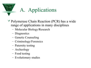
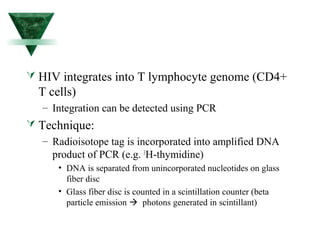
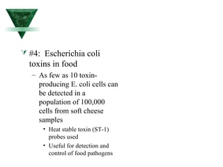
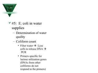
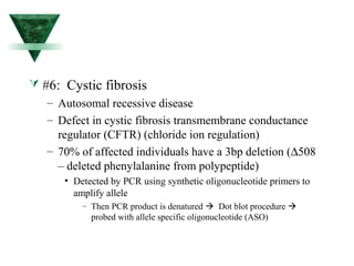
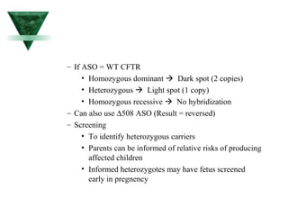
Applications of 
Recombinant DNA Technology 
© Sherry Fuller-Espie, 2002
I. Polymerase Chain Reaction
A. Applications 
 Polymerase Chain Reaction (PCR) has a wide 
range of applications in many disciplines 
– Molecular Biology/Research 
– Diagnostics 
– Genetic Counseling 
– Criminology/Forensics 
– Paternity testing 
– Archeology 
– Food testing 
– Evolutionary studies
B. Advantages over 
traditional methodologies 
 Fast and efficient amplification of specific DNA 
sequences 
 No requirement for cloning or subcloning 
 Tiny amounts of material are usually sufficient 
 Disease diagnoses will be greatly expedited by 
PCR to identify microorganisms in infected people 
who would prove falsely negative by other 
diagnostic procedures
 Drawbacks: 
– Can introduce errors in DNA during amplification 
process 
• Error-prone DNA polymerases 
• New enzymes are reducing this tendency 
– Vent polymerase vs. Taq polymerase 
– Contaminants can give rise to false positives or 
erroneous results 
• Numerous controls must be included 
• Controlled environment
C. Procedure 
 A specific DNA sequence is amplified using DNA 
polymerase and oligonucleotide primers using many cycles 
(25-30) 
 ~106-fold amplification achieved 
 Each cycle includes the following steps: 
– Denaturation (92oC) (separation of dsDNA) 
– Annealing (55oC) (primer binds to complementary regions) 
– Extension (72oC) (primer is elongated by DNA polymerase) 
 Exponential amplification of starting material achieved
 Required reagents for PCR: 
– Template DNA (can be DNA or RNA) 
• If RNA is used, an extra first step must be introduced 
involving reverse transcription of RNA to produce DNA 
template (= Reverse transcription PCR = RT-PCR) 
– Two flanking oligonucleotide primers (excess) 
– Nucleotides (excess) 
– Heat-stable DNA polymerase 
– Thermocycler to automate process (~6 minutes per 
cycle, ~3 hours for 30 cycles)
 HIV integrates into T lymphocyte genome (CD4+ 
T cells) 
– Integration can be detected using PCR 
 Technique: 
– Radioisotope tag is incorporated into amplified DNA 
product of PCR (e.g. 3H-thymidine) 
• DNA is separated from unincorporated nucleotides on glass 
fiber disc 
• Glass fiber disc is counted in a scintillation counter (beta 
particle emission  photons generated in scintillant)
Benefits: 
– Earlier treatment can begin 
– Especially useful for newborn determination of 
HIV status 
• Ab tests can’t rule out maternal IgG Abs 
– Maternal IgG transfer transplacentally 
– Can cause false positive result in newborn serum 
• Have to wait several months to determine if Ab are 
of fetal-origin
 #2: Lyme Disease – Detection of Borrelia 
burgdorferi 
– Caused by tick-borne spirochete 
– Arthropod transmission 
– Left untreated or not quickly enough: 
• Arthritis and neurological complications (can be fatal) 
– Problems: 
• Very difficult to cultivate spirochetes in lab media 
• Hard to see using conventional microscope 
– PCR can detect as few as 5 spirochetes in a sample!! 
• Can detect numerous strains (even if antigenic epitopes vary 
 serological limitations)
D. Examples 
#1: Detection of HIV in T-lymphocytes 
– Serological techniques reguire humoral 
immune responses to become activated for 
successful detection of anti-HIV antibodies 
(seroconversion) 
• Acquired immune responses can take 10-14 days 
before Ab titers reach maximum levels. 
• Individual may test negative and transmit HIV 
unknowingly (False negative)
#3: Human Papilloma Virus (HPV) 
– Causes genital warts and cervical cancer 
– Tissue sample from cervix used in pCR 
reaction 
– Treatment can begin earlier 
• Acyclovir 
• Gancyclovir
 #4: Escherichia coli 
toxins in food 
– As few as 10 toxin-producing 
E. coli cells can 
be detected in a 
population of 100,000 
cells from soft cheese 
samples 
• Heat stable toxin (ST-1) 
probes used 
• Useful for detection and 
control of food pathogens
– Other potential applications: 
• Clostridium botulinum 
• Campylobacter jejuni 
• Vibrio cholerae 
• Listeria monocytogenes 
• Salmonella spp. 
• Salmonella typhi 
• Shigella spp. 
• Giardia 
• Trichinosis
 #5: E. coli in water 
supplies 
– Determination of water 
quality 
– Coliform count 
• Filter water  Lyse 
cells to release DNA  
PCR 
• Primers specific for 
lactose utilization genes 
(DNAs from other 
coliforms do not 
respond to the primers)
 #6: Cystic fibrosis 
– Autosomal recessive disease 
– Defect in cystic fibrosis transmembrane conductance 
regulator (CFTR) (chloride ion regulation) 
– 70% of affected individuals have a 3bp deletion (D508 
– deleted phenylalanine from polypeptide) 
• Detected by PCR using synthetic oligonucleotide primers to 
amplify allele 
– Then PCR product is denatured  Dot blot procedure  
probed with allele specific oligonucleotide (ASO)
– If ASO = WT CFTR 
• Homozygous dominant  Dark spot (2 copies) 
• Heterozygous  Light spot (1 copy) 
• Homozygous recessive  No hybridization 
– Can also use D508 ASO (Result = reversed) 
– Screening 
• To identify heterozygous carriers 
• Parents can be informed of relative risks of producing 
affected children 
• Informed heterozygotes may have fetus screened 
early in pregnency
II. Transgenic Animals
A. Introduction 
 Transgenic animals: 
– Animals which have been genetically engineered to 
contain one or more genes from an exogenous source. 
– Transgenes are integrated into the genome. 
– Transgenes can be transmitted through the germline to 
progeny. 
– First transgenic animal produced = “Founder Animal”
B. Introduction of foreign genes 
into intact organisms 
 Procedure is basically the same regardless of 
which animal is involved. 
 Integration usually occurs prior to DNA 
replication in the fertilized oocyte. 
– Majority of transgenic animals carry the gene in all of 
their cells, including the germ cells. Transmission 
to next generation requires germline integration. 
– Some integration events occur subsequent to DNA 
replication giving rise to mosaic animals which may 
or may not contain the transgene in its germline.
C. Procedure for Producing 
Transgenic Mice 
Three different breeding pairs of mice are 
required.
 First Breeding Pair: 
– Fertile male + superovulated female 
• Fertile male = stud (changed regularly to ensure performance) 
• Superovulated female = immature female induced to 
superovulate 
– Pregnant mare’s serum (=FSH) on day 1 
– Human Chorionic Gonadotropin (=LH) on day 3 
• Mated on day 3 
• Fertilized oocytes microinjected on day 4 with foreign DNA 
construct. 
• Microinjected oocytes are transferred to the oviducts of 
surrogate mothers at end of day 4.
 Second breeding pair: 
– Sterile male + surrogate mother 
• Sterile male produced through vasectomy 
• Surrogate mother must mate to be suitable recipient of 
injected eggs 
• Mated on day 3 
• Microinjected oocytes from first breeding pair are 
transferred to oviducts on day 4 
• Embryos implant in uterine wall and are born 19 days later. 
• Southern blotting techniques confirm presence and copy 
number of transgenes.
D. Manipulation of Fertilized 
Oocytes 
See Slides
E. Gene Expression in 
Transgenic Mice 
 In order to discriminate the products of the 
injected gene from those of an endogenous 
counterpart, the injected gene must be marked in 
some way. 
– Mini-genes where exons are deleted of cDNA where 
introns are absent. 
– Modification by insertion/deletion/mutagenesis of a 
few nucleotides (e.g. the gain or loss of a restriction 
endonuclease site). 
– Hybrid genes where foreign epitopes are expressed on 
transgenic products.
F. Tissue-Specific Gene 
Expression 
 Generally, if a tissue-specific gene is expressed at all, then 
it is expressed appropriately, despite the fact that it has 
integrated at a different chromosomal location. 
– Trans-acting proteins involved in establishing tissue-specific 
expression are capable of finding their cognate sequences and 
activation transcription at various chromosomal locations. 
– Levels of expression vary between founder animals as 
chromosomal position can influence accessibility of the transgenes 
to these trans-acting transcription factors. 
– Some founders do not express the transgene at all owing to 
integration into heterochromatin domains where DNA is 
methylated heavily (silent).
 To study developmentally regulated genes
H. More Examples of Studies 
Utilizing Transgenic Mice 
“Pharm” animals (transgenic livestock) 
– Bioreactors whose cells have been engineered to 
synthesize marketable proteins 
– DNA constructs contain desired gene and appropriate 
regulatory sequences (tissue-specific promoters) 
– More economical than producing desired proteins in 
cell culture
I. Examples of Bioreactors 
 Naked human Hb from 
pigs 
 Human lactoferrin in 
cows’ milk 
 Alpha-1-antitrypsin in 
sheep 
 HGH in mouse urine 
(uroplakin promoters) 
 Human antibodies in mice 
(H and L chain tgenics  
hybridomas) 
 CfTCR in goats 
 Tissue plasminogen 
activator (TPA) in goats 
 Human antithrombin III in 
goats 
 Malaria antigens in goats 
(vaccine) 
 Alpha-glucosidase in 
rabbits (Pompe’s disease
J. Transgenic Pigs for the 
Production of Organs for 
Transplantation 
 Pig organs are rejected acutely due to the presence 
of human antibodies to pig antigens. 
 Once human antibodies are bound to pig organs, 
human complement is activated and triggers the 
complement cascade and organ destruction. 
 Transgenic pigs with complement inhibitors have 
been produced and are gaining feasibility as a 
source of xenogeneic organs for transplantation.
III. The Knockout Mouse
A. What is a Knockout 
Mouse? 
A really good-looking mouse? 
A mouse in which a very specific 
endogenous gene has been altered in such a 
way that interferes with normal expression, 
i.e. it has been knocked out.
B. Why Produce KO Mice? 
To study effects of gene products, 
biochemical pathways, alternative 
(compensatory) pathways, and 
developmental pathways 
To recreate human diseases in animals to 
establish models to test the beneficial 
effects of drugs or gene therapy.
C. Procedure for Generating 
a KO Mouse 
 Gene alteration in KO mice is targeted to very specific 
genes. 
 DNA must integrate at precise positions in the genome. 
 Integration of the altered gene takes place in embryonic 
stem cells ex vivo. 
 Verification of exact location of integration occurs before 
the ESC is introduced into blastocysts to become part of 
the developing embryo.
D. Pluripotent ES Cells 
 Pluripotent ES cells are undifferentiated early embryonic 
cells derived from the inner cell mass of mouse 
blastocysts. 
 In vitro ES cells must be grown on a feeder layer of 
fibroblasts to prevent them from differentiating. 
 Introduction of the transgene is achieved by 
electroporation of retroviral infection. 
 The transgene must integrate via recombination, not 
randomly. 
 Cells transfected successfully can be identified prior to 
injection into blastocysts.
E. Specific Gene Targeting in ES 
Cells 
 Gene targeting can be achieved using gene 
constructs designed for homologous 
recombination. This technique can be used to 
either: 
– Knockout functional genes to study their contribution 
to different developmental or disease processes (null 
mutations) 
• Genes encoding b2m, MHC class I and II. CD2, Ii, TCR, Ig, 
IL-4, IL-2, FceR, TAP1/2, RAG-2,and many more (>100)! 
– Replace a functional gene for a mutated/non-functional 
gene to restore wild type phenotype . 
• Gene encoding HGPRT in mice deficient for HGPRT (called 
Lesch-Nhyan syndrome in humans).
F. DNA Constructs for 
Recombination 
 DNA vectors contain the gene of interest which 
has been interrupted with an antibiotic resistance 
gene (hygromycin resistance, or G418 resistance). 
 To ensure targeted integration has occurred, the 
flanking DNA contains the thymidine kinase gene. 
If TK integrates (random insertion), then the 
transfected cells die when grown in selective 
media (gancyclovir).
G. Selection of Targeted ES 
Cells 
 Gancyclovir resistant and G418 resistant ES cells 
grow into small clumps on top of feeder cells. 
 The colonies of cells can be “picked” off and 
transferred to new wells (at 0.3 cells per well 
seeding density) containing feeder cells. 
 When sufficient numbers of cells are obtained, 
they are: 
– Frozen for safe storage 
– Analyzed by Southern blotting or PCR to determine 
nature of integration event 
– Microinjected into the blastocoel cavity of blastocysts.
III. Dolly and the Advancement 
of Animal Cloning
How Was Dolly Made?

Dna r tech.applications

  • 1.
    Applications of RecombinantDNA Technology © Sherry Fuller-Espie, 2002
  • 2.
  • 3.
    A. Applications Polymerase Chain Reaction (PCR) has a wide range of applications in many disciplines – Molecular Biology/Research – Diagnostics – Genetic Counseling – Criminology/Forensics – Paternity testing – Archeology – Food testing – Evolutionary studies
  • 4.
    B. Advantages over traditional methodologies  Fast and efficient amplification of specific DNA sequences  No requirement for cloning or subcloning  Tiny amounts of material are usually sufficient  Disease diagnoses will be greatly expedited by PCR to identify microorganisms in infected people who would prove falsely negative by other diagnostic procedures
  • 5.
     Drawbacks: –Can introduce errors in DNA during amplification process • Error-prone DNA polymerases • New enzymes are reducing this tendency – Vent polymerase vs. Taq polymerase – Contaminants can give rise to false positives or erroneous results • Numerous controls must be included • Controlled environment
  • 6.
    C. Procedure A specific DNA sequence is amplified using DNA polymerase and oligonucleotide primers using many cycles (25-30)  ~106-fold amplification achieved  Each cycle includes the following steps: – Denaturation (92oC) (separation of dsDNA) – Annealing (55oC) (primer binds to complementary regions) – Extension (72oC) (primer is elongated by DNA polymerase)  Exponential amplification of starting material achieved
  • 7.
     Required reagentsfor PCR: – Template DNA (can be DNA or RNA) • If RNA is used, an extra first step must be introduced involving reverse transcription of RNA to produce DNA template (= Reverse transcription PCR = RT-PCR) – Two flanking oligonucleotide primers (excess) – Nucleotides (excess) – Heat-stable DNA polymerase – Thermocycler to automate process (~6 minutes per cycle, ~3 hours for 30 cycles)
  • 8.
     HIV integratesinto T lymphocyte genome (CD4+ T cells) – Integration can be detected using PCR  Technique: – Radioisotope tag is incorporated into amplified DNA product of PCR (e.g. 3H-thymidine) • DNA is separated from unincorporated nucleotides on glass fiber disc • Glass fiber disc is counted in a scintillation counter (beta particle emission  photons generated in scintillant)
  • 9.
    Benefits: – Earliertreatment can begin – Especially useful for newborn determination of HIV status • Ab tests can’t rule out maternal IgG Abs – Maternal IgG transfer transplacentally – Can cause false positive result in newborn serum • Have to wait several months to determine if Ab are of fetal-origin
  • 10.
     #2: LymeDisease – Detection of Borrelia burgdorferi – Caused by tick-borne spirochete – Arthropod transmission – Left untreated or not quickly enough: • Arthritis and neurological complications (can be fatal) – Problems: • Very difficult to cultivate spirochetes in lab media • Hard to see using conventional microscope – PCR can detect as few as 5 spirochetes in a sample!! • Can detect numerous strains (even if antigenic epitopes vary  serological limitations)
  • 11.
    D. Examples #1:Detection of HIV in T-lymphocytes – Serological techniques reguire humoral immune responses to become activated for successful detection of anti-HIV antibodies (seroconversion) • Acquired immune responses can take 10-14 days before Ab titers reach maximum levels. • Individual may test negative and transmit HIV unknowingly (False negative)
  • 12.
    #3: Human PapillomaVirus (HPV) – Causes genital warts and cervical cancer – Tissue sample from cervix used in pCR reaction – Treatment can begin earlier • Acyclovir • Gancyclovir
  • 13.
     #4: Escherichiacoli toxins in food – As few as 10 toxin-producing E. coli cells can be detected in a population of 100,000 cells from soft cheese samples • Heat stable toxin (ST-1) probes used • Useful for detection and control of food pathogens
  • 14.
    – Other potentialapplications: • Clostridium botulinum • Campylobacter jejuni • Vibrio cholerae • Listeria monocytogenes • Salmonella spp. • Salmonella typhi • Shigella spp. • Giardia • Trichinosis
  • 15.
     #5: E.coli in water supplies – Determination of water quality – Coliform count • Filter water  Lyse cells to release DNA  PCR • Primers specific for lactose utilization genes (DNAs from other coliforms do not respond to the primers)
  • 16.
     #6: Cysticfibrosis – Autosomal recessive disease – Defect in cystic fibrosis transmembrane conductance regulator (CFTR) (chloride ion regulation) – 70% of affected individuals have a 3bp deletion (D508 – deleted phenylalanine from polypeptide) • Detected by PCR using synthetic oligonucleotide primers to amplify allele – Then PCR product is denatured  Dot blot procedure  probed with allele specific oligonucleotide (ASO)
  • 17.
    – If ASO= WT CFTR • Homozygous dominant  Dark spot (2 copies) • Heterozygous  Light spot (1 copy) • Homozygous recessive  No hybridization – Can also use D508 ASO (Result = reversed) – Screening • To identify heterozygous carriers • Parents can be informed of relative risks of producing affected children • Informed heterozygotes may have fetus screened early in pregnency
  • 18.
  • 19.
    A. Introduction Transgenic animals: – Animals which have been genetically engineered to contain one or more genes from an exogenous source. – Transgenes are integrated into the genome. – Transgenes can be transmitted through the germline to progeny. – First transgenic animal produced = “Founder Animal”
  • 20.
    B. Introduction offoreign genes into intact organisms  Procedure is basically the same regardless of which animal is involved.  Integration usually occurs prior to DNA replication in the fertilized oocyte. – Majority of transgenic animals carry the gene in all of their cells, including the germ cells. Transmission to next generation requires germline integration. – Some integration events occur subsequent to DNA replication giving rise to mosaic animals which may or may not contain the transgene in its germline.
  • 21.
    C. Procedure forProducing Transgenic Mice Three different breeding pairs of mice are required.
  • 22.
     First BreedingPair: – Fertile male + superovulated female • Fertile male = stud (changed regularly to ensure performance) • Superovulated female = immature female induced to superovulate – Pregnant mare’s serum (=FSH) on day 1 – Human Chorionic Gonadotropin (=LH) on day 3 • Mated on day 3 • Fertilized oocytes microinjected on day 4 with foreign DNA construct. • Microinjected oocytes are transferred to the oviducts of surrogate mothers at end of day 4.
  • 23.
     Second breedingpair: – Sterile male + surrogate mother • Sterile male produced through vasectomy • Surrogate mother must mate to be suitable recipient of injected eggs • Mated on day 3 • Microinjected oocytes from first breeding pair are transferred to oviducts on day 4 • Embryos implant in uterine wall and are born 19 days later. • Southern blotting techniques confirm presence and copy number of transgenes.
  • 24.
    D. Manipulation ofFertilized Oocytes See Slides
  • 25.
    E. Gene Expressionin Transgenic Mice  In order to discriminate the products of the injected gene from those of an endogenous counterpart, the injected gene must be marked in some way. – Mini-genes where exons are deleted of cDNA where introns are absent. – Modification by insertion/deletion/mutagenesis of a few nucleotides (e.g. the gain or loss of a restriction endonuclease site). – Hybrid genes where foreign epitopes are expressed on transgenic products.
  • 26.
    F. Tissue-Specific Gene Expression  Generally, if a tissue-specific gene is expressed at all, then it is expressed appropriately, despite the fact that it has integrated at a different chromosomal location. – Trans-acting proteins involved in establishing tissue-specific expression are capable of finding their cognate sequences and activation transcription at various chromosomal locations. – Levels of expression vary between founder animals as chromosomal position can influence accessibility of the transgenes to these trans-acting transcription factors. – Some founders do not express the transgene at all owing to integration into heterochromatin domains where DNA is methylated heavily (silent).
  • 27.
     To studydevelopmentally regulated genes
  • 28.
    H. More Examplesof Studies Utilizing Transgenic Mice “Pharm” animals (transgenic livestock) – Bioreactors whose cells have been engineered to synthesize marketable proteins – DNA constructs contain desired gene and appropriate regulatory sequences (tissue-specific promoters) – More economical than producing desired proteins in cell culture
  • 29.
    I. Examples ofBioreactors  Naked human Hb from pigs  Human lactoferrin in cows’ milk  Alpha-1-antitrypsin in sheep  HGH in mouse urine (uroplakin promoters)  Human antibodies in mice (H and L chain tgenics  hybridomas)  CfTCR in goats  Tissue plasminogen activator (TPA) in goats  Human antithrombin III in goats  Malaria antigens in goats (vaccine)  Alpha-glucosidase in rabbits (Pompe’s disease
  • 30.
    J. Transgenic Pigsfor the Production of Organs for Transplantation  Pig organs are rejected acutely due to the presence of human antibodies to pig antigens.  Once human antibodies are bound to pig organs, human complement is activated and triggers the complement cascade and organ destruction.  Transgenic pigs with complement inhibitors have been produced and are gaining feasibility as a source of xenogeneic organs for transplantation.
  • 31.
  • 32.
    A. What isa Knockout Mouse? A really good-looking mouse? A mouse in which a very specific endogenous gene has been altered in such a way that interferes with normal expression, i.e. it has been knocked out.
  • 33.
    B. Why ProduceKO Mice? To study effects of gene products, biochemical pathways, alternative (compensatory) pathways, and developmental pathways To recreate human diseases in animals to establish models to test the beneficial effects of drugs or gene therapy.
  • 34.
    C. Procedure forGenerating a KO Mouse  Gene alteration in KO mice is targeted to very specific genes.  DNA must integrate at precise positions in the genome.  Integration of the altered gene takes place in embryonic stem cells ex vivo.  Verification of exact location of integration occurs before the ESC is introduced into blastocysts to become part of the developing embryo.
  • 35.
    D. Pluripotent ESCells  Pluripotent ES cells are undifferentiated early embryonic cells derived from the inner cell mass of mouse blastocysts.  In vitro ES cells must be grown on a feeder layer of fibroblasts to prevent them from differentiating.  Introduction of the transgene is achieved by electroporation of retroviral infection.  The transgene must integrate via recombination, not randomly.  Cells transfected successfully can be identified prior to injection into blastocysts.
  • 36.
    E. Specific GeneTargeting in ES Cells  Gene targeting can be achieved using gene constructs designed for homologous recombination. This technique can be used to either: – Knockout functional genes to study their contribution to different developmental or disease processes (null mutations) • Genes encoding b2m, MHC class I and II. CD2, Ii, TCR, Ig, IL-4, IL-2, FceR, TAP1/2, RAG-2,and many more (>100)! – Replace a functional gene for a mutated/non-functional gene to restore wild type phenotype . • Gene encoding HGPRT in mice deficient for HGPRT (called Lesch-Nhyan syndrome in humans).
  • 37.
    F. DNA Constructsfor Recombination  DNA vectors contain the gene of interest which has been interrupted with an antibiotic resistance gene (hygromycin resistance, or G418 resistance).  To ensure targeted integration has occurred, the flanking DNA contains the thymidine kinase gene. If TK integrates (random insertion), then the transfected cells die when grown in selective media (gancyclovir).
  • 38.
    G. Selection ofTargeted ES Cells  Gancyclovir resistant and G418 resistant ES cells grow into small clumps on top of feeder cells.  The colonies of cells can be “picked” off and transferred to new wells (at 0.3 cells per well seeding density) containing feeder cells.  When sufficient numbers of cells are obtained, they are: – Frozen for safe storage – Analyzed by Southern blotting or PCR to determine nature of integration event – Microinjected into the blastocoel cavity of blastocysts.
  • 39.
    III. Dolly andthe Advancement of Animal Cloning
  • 40.