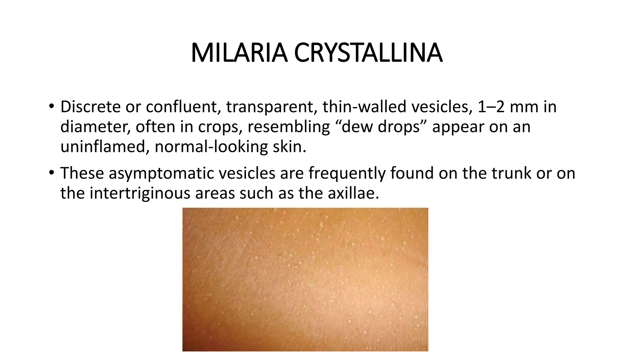
DISORDER OF SWEAT GLAND
PRESENTER : DR. ALOK JINDAL
• Sweat glands can be distinguished into 3 types on the basis of
morphology and structures
1. Eccrine
2. Apocrine
3. Apoeccrine glands
ECCRINE GLAND
• Present almost everywhere on the human skin
• Absent on the vermilion border of the lips, nail
bed, external ear canal, clitoris, and labia minora.
• On an average, there are 1.6–4 million sweat
glands on the body.
• Back: 64 glands/cm2
• Forearm: 108 glands/cm2
• Forehead: 181 glands/cm2
• Palms and soles: 600–700 glands/cm2
• Eccrine glands are merocrine gland in that no breakdown of cellular
material occur, and the secretion is discharged by the fusion of walls
of the secretory vesicles with the plasma membrane and content of
the vesicles are released.
• Eccrine glands are called Atrichial as they develop from the superficial
epidermis and remain independent of the hair follicle.
Functions of sweat
• Thermoregulation
• Moisten the skin of the palms and soles during periods of
activity, thus improving their grip
• It also acts as lesser organs of excretion (Drugs)
• Plays a pivotal role in wound healing i.e Lactate in sweat may
regulate desquamation of stratum corneum
• Mitogenic effects
• Proinflammatory action
APOCRINE GLAND
• Apocrine glands are commonly seen in the
axillae, areolae (Montgomery tubercles),
periumbilical, perineal and circumoral areas.
• Apocrine glands are larger than eccrine
glands and contain a secretory and a ductal
portion
• The coiled secretory portion is present in
lower dermis or in the subcutaneous fat and
a straight excretory duct empties into the
infundibulum of the hair follicle slightly
above the entrance of the sebaceous duct
• Apocrine glands are called epitrichial as they develop from the
external root sheath of the hair follicle and usually remain attached to
it
• Human apocrine sweat is a protein-rich, milky, or viscid, colorless
secretion when it is first formed.
• In the classical apocrine type of secretion an apical cap and a dividing
membrane are formed initially and apical cap is then detached and
discharged into the lumen of the gland by a process called
“Apocopation”.
• Respond to emotional stimuli that promote sympathetic discharge.
• With aging, apocrine glands tend to accumulate lipofuscin and
undergo attenuation.
APOECCRINE GLAND
• Third type of sweat glands, called the apoeccrine or mixed glands, is
found in the adult human axillae.
• Share some of the morphological and functional features of both.
• They are found in all levels of the dermis and their size are variable.
• The apoeccrine gland has a long duct that opens directly on to the
skin surface.
• The glands function like eccrine glands and significantly contribute to
the overall axillary sweating in adults
PHYSIOLOGY OF SWEATING
• Sweat gland activity is controlled by three principal physiological
stimuli thermal, emotional, and gustatory.
• Thermoregulatory sweating is controlled by thermosensitive neurons
in the hypothalamus.
• Emotional stimuli produce sweating on the palms and soles.
• Gustatory stimuli induce sweating on the lips, forehead, and nose,
and such sweating is believed to be controlled by medullary nuclei.
• Sweat glands are innervated by postganglionic sympathetic fibers.
• Acetylcholine (ACH) is the principal terminal neurotransmitter.
• Eccrine sweating is the result of sympathetic activity.
COMPOSITION OF SWEAT
• Sodium
• Potassium
• Chloride
• Bicarbonate
• Lactate
• Urea
• Ammonia
• Free amino acids
• Proteins 20 mg/dL
• Glucose 0.2–1.5 mg/dL
• Pyruvate
• Proteolytic enzymes
ALTERATION IN COMPOSITION OF SWEAT IN
DISEASE
ADRENOCORTICAL DISORDER
• Cushing syndrome and hyperaldosteronism in which there is excessive
secretion of adrenal hormones with mineralocorticoid activity, the
concentration of sodium and chloride in sweat decreases and that of
potassium increases
CYSTIC FIBROSIS
• Autosomal recessively inherited genetic disorder affects all the
exocrine glands
• Show a marked elevation of electrolytes in their sweat secretion.
• Elevated sweat sodium concentration (65–120 mmol)is the diagnostic
test for Cystic Fibrosis.
DISORDERS OF SWEAT GLAND
Disorders of Eccrine sweat glands
• HYPERHYDROSIS
• ANHIDROSIS/HYPOHIDROSIS
HYPERHYDROSIS
• Excessive sweating
• It is diificult to define when sweating is
excessive.
• when sweating is clinically noticeable
under conditions where it would not be
normally expected to occur or where it is
excess.
PRIMARY FOCAL HYPERHYDROSIS
• Excessive, bilateral, and relatively symmetric sweating occurring in
axillae, palms, soles, or the craniofacial region.
• Criteria for diagnosis is ::
Focal, visible, excessive sweating of at least 6 months duration without
apparent cause with at least two of the following characteristics:
1. Bilateral and relatively symmetrical involvement
2. Impairment of daily activities
3. Minimum one occurrence per week
4. Age of onset less than 25 years
5. Familial history
6. No sweating while asleep
VOLAR HYPERHYDROSIS
• Excessive sweating of the palms and
soles
• May literally drip with sweat and can
be a source of disability at work or
studies.
• Volar hyperhidrosis may sometimes
be associated with excessive axillary
sweating.
• Disease may persist indefinitely,
although there is a tendency for
spontaneous improvement with age.
Palmoplantar hyperhidrosis is associated with syndromes :
• Palmoplantar keratoderma with clinodactyly,
• Nail-patella syndrome,
• Raynaud disease,
• Erythromelalgia,
• Arteriovenous fistula,
• Cold injury,
• Rheumatoid arthritis.
AXILLARY HYPERHYDROSIS
• Axillary hyperhidrosis is essentially a postpubertal problem, usually
beginning between 15 and 18 years of age.
• Commonly phasic or episodic but can be at times continuous.
• Worse in a hot and humid environment.
CRANIOFACIAL HYPERHIDROSIS
• Craniofacial hyperhidrosis is generally phasic and occurs in middle
age.
• Heat, exercise, and sometimes eating certain foods can trigger
episodes of sweating.
GUSTATORY HYPERHIDROSIS
• Generally unilateral, involves the
preauricular or infraauricular areas of the
face frequently, and can be mild or
profuse.
• Largely the result of damage to the
sympathetic nerves around the head and
neck with subsequent aberrant
regeneration and synapsing.
Thus, pathological gustatory sweating results from
• (1) upper dorsal sympathectomy
• (2) involvement of cervical sympathetic trunk by a tumor as in
Pancoast syndrome
• (3)sympathetic hyperactivity following encephalitis or syringomyelia
• (4) peripheral autonomic or sensory neuropathy as in diabetes
mellitus
• (5) Parotid gland abscesses or parotitis
AURICULOTEMPORAL SYNDROME
• AKA FREY SYNDROME
• The auriculotemporal nerve
contains afferent sensory
fibers from the skin of the
preauricular area, efferent
parasympathetic
secretomotor to parotid
gland, sympathetic secretory
fibers to the sweat glands.
• Injury to the nerve, regenerating parasympathetic fibers in the
auriculotemporal nerve, by a process of misdirection, migrate and
join with the distal sympathetic fibers innervating the sweat glands
and the blood vessels.
• Consequently, masticatory stimuli (drinking, chewing, eating, etc.) or
sometime tactile stimuli that would normally activate secretory
impulses to the parotid gland, instead activate the sweat glands and
the blood vessels to cause pain, flushing, and sweating in the
distribution of the auriculotemporal nerve.
INVESTIGATION
• Hyperhidrosis disease severity scale (HDSS) :
1. The sweating is never evident and does not affect daily routine.
2. The sweating can be tolerable and sometimes affects daily routine.
3. The sweating can be barely tolerated and often affects daily routine.
4. The sweating is never tolerable and always affects daily routine.
Score of 1: mild, 2: moderate, 3–4: severe.
TREATMENT
• Anticholinergic drugs such as atropine or atropine analogs are used
• But not proffered because they can cause dryness of the mouth,
vision disturbances, glaucoma, hyperthermia, or convulsions.
• Ganglion-blocking agents also inhibit sweating but hypotension is
side effect.
• Calcium channel blockers, such as diltiazem is helpful
• Propantheline bromide (15 mg t.i.d.) is a useful agent in volar
hyperhidrosis lasting for about 4 - 6 hour.
TOPICAL TREATMENT
• Solution of 2% formaldehyde, 10% glutaraldehyde, 5–10% methenamine
gel, or 10% tannic acid in 70% alcohol is used for plantar hyperhidrosis.
• Palmar hyperhidrosis, 1–2% formaldehyde or 2% glutaraldehyde is used.
• Axillary hyperhidrosis respond to topical antiperspirants. Most
antiperspirants contain acidic salts of aluminum or zinc.
• Twenty percent aluminum chloride hexahydrate in anhydrous ethyl alcohol,
with or without occlusion, applied at night.
• Combining aluminum chloride with triethanolamine may reduce irritation.
• Aluminum chloride 25–50% in a 4–6% salicylic acid gel has been reported
to be effective and also well tolerated in hyperhidrosis of palms and soles.
Iontophoresis
• Tap water iontophoresis is an
effective, safe, and inexpensive for
patients of palmoplantar
hyperhidrosis and to a lesser
extent, for those suffering from
axillary hyperhidrosis.
• Treatment of each palm or sole
with 15–20 mA for 30 minutes daily
leads to cessation of sweating
within a week.
• Once control has been achieved, a
single treatment may prove
effective for several weeks.
BOTULINUM TOXIN
• BTX A is the most potent and is most used.
• Believed to have greater antisecretory activity and should have
particular application in conditions like hyperhidrosis
LASER
• Long-pulsed laser hair reduction using Nd:YAG 1064 nm laser lead to
reduction in sweating, probably by causing damage to the structures
around hair follicles including eccrine and apocrine glands.
Microwave Thermolysis
• Creating thermolysis of sweat glands where microwave absorption was
maximized near the dermo-hypodermal interface so that the sweat gland is
destroyed.
Ultrasound Therapy
• Micro-focused ultrasound plus visualization is found to be an efficient
treatment modality for axillary hyperhidrosis.
Thoracoscopic Sympathectomy
• Much less invasive and allows for a quick recovery from surgery.
• 5–10 mm incisions below the armpit and then a small fiber optic television
camera is inserted into the chest cavity to find the sympathetic chain.
• This procedure is very effective and operator dependent, and can be used
for axillary and palmar hyperhidrosis.
TYPE 1ST LINE 2ND LINE LAST RESORT
PALMAR TOPICAL THERAPY
TAP WATER IONTOPHORESIS
BTX A ENDOSCOPIC THORACIC
SYMPATHECTOMY
PLANTAR TOPICAL THERAPY
TAP WATER IONTOPHORESIS
BTX A
AXILLARY TOPICAL THERAPY
TAP WATER IONTOPHORESIS
BTX A
BTX A + TOPICAL THERAPY
ENDOSCOPIC THORACIC
SYMPATHECTOMY
CRANIOFACIAL TOPICAL THERAPY BTX A ENDOSCOPIC THORACIC
SYMPATHECTOMY
HYPOHYDROSIS/ANHIDROSIS
• Hypohidrosis is reduced sweating and anhidrosis is complete absence
of sweating.
• Inability to produce and deliver sweat to the skin surface, even in the
presence of appropriate stimuli.
SWEAT RETENTION SYNDROME
Classification of sweat retention syndromes is as follows:
1. Miliaria
a. Miliaria crystallina
b. Miliaria rubra
c. Miliaria profunda
2. Tropical anhidrotic asthenia
3. Transient acantholytic dermatosis
MILIARIA
• It is a group of disorders in which there is obstruction to the egress of
sweat.
• Occlusion of sweat duct pores by keratin in response to epidermal
injury may lead to rupture of sweat glands.
CLASSIFICATION LEVEL
Miliaria crystallina Stratum corneum
Miliaria rubra Stratum granulosum
Miliaria profunda Dermoepidermal junction
CLASSIFICATION LEVEL
Miliaria crystallina Stratum corneum
Miliaria rubra Stratum granulosum
Miliaria profunda Dermoepidermal junction
MILARIA CRYSTALLINA
• Discrete or confluent, transparent, thin-walled vesicles, 1–2 mm in
diameter, often in crops, resembling “dew drops” appear on an
uninflamed, normal-looking skin.
• These asymptomatic vesicles are frequently found on the trunk or on
the intertriginous areas such as the axillae.
MILARIA RUBRA
• Small, discrete non follicular
erythematous macules or papules that
sometimes may contain a minute
vesicle.
• large areas of closely studded papules
appear forming “sheets of erythema”.
• paroxysmal stinging or pricking
sensation or pruritus.
• Eruption frequently affects the flexural
areas, face, and the scalp.
• On dermascopy, “large white globules” are seen, in contradiction to
the usual white dots seen of the normal skin which indicate eccrine
pores.
• These white globules have a central white area which is bounded by a
darker halo resembling a white bull’s-eye.
• Confirmed by skin biopsy.
MILARIA PROFUNDA
• The eruption of consists of pale or flesh
colored, 1–3-mm-sized papules that
resemble gooseflesh.
• More transient
• generally asymptomatic.
• Biopsy showed plugged and hyperplastic
dilated acrosyringia and deep dermal
ducts along with granulomatous
inflammatory reaction.
COURSE AND TREATMENT
• Remits spontaneously once the causal factors are removed.
• But on continued exposure to hot and humid weather, the disorder
may extend to affect large body areas.
• most effective for miliaria is to place the patient in cool environment
• Avoidance of excessive exposure to heat, humidity, exercise, and use
of tight or occlusive clothing are frequently curative.
• A cool bath often helps in alleviating pruritus.
• Emollients-like anhydrous Lanolin or Calamine liniment for
smoothening of skin.
• Isotretinoin was successful in a recalcitrant case of miliaria profunda
Tropical Anhidrotic Asthenia
• State of heat intolerance and exhaustion.
• The primary event is a generalized anhidrosis due to widespread poral
occlusion and miliaria.
• Central or peripheral neurological damage or exhaustion of the
eccrine sweat apparatus.
• Rest in cool environment leads to recovery
• Hidromeiosis
• Decrease in the rate of thermally induced sweating that occurs after
several hours of very active sweating in a humid environment.
• Due to poral occlusion from an overhydrated stratum corneum.
• Granulosis Rubra Nasi
• Rare & genetic disorder
• Diffuse erythema of the nose, cheeks, upper
lip, and chin is a constant feature.
• Small dark red papules that disappear on
diascopy appear on the affected areas along
with small beads of sweat.
• Usually subsides spontaneously.
Hidrocystoma
• Rare disorder and is thought to result from malformations of the
eccrine ducts leading to permanent or temporary retention of sweat.
• Asymptomatic, vesicles or clear cysts, or shiny papules and are mostly
seen over the face and can occasionally be pigmented.
• Solitary lesions treated with excision or electrocautery. For multiple
lesions intracutaneous injections of BTX A
DISORDER OF APOCRINE GLAND
• Bacterial action on apocrine sweat is responsible for the axillary odor.
• Corynebacteria are chiefly responsible for the degradation of
apocrine sweat.
• Certain inheritable aminoacidurias, from excretion of specific
substances, also produce characteristic skin odors
BROMHIDROSIS
• Malodorous or offensive sweating.
• It may be apocrine or eccrine in origin.
• following are the types of bromhidrosis :
1. Apocrine: Axillary
2. Eccrine
a. Keratinogenic (plantar, intertriginous)
b. Metabolic (inherited aminoacidurias)
c. Exogenous (foods, drugs, chemicals)
Apocrine Bromhidrosis
• Apocrine sweat is sterile and non odorous
• secretion is acted on by gram-positive bacteria
• axillary microflora convert androstadienone and androstadienol that
are present in apocrine sweat to 5-α androstenone and 3-α and 3-β
androstenols that are responsible for the urine-like and musk-like
odour of the human underarm sweat.
• axillary skin Is clinically normal. No histopathological alterations.
• Apocrine bromhidrosis is a chronic disease and spontaneous
remission is very uncommon.
Treatment of Apocrine Bromhidrosis
• The principles of treatment are
(1) removal of the apocrine sweat
(2) alteration of odorogenic substances that accumulate
(3) reduction of bacterial flora,
(4) masking of odor
• Shaving off the axillary hair is also a useful.
• Aluminum chloride 2% in absolute alcohol at bedtime aids in odor control
• Encouraged to use perfumes and scents to mask the odor
Eccrine Bromhidrosis
• In keratinogenic bromhidrosis, the soles of feet and intertriginous skin, are
the common sources of malodor.
• it results from bacterial action on the eccrine-sweat-macerated stratum
corneum.
• Exogenous eccrine bromhidrosis is due to ingestion of foods like onion and
garlic and also odors emanating from excretion of drugs and chemicals
• Variety of inherited metabolic disorders impart a distinctive smell to the
eccrine sweat due to the excretion of certain amino acids in the sweat.
TREATMENT
• Inhibition of regional bacterial flora and reduction of local eccrine
sweating.
• Frequent use of dusting powder or any adsorbent powder to absorb
the excessive sweat and application of topical antibacterial agents to
reduce the bacterial population.
APOCRINE CHROMHIDROSIS
• Secretion of colored apocrine sweat and involves
either face or axilla.
• The chromogens responsible for the colored
sweat are normally occurring lipofuscins that are
found in a higher concentration or in a higher
state of oxidation in the sweat of patients with
apocrine chromhidrosis.
• Facial chromhidrosis sweating is episodic and
appears over the cheeks and malar eminences
and may involves forehead or eyelids.
• The color is usually quite dark or it is blue or
black
• The diagnosis is made from history of patient and physical examination.
• Intradermal injection of epinephrine, norepinephrine, or oxytocin induces
the characteristic secretion.
• Microscopic examination of the affected skin reveals the chromhidrotic
apocrine glands.
• No safe or effective treatment for chromhidrosis.
• Manual or pharmacological emptying of the affected glands may provide a
48–72 hour symptom-free period.
• Facial chromhidrosis successfully treated with intradermal injections of
BTX A.
FOX FORDYCE SPOTS
• Apocrine miliaria is a chronic, pruritic papular
eruption of the apocrine areas, principally the
axillae and the pubis.
• Discrete, small, firm, skin colored or slightly
pigmented pruritic follicular papules.
• Itching is a constant feature and is often
paroxysmal and is precipitated by emotional or
other stimuli that induce apocrine sweating
• Apocrine sweating is absent in the affected sites
and the normal apocrine odor is minimal or
absent
DISORDERS OF SWEAT GLANDS

DISORDERS OF SWEAT GLANDS

  • 1.
    DISORDER OF SWEATGLAND PRESENTER : DR. ALOK JINDAL
  • 2.
    • Sweat glandscan be distinguished into 3 types on the basis of morphology and structures 1. Eccrine 2. Apocrine 3. Apoeccrine glands
  • 3.
    ECCRINE GLAND • Presentalmost everywhere on the human skin • Absent on the vermilion border of the lips, nail bed, external ear canal, clitoris, and labia minora. • On an average, there are 1.6–4 million sweat glands on the body. • Back: 64 glands/cm2 • Forearm: 108 glands/cm2 • Forehead: 181 glands/cm2 • Palms and soles: 600–700 glands/cm2
  • 4.
    • Eccrine glandsare merocrine gland in that no breakdown of cellular material occur, and the secretion is discharged by the fusion of walls of the secretory vesicles with the plasma membrane and content of the vesicles are released. • Eccrine glands are called Atrichial as they develop from the superficial epidermis and remain independent of the hair follicle.
  • 5.
    Functions of sweat •Thermoregulation • Moisten the skin of the palms and soles during periods of activity, thus improving their grip • It also acts as lesser organs of excretion (Drugs) • Plays a pivotal role in wound healing i.e Lactate in sweat may regulate desquamation of stratum corneum • Mitogenic effects • Proinflammatory action
  • 6.
    APOCRINE GLAND • Apocrineglands are commonly seen in the axillae, areolae (Montgomery tubercles), periumbilical, perineal and circumoral areas. • Apocrine glands are larger than eccrine glands and contain a secretory and a ductal portion • The coiled secretory portion is present in lower dermis or in the subcutaneous fat and a straight excretory duct empties into the infundibulum of the hair follicle slightly above the entrance of the sebaceous duct
  • 7.
    • Apocrine glandsare called epitrichial as they develop from the external root sheath of the hair follicle and usually remain attached to it • Human apocrine sweat is a protein-rich, milky, or viscid, colorless secretion when it is first formed. • In the classical apocrine type of secretion an apical cap and a dividing membrane are formed initially and apical cap is then detached and discharged into the lumen of the gland by a process called “Apocopation”. • Respond to emotional stimuli that promote sympathetic discharge. • With aging, apocrine glands tend to accumulate lipofuscin and undergo attenuation.
  • 8.
    APOECCRINE GLAND • Thirdtype of sweat glands, called the apoeccrine or mixed glands, is found in the adult human axillae. • Share some of the morphological and functional features of both. • They are found in all levels of the dermis and their size are variable. • The apoeccrine gland has a long duct that opens directly on to the skin surface. • The glands function like eccrine glands and significantly contribute to the overall axillary sweating in adults
  • 10.
    PHYSIOLOGY OF SWEATING •Sweat gland activity is controlled by three principal physiological stimuli thermal, emotional, and gustatory. • Thermoregulatory sweating is controlled by thermosensitive neurons in the hypothalamus. • Emotional stimuli produce sweating on the palms and soles. • Gustatory stimuli induce sweating on the lips, forehead, and nose, and such sweating is believed to be controlled by medullary nuclei.
  • 11.
    • Sweat glandsare innervated by postganglionic sympathetic fibers. • Acetylcholine (ACH) is the principal terminal neurotransmitter. • Eccrine sweating is the result of sympathetic activity.
  • 12.
    COMPOSITION OF SWEAT •Sodium • Potassium • Chloride • Bicarbonate • Lactate • Urea • Ammonia • Free amino acids • Proteins 20 mg/dL • Glucose 0.2–1.5 mg/dL • Pyruvate • Proteolytic enzymes
  • 13.
    ALTERATION IN COMPOSITIONOF SWEAT IN DISEASE ADRENOCORTICAL DISORDER • Cushing syndrome and hyperaldosteronism in which there is excessive secretion of adrenal hormones with mineralocorticoid activity, the concentration of sodium and chloride in sweat decreases and that of potassium increases
  • 14.
    CYSTIC FIBROSIS • Autosomalrecessively inherited genetic disorder affects all the exocrine glands • Show a marked elevation of electrolytes in their sweat secretion. • Elevated sweat sodium concentration (65–120 mmol)is the diagnostic test for Cystic Fibrosis.
  • 15.
    DISORDERS OF SWEATGLAND Disorders of Eccrine sweat glands • HYPERHYDROSIS • ANHIDROSIS/HYPOHIDROSIS
  • 16.
    HYPERHYDROSIS • Excessive sweating •It is diificult to define when sweating is excessive. • when sweating is clinically noticeable under conditions where it would not be normally expected to occur or where it is excess.
  • 17.
    PRIMARY FOCAL HYPERHYDROSIS •Excessive, bilateral, and relatively symmetric sweating occurring in axillae, palms, soles, or the craniofacial region. • Criteria for diagnosis is :: Focal, visible, excessive sweating of at least 6 months duration without apparent cause with at least two of the following characteristics:
  • 18.
    1. Bilateral andrelatively symmetrical involvement 2. Impairment of daily activities 3. Minimum one occurrence per week 4. Age of onset less than 25 years 5. Familial history 6. No sweating while asleep
  • 20.
    VOLAR HYPERHYDROSIS • Excessivesweating of the palms and soles • May literally drip with sweat and can be a source of disability at work or studies. • Volar hyperhidrosis may sometimes be associated with excessive axillary sweating. • Disease may persist indefinitely, although there is a tendency for spontaneous improvement with age.
  • 21.
    Palmoplantar hyperhidrosis isassociated with syndromes : • Palmoplantar keratoderma with clinodactyly, • Nail-patella syndrome, • Raynaud disease, • Erythromelalgia, • Arteriovenous fistula, • Cold injury, • Rheumatoid arthritis.
  • 22.
    AXILLARY HYPERHYDROSIS • Axillaryhyperhidrosis is essentially a postpubertal problem, usually beginning between 15 and 18 years of age. • Commonly phasic or episodic but can be at times continuous. • Worse in a hot and humid environment.
  • 23.
    CRANIOFACIAL HYPERHIDROSIS • Craniofacialhyperhidrosis is generally phasic and occurs in middle age. • Heat, exercise, and sometimes eating certain foods can trigger episodes of sweating.
  • 24.
    GUSTATORY HYPERHIDROSIS • Generallyunilateral, involves the preauricular or infraauricular areas of the face frequently, and can be mild or profuse. • Largely the result of damage to the sympathetic nerves around the head and neck with subsequent aberrant regeneration and synapsing.
  • 25.
    Thus, pathological gustatorysweating results from • (1) upper dorsal sympathectomy • (2) involvement of cervical sympathetic trunk by a tumor as in Pancoast syndrome • (3)sympathetic hyperactivity following encephalitis or syringomyelia • (4) peripheral autonomic or sensory neuropathy as in diabetes mellitus • (5) Parotid gland abscesses or parotitis
  • 26.
    AURICULOTEMPORAL SYNDROME • AKAFREY SYNDROME • The auriculotemporal nerve contains afferent sensory fibers from the skin of the preauricular area, efferent parasympathetic secretomotor to parotid gland, sympathetic secretory fibers to the sweat glands.
  • 27.
    • Injury tothe nerve, regenerating parasympathetic fibers in the auriculotemporal nerve, by a process of misdirection, migrate and join with the distal sympathetic fibers innervating the sweat glands and the blood vessels. • Consequently, masticatory stimuli (drinking, chewing, eating, etc.) or sometime tactile stimuli that would normally activate secretory impulses to the parotid gland, instead activate the sweat glands and the blood vessels to cause pain, flushing, and sweating in the distribution of the auriculotemporal nerve.
  • 28.
    INVESTIGATION • Hyperhidrosis diseaseseverity scale (HDSS) : 1. The sweating is never evident and does not affect daily routine. 2. The sweating can be tolerable and sometimes affects daily routine. 3. The sweating can be barely tolerated and often affects daily routine. 4. The sweating is never tolerable and always affects daily routine. Score of 1: mild, 2: moderate, 3–4: severe.
  • 29.
    TREATMENT • Anticholinergic drugssuch as atropine or atropine analogs are used • But not proffered because they can cause dryness of the mouth, vision disturbances, glaucoma, hyperthermia, or convulsions. • Ganglion-blocking agents also inhibit sweating but hypotension is side effect. • Calcium channel blockers, such as diltiazem is helpful • Propantheline bromide (15 mg t.i.d.) is a useful agent in volar hyperhidrosis lasting for about 4 - 6 hour.
  • 30.
    TOPICAL TREATMENT • Solutionof 2% formaldehyde, 10% glutaraldehyde, 5–10% methenamine gel, or 10% tannic acid in 70% alcohol is used for plantar hyperhidrosis. • Palmar hyperhidrosis, 1–2% formaldehyde or 2% glutaraldehyde is used. • Axillary hyperhidrosis respond to topical antiperspirants. Most antiperspirants contain acidic salts of aluminum or zinc. • Twenty percent aluminum chloride hexahydrate in anhydrous ethyl alcohol, with or without occlusion, applied at night. • Combining aluminum chloride with triethanolamine may reduce irritation. • Aluminum chloride 25–50% in a 4–6% salicylic acid gel has been reported to be effective and also well tolerated in hyperhidrosis of palms and soles.
  • 31.
    Iontophoresis • Tap wateriontophoresis is an effective, safe, and inexpensive for patients of palmoplantar hyperhidrosis and to a lesser extent, for those suffering from axillary hyperhidrosis. • Treatment of each palm or sole with 15–20 mA for 30 minutes daily leads to cessation of sweating within a week. • Once control has been achieved, a single treatment may prove effective for several weeks.
  • 32.
    BOTULINUM TOXIN • BTXA is the most potent and is most used. • Believed to have greater antisecretory activity and should have particular application in conditions like hyperhidrosis LASER • Long-pulsed laser hair reduction using Nd:YAG 1064 nm laser lead to reduction in sweating, probably by causing damage to the structures around hair follicles including eccrine and apocrine glands.
  • 33.
    Microwave Thermolysis • Creatingthermolysis of sweat glands where microwave absorption was maximized near the dermo-hypodermal interface so that the sweat gland is destroyed. Ultrasound Therapy • Micro-focused ultrasound plus visualization is found to be an efficient treatment modality for axillary hyperhidrosis. Thoracoscopic Sympathectomy • Much less invasive and allows for a quick recovery from surgery. • 5–10 mm incisions below the armpit and then a small fiber optic television camera is inserted into the chest cavity to find the sympathetic chain. • This procedure is very effective and operator dependent, and can be used for axillary and palmar hyperhidrosis.
  • 34.
    TYPE 1ST LINE2ND LINE LAST RESORT PALMAR TOPICAL THERAPY TAP WATER IONTOPHORESIS BTX A ENDOSCOPIC THORACIC SYMPATHECTOMY PLANTAR TOPICAL THERAPY TAP WATER IONTOPHORESIS BTX A AXILLARY TOPICAL THERAPY TAP WATER IONTOPHORESIS BTX A BTX A + TOPICAL THERAPY ENDOSCOPIC THORACIC SYMPATHECTOMY CRANIOFACIAL TOPICAL THERAPY BTX A ENDOSCOPIC THORACIC SYMPATHECTOMY
  • 35.
    HYPOHYDROSIS/ANHIDROSIS • Hypohidrosis isreduced sweating and anhidrosis is complete absence of sweating. • Inability to produce and deliver sweat to the skin surface, even in the presence of appropriate stimuli.
  • 36.
    SWEAT RETENTION SYNDROME Classificationof sweat retention syndromes is as follows: 1. Miliaria a. Miliaria crystallina b. Miliaria rubra c. Miliaria profunda 2. Tropical anhidrotic asthenia 3. Transient acantholytic dermatosis
  • 37.
    MILIARIA • It isa group of disorders in which there is obstruction to the egress of sweat. • Occlusion of sweat duct pores by keratin in response to epidermal injury may lead to rupture of sweat glands. CLASSIFICATION LEVEL Miliaria crystallina Stratum corneum Miliaria rubra Stratum granulosum Miliaria profunda Dermoepidermal junction
  • 38.
    CLASSIFICATION LEVEL Miliaria crystallinaStratum corneum Miliaria rubra Stratum granulosum Miliaria profunda Dermoepidermal junction
  • 39.
    MILARIA CRYSTALLINA • Discreteor confluent, transparent, thin-walled vesicles, 1–2 mm in diameter, often in crops, resembling “dew drops” appear on an uninflamed, normal-looking skin. • These asymptomatic vesicles are frequently found on the trunk or on the intertriginous areas such as the axillae.
  • 40.
    MILARIA RUBRA • Small,discrete non follicular erythematous macules or papules that sometimes may contain a minute vesicle. • large areas of closely studded papules appear forming “sheets of erythema”. • paroxysmal stinging or pricking sensation or pruritus. • Eruption frequently affects the flexural areas, face, and the scalp.
  • 41.
    • On dermascopy,“large white globules” are seen, in contradiction to the usual white dots seen of the normal skin which indicate eccrine pores. • These white globules have a central white area which is bounded by a darker halo resembling a white bull’s-eye. • Confirmed by skin biopsy.
  • 42.
    MILARIA PROFUNDA • Theeruption of consists of pale or flesh colored, 1–3-mm-sized papules that resemble gooseflesh. • More transient • generally asymptomatic. • Biopsy showed plugged and hyperplastic dilated acrosyringia and deep dermal ducts along with granulomatous inflammatory reaction.
  • 43.
    COURSE AND TREATMENT •Remits spontaneously once the causal factors are removed. • But on continued exposure to hot and humid weather, the disorder may extend to affect large body areas. • most effective for miliaria is to place the patient in cool environment • Avoidance of excessive exposure to heat, humidity, exercise, and use of tight or occlusive clothing are frequently curative. • A cool bath often helps in alleviating pruritus. • Emollients-like anhydrous Lanolin or Calamine liniment for smoothening of skin. • Isotretinoin was successful in a recalcitrant case of miliaria profunda
  • 44.
    Tropical Anhidrotic Asthenia •State of heat intolerance and exhaustion. • The primary event is a generalized anhidrosis due to widespread poral occlusion and miliaria. • Central or peripheral neurological damage or exhaustion of the eccrine sweat apparatus. • Rest in cool environment leads to recovery
  • 45.
    • Hidromeiosis • Decreasein the rate of thermally induced sweating that occurs after several hours of very active sweating in a humid environment. • Due to poral occlusion from an overhydrated stratum corneum. • Granulosis Rubra Nasi • Rare & genetic disorder • Diffuse erythema of the nose, cheeks, upper lip, and chin is a constant feature. • Small dark red papules that disappear on diascopy appear on the affected areas along with small beads of sweat. • Usually subsides spontaneously.
  • 46.
    Hidrocystoma • Rare disorderand is thought to result from malformations of the eccrine ducts leading to permanent or temporary retention of sweat. • Asymptomatic, vesicles or clear cysts, or shiny papules and are mostly seen over the face and can occasionally be pigmented. • Solitary lesions treated with excision or electrocautery. For multiple lesions intracutaneous injections of BTX A
  • 47.
    DISORDER OF APOCRINEGLAND • Bacterial action on apocrine sweat is responsible for the axillary odor. • Corynebacteria are chiefly responsible for the degradation of apocrine sweat. • Certain inheritable aminoacidurias, from excretion of specific substances, also produce characteristic skin odors
  • 48.
    BROMHIDROSIS • Malodorous oroffensive sweating. • It may be apocrine or eccrine in origin. • following are the types of bromhidrosis : 1. Apocrine: Axillary 2. Eccrine a. Keratinogenic (plantar, intertriginous) b. Metabolic (inherited aminoacidurias) c. Exogenous (foods, drugs, chemicals)
  • 49.
    Apocrine Bromhidrosis • Apocrinesweat is sterile and non odorous • secretion is acted on by gram-positive bacteria • axillary microflora convert androstadienone and androstadienol that are present in apocrine sweat to 5-α androstenone and 3-α and 3-β androstenols that are responsible for the urine-like and musk-like odour of the human underarm sweat. • axillary skin Is clinically normal. No histopathological alterations. • Apocrine bromhidrosis is a chronic disease and spontaneous remission is very uncommon.
  • 50.
    Treatment of ApocrineBromhidrosis • The principles of treatment are (1) removal of the apocrine sweat (2) alteration of odorogenic substances that accumulate (3) reduction of bacterial flora, (4) masking of odor • Shaving off the axillary hair is also a useful. • Aluminum chloride 2% in absolute alcohol at bedtime aids in odor control • Encouraged to use perfumes and scents to mask the odor
  • 51.
    Eccrine Bromhidrosis • Inkeratinogenic bromhidrosis, the soles of feet and intertriginous skin, are the common sources of malodor. • it results from bacterial action on the eccrine-sweat-macerated stratum corneum. • Exogenous eccrine bromhidrosis is due to ingestion of foods like onion and garlic and also odors emanating from excretion of drugs and chemicals • Variety of inherited metabolic disorders impart a distinctive smell to the eccrine sweat due to the excretion of certain amino acids in the sweat.
  • 52.
    TREATMENT • Inhibition ofregional bacterial flora and reduction of local eccrine sweating. • Frequent use of dusting powder or any adsorbent powder to absorb the excessive sweat and application of topical antibacterial agents to reduce the bacterial population.
  • 53.
    APOCRINE CHROMHIDROSIS • Secretionof colored apocrine sweat and involves either face or axilla. • The chromogens responsible for the colored sweat are normally occurring lipofuscins that are found in a higher concentration or in a higher state of oxidation in the sweat of patients with apocrine chromhidrosis. • Facial chromhidrosis sweating is episodic and appears over the cheeks and malar eminences and may involves forehead or eyelids. • The color is usually quite dark or it is blue or black
  • 54.
    • The diagnosisis made from history of patient and physical examination. • Intradermal injection of epinephrine, norepinephrine, or oxytocin induces the characteristic secretion. • Microscopic examination of the affected skin reveals the chromhidrotic apocrine glands. • No safe or effective treatment for chromhidrosis. • Manual or pharmacological emptying of the affected glands may provide a 48–72 hour symptom-free period. • Facial chromhidrosis successfully treated with intradermal injections of BTX A.
  • 55.
    FOX FORDYCE SPOTS •Apocrine miliaria is a chronic, pruritic papular eruption of the apocrine areas, principally the axillae and the pubis. • Discrete, small, firm, skin colored or slightly pigmented pruritic follicular papules. • Itching is a constant feature and is often paroxysmal and is precipitated by emotional or other stimuli that induce apocrine sweating • Apocrine sweating is absent in the affected sites and the normal apocrine odor is minimal or absent

Editor's Notes

  • #11 The formation of sweat within the eccrine sweat gland is a two-step process and consists of (1) elaboration of a nearly isotonic, primary, or precursor sweat from the interstitial fluid by the secretory coil through active transportation of Electrolytes. (2) modification of the primary sweat through partial reabsorption of NaCl– by the sweat duct to produce the ultimate secretory product, the hypotonic eccrine sweat.