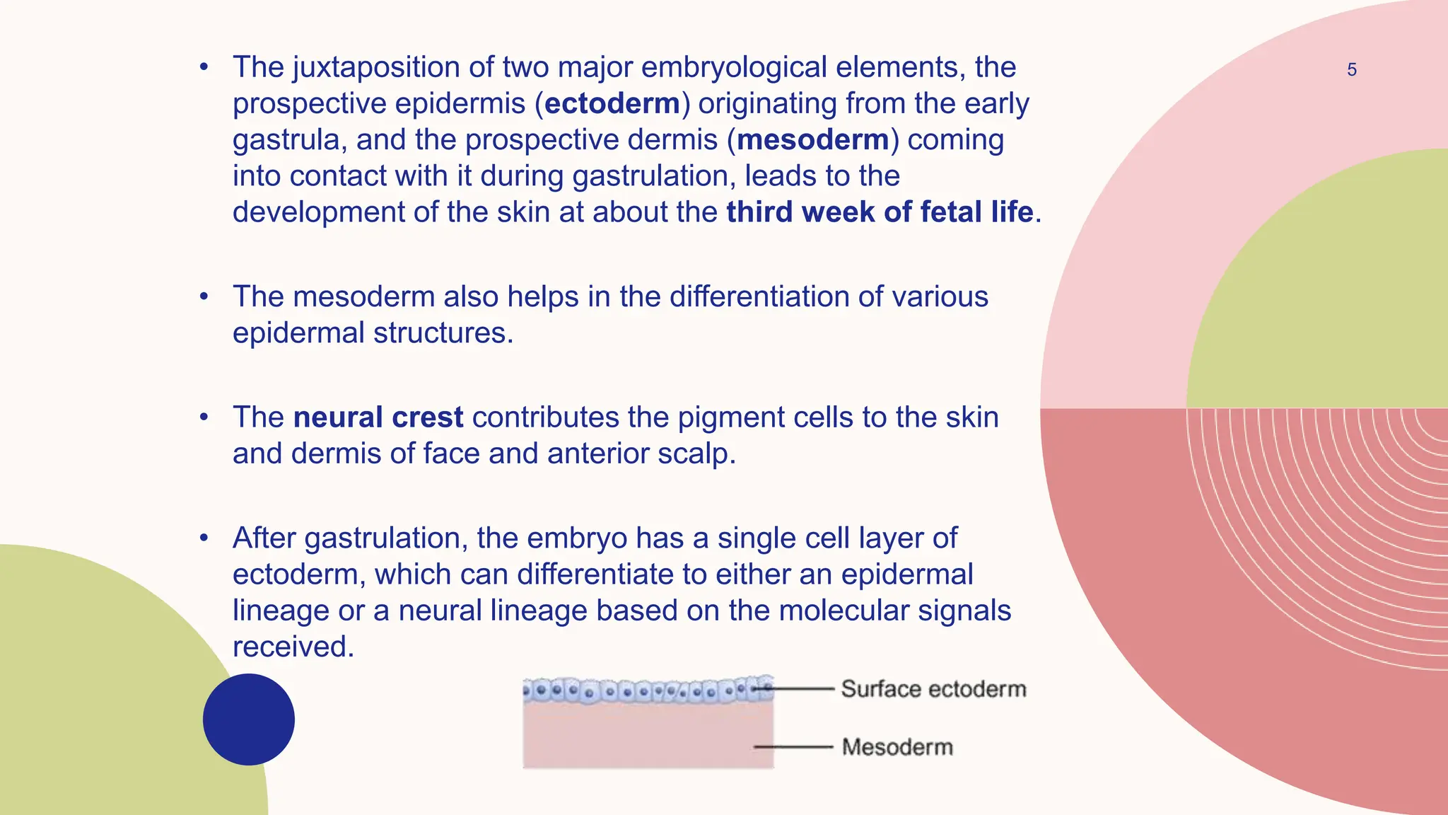
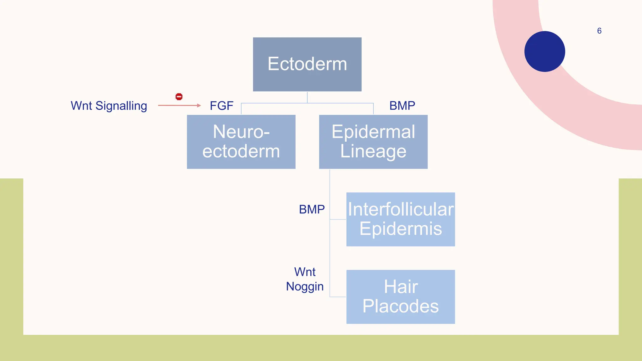
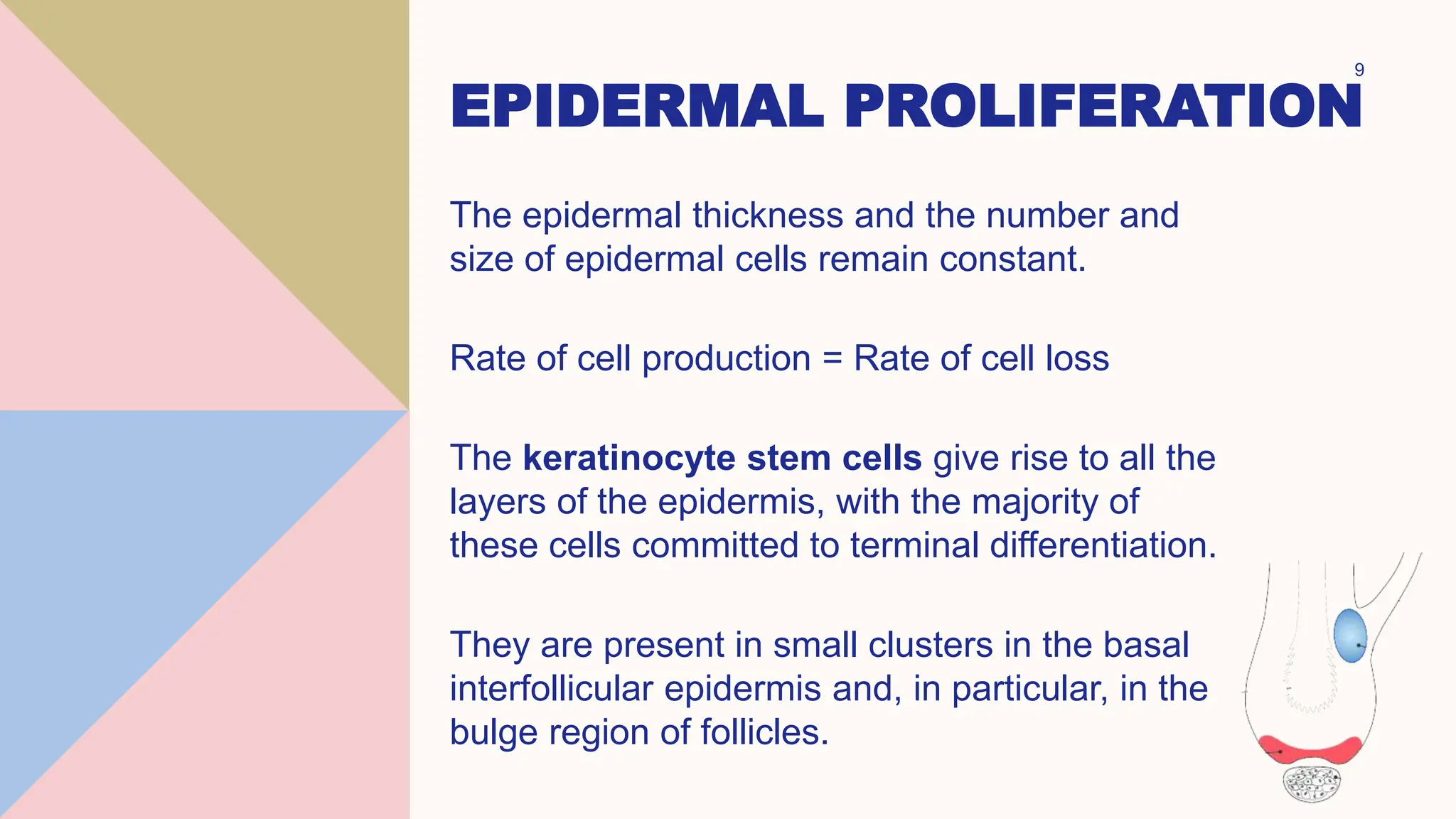
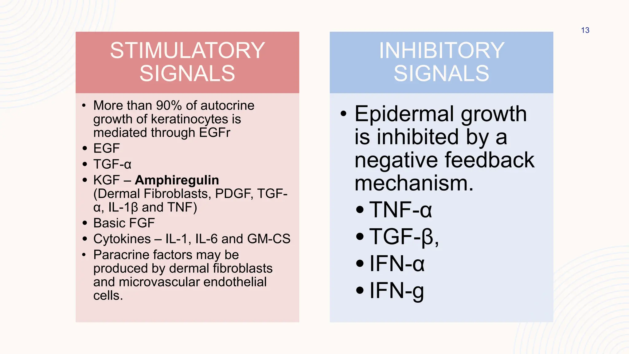
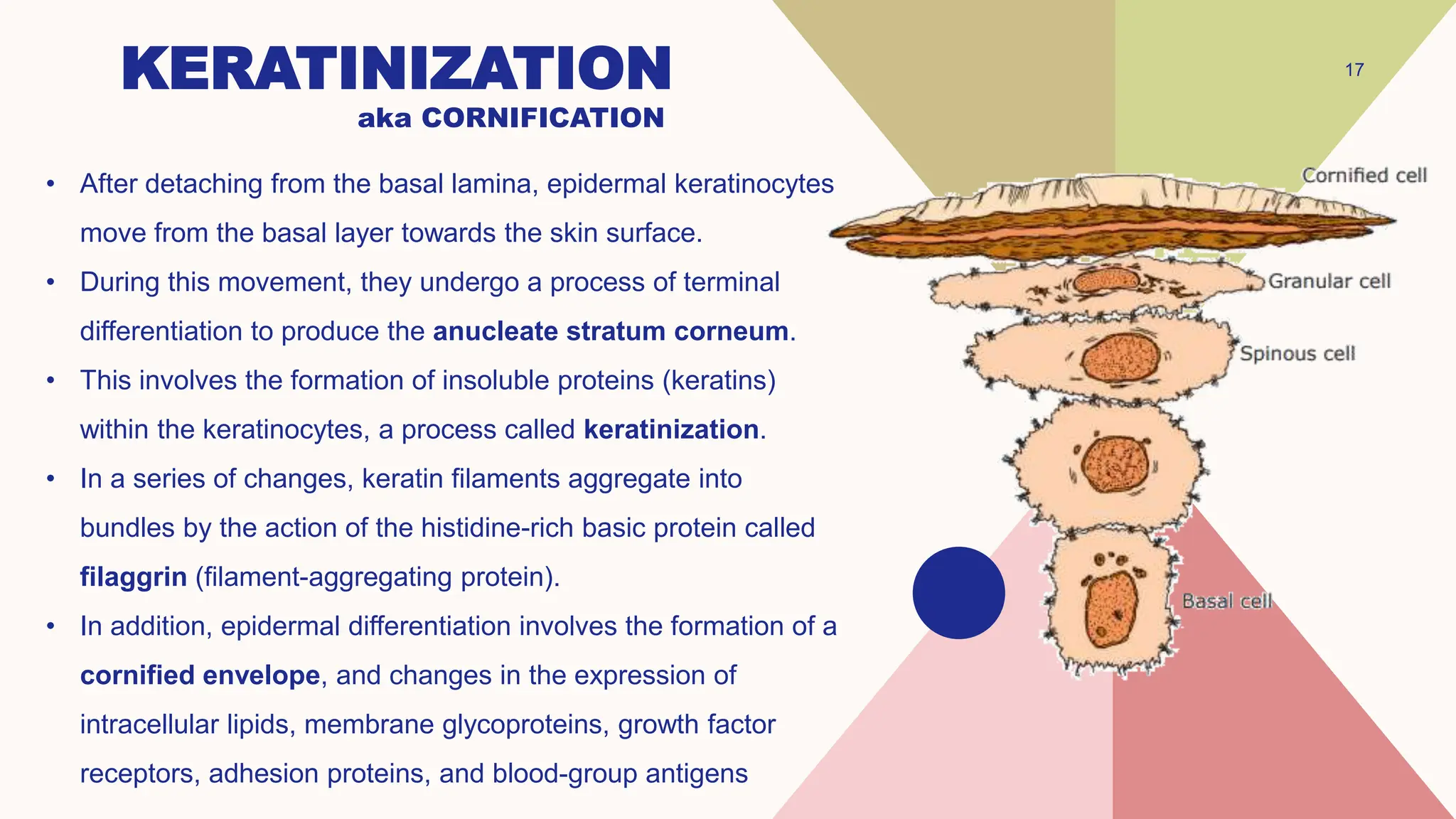
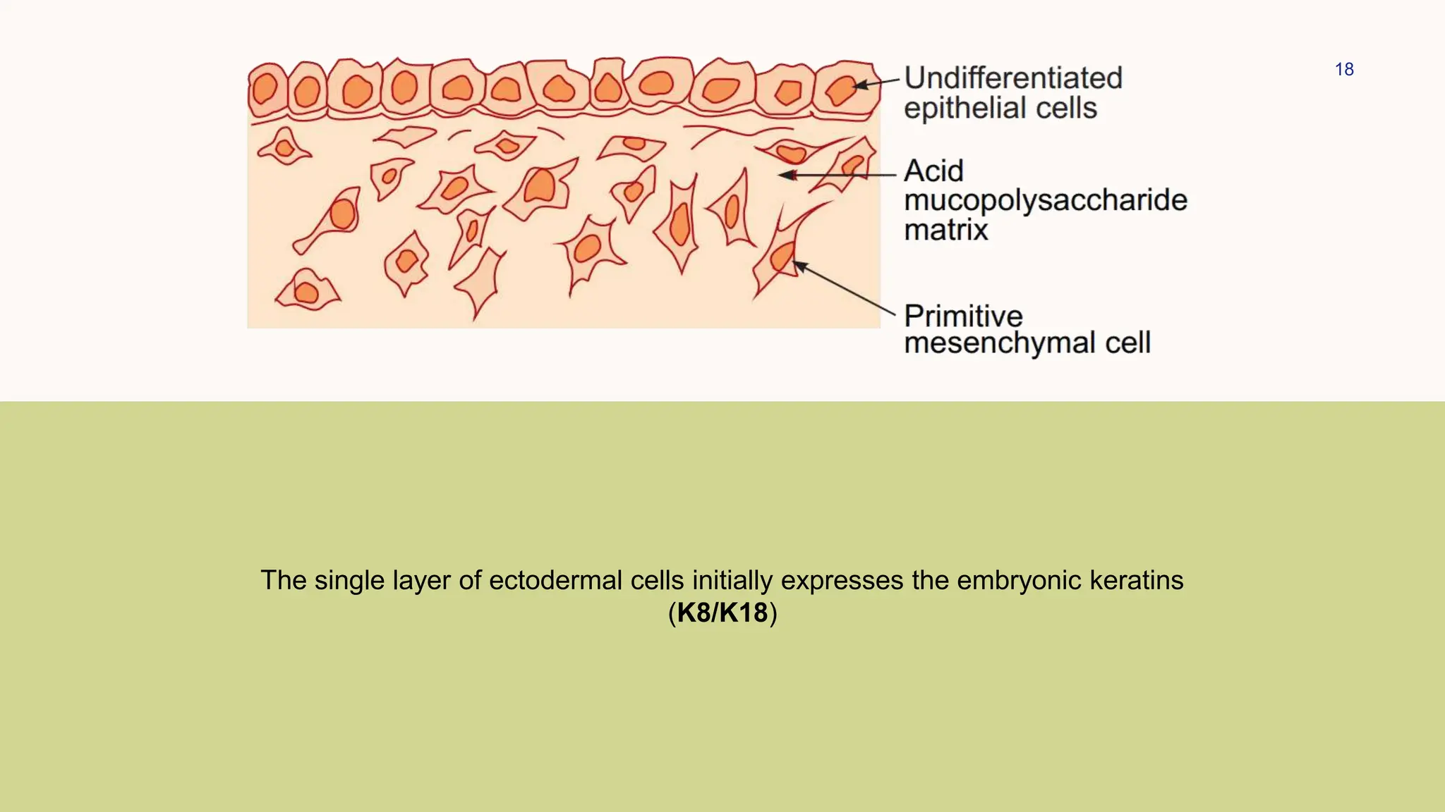
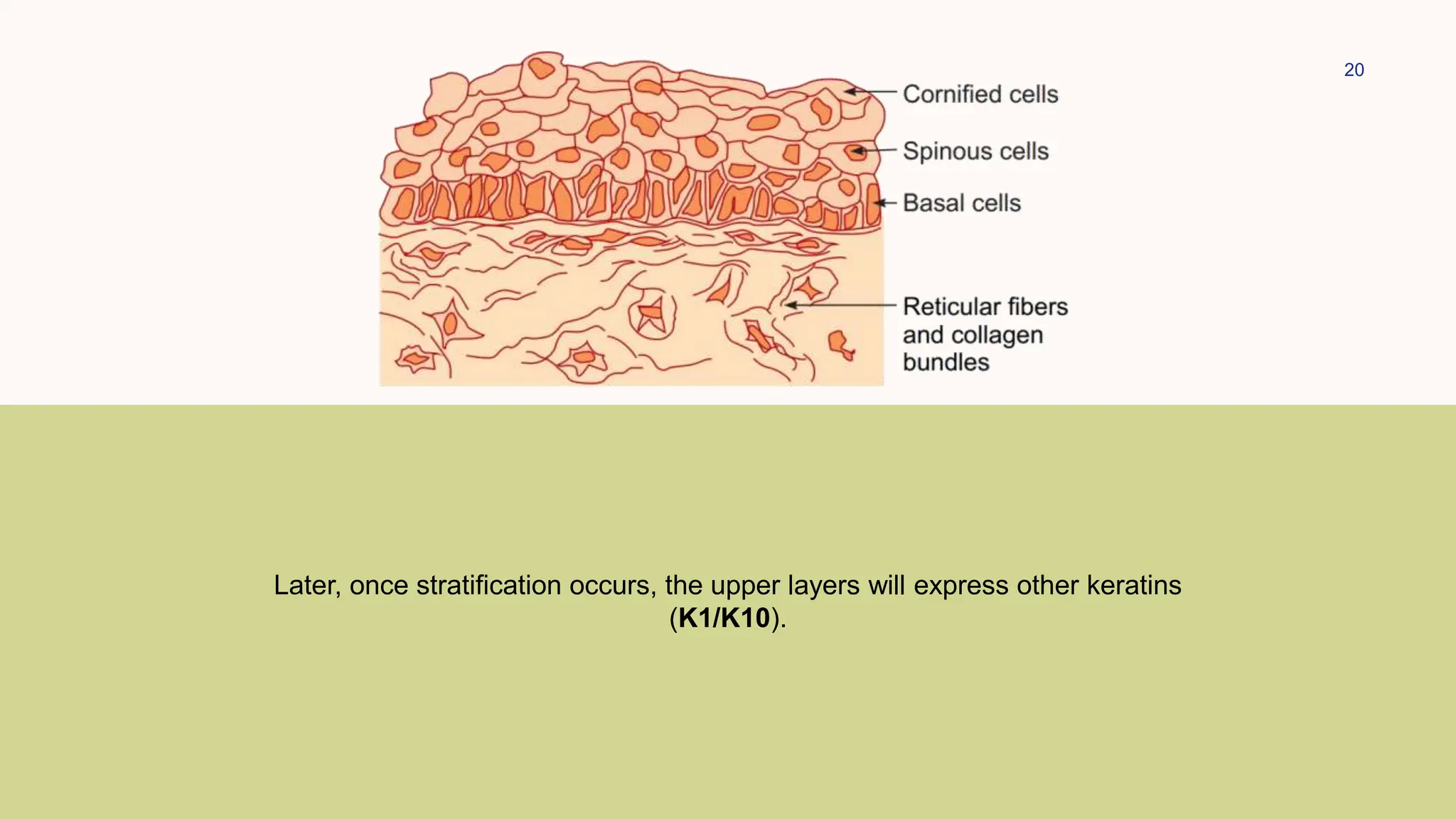
EPIDERMOPOIESIS
Nishkarsh Chugh
PGJR 1 Dermatology
EPIDERMO POIESIS
stratified squamous epithelium
that is terminally differentiated,
which renews continuously, and
forms appendages
Nishkarsh Chugh
PGJR 1 Dermatology
formation or production
AGENDA
• Origin and Development of the Skin
• Milestones in Embryonic Development of
Epidermis
• Epidermal Kinetics
• Keratinization
3
ORIGIN AND
DEVELOPMENT OF
SKIN
4
Specification
• 0-2 months
• ectoderm lateral to the neural plate is committed to become the epidermis and the
subsets of mesenchymal and neural crest cells are committed to form the dermis
Morphogenesis
• 2-5 months
• committed tissues begin to form their specialized structures including epidermal
stratification, epidermal appendage formation, subdivision between dermis and
subcutis, and vascular formation
Differentiation
• 5-9 months
• the newly specialized tissues further develop and assume their functionally mature
forms
5
• The juxtaposition of two major embryological elements, the
prospective epidermis (ectoderm) originating from the early
gastrula, and the prospective dermis (mesoderm) coming
into contact with it during gastrulation, leads to the
development of the skin at about the third week of fetal life.
• The mesoderm also helps in the differentiation of various
epidermal structures.
• The neural crest contributes the pigment cells to the skin
and dermis of face and anterior scalp.
• After gastrulation, the embryo has a single cell layer of
ectoderm, which can differentiate to either an epidermal
lineage or a neural lineage based on the molecular signals
received.
6
Ectoderm
Neuro-
ectoderm
Epidermal
Lineage
Interfollicular
Epidermis
Hair
Placodes
FGF BMP
Wnt Signalling
Wnt
Noggin
BMP
MILESTONES IN EMBRYONIC
DEVELOPMENT OF EPIDERMIS
Gestational Age Stages in development of the epidermis
3rd week Single layer of glycogen filled cells - Germinative or Basal Cells
6th week Double cell layer - Periderm or Epitrachial Layer
8-11 weeks Intermediate Cell Layer formed by the division of basal layer
Few microvilli appear on surface of epidermis
10th week Hemidesmosomes and Desmosomes appear with the formation of Basement
Membrane Zone
14th week Cells in the intermediate layer mature into the spinous layer
K1/K10 are expressed
21st week The cells in turn mature into the granular layer of flattened cells and
keratohyaline granules appear in upper layers
22-24 weeks Epidermal keratinization
24 weeks Periderm is cast off into the amniotic fluid along with the shed lanugo and sebum,
forming the vernix caseosa
EPIDERMAL
KINETICS
EPIDERMAL PROLIFERATION
The epidermal thickness and the number and
size of epidermal cells remain constant.
Rate of cell production = Rate of cell loss
The keratinocyte stem cells give rise to all the
layers of the epidermis, with the majority of
these cells committed to terminal differentiation.
They are present in small clusters in the basal
interfollicular epidermis and, in particular, in the
bulge region of follicles.
9
M
Phase
G1
Phase
S
Phase
G2
Phase
G0
Phase
50-57 hours
18-19 days
52-75 days
12-19 days
14 days
• Cell cycle for normal epidermis
• Mitotic divisions in the keratinocytes
• Epidermal turnover time
• Transit time from the basal layer to the stratum corneum
• Through the stratum corneum
REGULATION OF
EPIDERMAL
PROLIFERATION
13
STIMULATORY
SIGNALS
• More than 90% of autocrine
growth of keratinocytes is
mediated through EGFr
• EGF
• TGF-α
• KGF – Amphiregulin
(Dermal Fibroblasts, PDGF, TGF-
α, IL-1β and TNF)
• Basic FGF
• Cytokines – IL-1, IL-6 and GM-CS
• Paracrine factors may be
produced by dermal fibroblasts
and microvascular endothelial
cells.
INHIBITORY
SIGNALS
• Epidermal growth
is inhibited by a
negative feedback
mechanism.
• TNF-α
• TGF-β,
• IFN-α
• IFN-g
14
APOPTOSIS
• Programmed cell death is a
major cellular homeostatic
mechanism in the skin.
• Terminal differentiation of
epidermal keratinocytes
occurs by modified
apoptotic programs.
• Apoptosis also plays an
important role in the hair
follicle growth cycle.
SIGNAL TRANSDUCTION
PATHWAYS
• Signals from outside the
cell such as hormones
combine with cell receptors
to act intracellularly to
regulate the growth and the
differentiation of epidermal
cells.
• Some of these pathways
also include growth factors,
cyclic 3,5-adenosine
monophosphate (cAMP),
protein kinase C, inositol
phosphate, and protein
tyrosine kinase.
15
INTEGRINS
• Integrins are most evident
on basal keratinocytes at
the site of focal adhesions
and HDs.
• They serve as a physical
link between matrix
molecules (collagen,
laminin, fibronectin) and the
cytoskeleton of
keratinocytes, and act as
the route for bidirectional
communication that can
result in a change in gene
expression, pH, and
calcium fluxes
OTHERS
• Vitamin A
• Vitamin D
• ⬆️Calcium
• ⬇️Calcium
Differentiation
Differentiation
Proliferation
Proliferation
Proliferation
Differentiation
16
• The epidermis, like the mucosa, is maintained by cell
division within the stratum basale.
• This unique ability to replace lost cells is in contrast to
nerves and skeletal muscles, which have no cell division at
all, and the liver, where cell division can only occur in
response to injury.
• The basal layer produces, secretes, and assembles an
extracellular matrix (ECM), which constitutes much of the
underlying basement membrane that separates the
epidermis from the dermis.
• The most prominent basal ECM protein is laminin 5, which
uses α3β1-integrin for its assembly.
• As cells leave the basal layer and move outward towards
the skin surface, they withdraw from the cell cycle, switch
off integrin and laminin expression, and execute a
terminal differentiation program.
KERATINIZATION
• After detaching from the basal lamina, epidermal keratinocytes
move from the basal layer towards the skin surface.
• During this movement, they undergo a process of terminal
differentiation to produce the anucleate stratum corneum.
• This involves the formation of insoluble proteins (keratins)
within the keratinocytes, a process called keratinization.
• In a series of changes, keratin filaments aggregate into
bundles by the action of the histidine-rich basic protein called
filaggrin (filament-aggregating protein).
• In addition, epidermal differentiation involves the formation of a
cornified envelope, and changes in the expression of
intracellular lipids, membrane glycoproteins, growth factor
receptors, adhesion proteins, and blood-group antigens
17
aka CORNIFICATION
18
The single layer of ectodermal cells initially expresses the embryonic keratins
(K8/K18)
19
Once committed to the epidermal lineage these cells start to express K5/K14
instead.
20
Later, once stratification occurs, the upper layers will express other keratins
(K1/K10).
21
• Development of the epidermis and its appendages is
governed by various signalling pathways such as the Wnt
and Shh pathways.
• The development of the follicle and dermal papilla proceeds
by cross-talk of molecules secreted by the developing
follicular epidermis and the dermal condensate.
• Thereafter, the epidermis differentiates into follicular and
inter-follicular epidermis by the balanced action of follicular
stimulating and follicular inhibiting signals.
• The development of the dermal papillae and also conversion
of the hair placode to hair peg stage is regulated by the Shh
signalling and by the expression of Wnt5a (a target of Shh)
in the dermal papilla.
22
Ectoderm
Neuro-
ectoderm
Epidermal
Lineage
Interfollicular
Epidermis
Hair
Placodes
FGF BMP
Wnt Signalling
Wnt
Noggin
BMP
THANK YOU
Dr. Nishkarsh Chugh
PGJR1 Dermatology

Epidermopoises

  • 1.
  • 2.
    EPIDERMO POIESIS stratified squamousepithelium that is terminally differentiated, which renews continuously, and forms appendages Nishkarsh Chugh PGJR 1 Dermatology formation or production
  • 3.
    AGENDA • Origin andDevelopment of the Skin • Milestones in Embryonic Development of Epidermis • Epidermal Kinetics • Keratinization 3
  • 4.
    ORIGIN AND DEVELOPMENT OF SKIN 4 Specification •0-2 months • ectoderm lateral to the neural plate is committed to become the epidermis and the subsets of mesenchymal and neural crest cells are committed to form the dermis Morphogenesis • 2-5 months • committed tissues begin to form their specialized structures including epidermal stratification, epidermal appendage formation, subdivision between dermis and subcutis, and vascular formation Differentiation • 5-9 months • the newly specialized tissues further develop and assume their functionally mature forms
  • 5.
    5 • The juxtapositionof two major embryological elements, the prospective epidermis (ectoderm) originating from the early gastrula, and the prospective dermis (mesoderm) coming into contact with it during gastrulation, leads to the development of the skin at about the third week of fetal life. • The mesoderm also helps in the differentiation of various epidermal structures. • The neural crest contributes the pigment cells to the skin and dermis of face and anterior scalp. • After gastrulation, the embryo has a single cell layer of ectoderm, which can differentiate to either an epidermal lineage or a neural lineage based on the molecular signals received.
  • 6.
  • 7.
    MILESTONES IN EMBRYONIC DEVELOPMENTOF EPIDERMIS Gestational Age Stages in development of the epidermis 3rd week Single layer of glycogen filled cells - Germinative or Basal Cells 6th week Double cell layer - Periderm or Epitrachial Layer 8-11 weeks Intermediate Cell Layer formed by the division of basal layer Few microvilli appear on surface of epidermis 10th week Hemidesmosomes and Desmosomes appear with the formation of Basement Membrane Zone 14th week Cells in the intermediate layer mature into the spinous layer K1/K10 are expressed 21st week The cells in turn mature into the granular layer of flattened cells and keratohyaline granules appear in upper layers 22-24 weeks Epidermal keratinization 24 weeks Periderm is cast off into the amniotic fluid along with the shed lanugo and sebum, forming the vernix caseosa
  • 8.
  • 9.
    EPIDERMAL PROLIFERATION The epidermalthickness and the number and size of epidermal cells remain constant. Rate of cell production = Rate of cell loss The keratinocyte stem cells give rise to all the layers of the epidermis, with the majority of these cells committed to terminal differentiation. They are present in small clusters in the basal interfollicular epidermis and, in particular, in the bulge region of follicles. 9
  • 10.
  • 11.
    50-57 hours 18-19 days 52-75days 12-19 days 14 days • Cell cycle for normal epidermis • Mitotic divisions in the keratinocytes • Epidermal turnover time • Transit time from the basal layer to the stratum corneum • Through the stratum corneum
  • 12.
  • 13.
    13 STIMULATORY SIGNALS • More than90% of autocrine growth of keratinocytes is mediated through EGFr • EGF • TGF-α • KGF – Amphiregulin (Dermal Fibroblasts, PDGF, TGF- α, IL-1β and TNF) • Basic FGF • Cytokines – IL-1, IL-6 and GM-CS • Paracrine factors may be produced by dermal fibroblasts and microvascular endothelial cells. INHIBITORY SIGNALS • Epidermal growth is inhibited by a negative feedback mechanism. • TNF-α • TGF-β, • IFN-α • IFN-g
  • 14.
    14 APOPTOSIS • Programmed celldeath is a major cellular homeostatic mechanism in the skin. • Terminal differentiation of epidermal keratinocytes occurs by modified apoptotic programs. • Apoptosis also plays an important role in the hair follicle growth cycle. SIGNAL TRANSDUCTION PATHWAYS • Signals from outside the cell such as hormones combine with cell receptors to act intracellularly to regulate the growth and the differentiation of epidermal cells. • Some of these pathways also include growth factors, cyclic 3,5-adenosine monophosphate (cAMP), protein kinase C, inositol phosphate, and protein tyrosine kinase.
  • 15.
    15 INTEGRINS • Integrins aremost evident on basal keratinocytes at the site of focal adhesions and HDs. • They serve as a physical link between matrix molecules (collagen, laminin, fibronectin) and the cytoskeleton of keratinocytes, and act as the route for bidirectional communication that can result in a change in gene expression, pH, and calcium fluxes OTHERS • Vitamin A • Vitamin D • ⬆️Calcium • ⬇️Calcium Differentiation Differentiation Proliferation Proliferation Proliferation Differentiation
  • 16.
    16 • The epidermis,like the mucosa, is maintained by cell division within the stratum basale. • This unique ability to replace lost cells is in contrast to nerves and skeletal muscles, which have no cell division at all, and the liver, where cell division can only occur in response to injury. • The basal layer produces, secretes, and assembles an extracellular matrix (ECM), which constitutes much of the underlying basement membrane that separates the epidermis from the dermis. • The most prominent basal ECM protein is laminin 5, which uses α3β1-integrin for its assembly. • As cells leave the basal layer and move outward towards the skin surface, they withdraw from the cell cycle, switch off integrin and laminin expression, and execute a terminal differentiation program.
  • 17.
    KERATINIZATION • After detachingfrom the basal lamina, epidermal keratinocytes move from the basal layer towards the skin surface. • During this movement, they undergo a process of terminal differentiation to produce the anucleate stratum corneum. • This involves the formation of insoluble proteins (keratins) within the keratinocytes, a process called keratinization. • In a series of changes, keratin filaments aggregate into bundles by the action of the histidine-rich basic protein called filaggrin (filament-aggregating protein). • In addition, epidermal differentiation involves the formation of a cornified envelope, and changes in the expression of intracellular lipids, membrane glycoproteins, growth factor receptors, adhesion proteins, and blood-group antigens 17 aka CORNIFICATION
  • 18.
    18 The single layerof ectodermal cells initially expresses the embryonic keratins (K8/K18)
  • 19.
    19 Once committed tothe epidermal lineage these cells start to express K5/K14 instead.
  • 20.
    20 Later, once stratificationoccurs, the upper layers will express other keratins (K1/K10).
  • 21.
    21 • Development ofthe epidermis and its appendages is governed by various signalling pathways such as the Wnt and Shh pathways. • The development of the follicle and dermal papilla proceeds by cross-talk of molecules secreted by the developing follicular epidermis and the dermal condensate. • Thereafter, the epidermis differentiates into follicular and inter-follicular epidermis by the balanced action of follicular stimulating and follicular inhibiting signals. • The development of the dermal papillae and also conversion of the hair placode to hair peg stage is regulated by the Shh signalling and by the expression of Wnt5a (a target of Shh) in the dermal papilla.
  • 22.
  • 23.
    THANK YOU Dr. NishkarshChugh PGJR1 Dermatology

Editor's Notes

  • #5 The fetal skin development can be divided into three distinct but temporarily overlapping stages
  • #7 Activation of Wnt signalling inhibits the effect of fibroblast growth factor on the ectoderm. This promotes the expression of bone morphogenic proteins which causes the cells to commit to the epidermal or keratinocytic lineage.
  • #8 3rd wk- Embryonal basal cells are more columnar than fetal basal cells and they have not yet formed hemidesmosomes. 6th wk- Periderm is a transient layer which protects the underlying cells from continued exposure to the amniotic fluid. 8-11 wks- The cells in the intermediate layers contain mitochondria, Golgi complexes, and tonofilaments within and around them. 14th wk- Activation of the Wnt pathway in the underlying mesenchyme and P63 has been found to regulate the formation of the spinous layer and the stratification of the epidermis.
  • #9 Epidermal Kinetics i.e. Epidermal Proliferation and Differentiation
  • #10 Apart from stem cells, the basal layer also contains 2 more types of cells: Post Mitotic Cells which don’t undergo mitosis Transient Amplifying Cells which can undergo a limited i.e. 5-6 mitotic divisions
  • #11 The proliferative cells go through a cell cycle, which is defined as the time interval between two successive mitoses. It has 4 phases: M phase or Mitosis Phase G1 phase or Interphase or Postmitotic Growth Phase S Phase which is a phase of active DNA synthesis G2 Phase or premitotic phase or short resting phase Some basal cells may remain quiescent in the so-called G0 phase, which permits them to reenter the cell cycle under appropriate stimuli.
  • #12 Mitotic Index is the fraction of basal cells in mitotic phase at any time Labelling Index is fraction of basal cells in DNA synthesis Epidermal turnover time is the time taken by keratinocytes to pass from the basal layer to the stratum corneum and the skin surface.
  • #14 EGF - Human epidermal growth factor is a 6 kDa polypeptide that stimulates cell proliferation and differentiation in a wide variety of tissues including keratinocytes. It is, however, not synthesized in the epidermis. TGF-α - The transforming growth factor-α is a polypeptide synthesized by epidermal keratinocytes, stimulates growth of keratinocytes by an autocrine method after binding to the receptor Amphiregulin- It is the major autocrine keratinocyte growth factor (KGF) regulated by cellular glycosaminoglycans (GAGs) and upregulated by EGF and TGF-α. The protein derivative of the amphiregulin gene binds to the receptor. Dermal Fibroblasts, PDGF, TGF-α, IL-1β and TNF induce KFG gene.
  • #18 22-24 wks- It begins first on the head, face, palms, and soles
  • #23 Expression of BMPs in the epidermis and EGFR signalling are follicular inhibitors. These are active in the inter-follicular epidermis Wnt/βcatenin signalling and BMP inhibitors like noggin from the dermal condensate, and the Wnt10b in the hair placode are follicular promoters. .