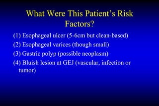
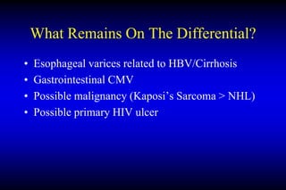
CPC
01.12.10
John O. Clarke, M.D.
Assistant Professor of Medicine
Director of Esophageal Motility
Johns Hopkins University
Questions to Address
(1)What were the patient’s risk factors for
bleeding?
(2)How does HIV affect the differential?
(3)What is the most likely diagnosis?
Differential Diagnosis of Upper GI
Bleed
• Ulcerative or erosive
– Idiopathic
– Drug-induced
– H. pylori
– Other Infectious
– Stress-induced
– Zollinger-Ellison Syndrome
• Esophagitis
– Peptic
– Infectious
– Pill-induced
• Portal hypertension
– Varices
– Portal hypertensive gastropathy
• Vascular
– Idiopathic angiomas
– Osler-Weber-Rendu syndrome
– Dieulafoy’s lesion
– Gastric antral vascular ectasia
– Radiation-induced telangectasia
– Blue rubber bleb nevus syndrome
• Traumatic or post-surgical
– Mallory-Weiss tear
– Surgical anastomosis
– Aortoenteric fistula
– Post-polypectomy
• Tumors
DDx of Massive Upper GI Bleed
• Ulcer
– Idiopathic
– Drug-induced
– H. pylori
– Other infections
• Esophagitis
– Infectious
• Portal hypertension
– Varices
• Vascular
– Dieulafoy’s lesion
• Traumatic
– Mallory-Weiss tear
– Aortoenteric fistula
• Tumors
What affect does HIV have?
• All items on the prior slide are still possible
• Infection and tumors become more likely
Relationship of CD4 Count to
Infection & Tumor
Wilcox C M. Gut 2008;57:861-870
What Were This Patient’s Risk
Factors?
(1) Esophageal ulcer (5-6cm but clean-based)
(2) Esophageal varices (though small)
(3) Gastric polyp (possible neoplasm)
(4) Bluish lesion at GEJ (vascular, infection or
tumor)
Esophageal Ulcer
“An ulcer was found in the mid esophagus 5-6cm in length . . . not bleeding”
• What is the differential diagnosis?
(1) Pill-induced
(2) Infectious
- HSV (IgG +)
- CMV (PCR +)
- Primary HIV
- Other (no indication from history)
(3) Neoplasm
- Adenocarcinoma
- Squamous cell
- Lymphoma
- Kaposi’s sarcoma
• Unlikely to be the primary source unless it eroded into a varix/artery or rapidly grew in size
Esophageal Varices
“There were 2-3 small associated esophageal varices . . . 2 bands were
placed”
• Esophageal varices are indicative of portal hypertension, either from
cirrhosis and/or portal vein thrombosis
• The patient clearly had cirrhosis based on history, exam, blood labwork
(prothrombin time, albumin, bilirubin, platelets, ammonia), and ascitic
fluid analysis
• Mortality from variceal hemorrhage remains approximately 35%
• On a side note, he should have had antibacterial therapy given an active
GI bleed in the context of cirrhosis and known ascites . . .
• Could be the source of bleeding but slightly atypical for small varices to
result in catastrophic hemorrhage
Gastric polyp
“A 2-cm polyp was seen near the angularis. This lesion was
not biopsied”
• DDx
– Benign (hyperplastic, fundic gland polyp, adenoma)
– Primary cancer
• Gastric adenocarcinoma
– Metastatic or diffuse
• Lymphoma
• Kaposi’s sarcoma
• Unlikely to be the source of bleeding
Bluish Lesion at GEJ
“A 1.5 cm bluish ‘bean-like’ lesion was seen at the GEJ. This appeared to
be a vermiform lesion rather than a visible vessel”
• What is this?
– Tumor
• Kaposi’s sarcoma
• Lymphoma
• Metastatic
– Vascular
• Dieulafoy’s lesion (atypical to be at GEJ)
• Gastric varix (atypical description)
– Infection (atypical)
• Unlikely to be primary source of bleeding (unless eroded into a varix)
Other Factors to Consider
(1)The stomach was not cleared during endoscopy
(2)Ascitic fluid represents portal hypertension;
however, infection/neoplasm could be
superimposed (SAAG less accurate)
(3) Travel to Asia & Mexico may have led to
atypical exposure not listed above
(4) Left pleural effusion less common with ascites
than right (and may be masking another process)
Specific Topics To Cover
Gastrointestinal CMV
• Occurs in up to 5% of untreated patients with AIDS
• Most cases occur when CD4 < 50
• Prior to HAART, universally fatal on diagnosis
– Colitis: 4 months
– Esophagitis: 8 months
• Often complicated by hemorrhage or perforation
• Can occur anywhere throughout the GI tract
• Multiple large ulcers
• Multiple biopsies required to make diagnosis
Gastrointestinal HSV
• Less common than CMV in patients with
HIV/AIDS
• Occurs in 3-5% of HIV patients with esophagitis
• Rarely causes ulcers larger than 2cm
• Usually causes multiple ulcers
Primary HIV Ulcers
• Mechanism poorly understood
• May occur in 4-12% of patient with HIV/AIDS
with esophageal symptoms
• Typically very large in size; often solitary
• Can be associated with bleeding or perforation
• Biopsies are routinely non-diagnostic
• Responds to prednisone or thalidomide
Kaposi’s Sarcoma
• Low-grade vascular tumor a/w HHV-8
• 20,000 times more frequent in AIDS patients than general
population
• GI involvement in 40% of patients
• Appear as hemorrhagic nodules on endoscopy
• Usually associated with skin findings (not reported in this
case)
• HH8 also associated with primary effusion lymphoma
(theoretically possible in this case but less likely)
Non-Hodgkin Lymphoma
• 25-40% of HIV patients eventually develop
malignancy; 10% are NHL
• Risk of NHL 60-165 fold greater if HIV
• CD4 count usually < 100
• Can present as esophageal ulcers, polyps or
nodules in the GI tract
• Can be associated with hemorrhage (although not
classic)
What Remains On The Differential?
• Esophageal varices related to HBV/Cirrhosis
• Gastrointestinal CMV
• Possible malignancy (Kaposi’s Sarcoma > NHL)
• Possible primary HIV ulcer
Clinical Diagnosis
• Kaposi’s Sarcoma
• Gastrointestinal CMV
• Cirrhosis re: HBV with esophageal varices
• HIV/AIDS

Discussant_Slides.ppt

  • 1.
    CPC 01.12.10 John O. Clarke,M.D. Assistant Professor of Medicine Director of Esophageal Motility Johns Hopkins University
  • 2.
    Questions to Address (1)Whatwere the patient’s risk factors for bleeding? (2)How does HIV affect the differential? (3)What is the most likely diagnosis?
  • 3.
    Differential Diagnosis ofUpper GI Bleed • Ulcerative or erosive – Idiopathic – Drug-induced – H. pylori – Other Infectious – Stress-induced – Zollinger-Ellison Syndrome • Esophagitis – Peptic – Infectious – Pill-induced • Portal hypertension – Varices – Portal hypertensive gastropathy • Vascular – Idiopathic angiomas – Osler-Weber-Rendu syndrome – Dieulafoy’s lesion – Gastric antral vascular ectasia – Radiation-induced telangectasia – Blue rubber bleb nevus syndrome • Traumatic or post-surgical – Mallory-Weiss tear – Surgical anastomosis – Aortoenteric fistula – Post-polypectomy • Tumors
  • 4.
    DDx of MassiveUpper GI Bleed • Ulcer – Idiopathic – Drug-induced – H. pylori – Other infections • Esophagitis – Infectious • Portal hypertension – Varices • Vascular – Dieulafoy’s lesion • Traumatic – Mallory-Weiss tear – Aortoenteric fistula • Tumors
  • 5.
    What affect doesHIV have? • All items on the prior slide are still possible • Infection and tumors become more likely
  • 6.
    Relationship of CD4Count to Infection & Tumor Wilcox C M. Gut 2008;57:861-870
  • 7.
    What Were ThisPatient’s Risk Factors? (1) Esophageal ulcer (5-6cm but clean-based) (2) Esophageal varices (though small) (3) Gastric polyp (possible neoplasm) (4) Bluish lesion at GEJ (vascular, infection or tumor)
  • 8.
    Esophageal Ulcer “An ulcerwas found in the mid esophagus 5-6cm in length . . . not bleeding” • What is the differential diagnosis? (1) Pill-induced (2) Infectious - HSV (IgG +) - CMV (PCR +) - Primary HIV - Other (no indication from history) (3) Neoplasm - Adenocarcinoma - Squamous cell - Lymphoma - Kaposi’s sarcoma • Unlikely to be the primary source unless it eroded into a varix/artery or rapidly grew in size
  • 9.
    Esophageal Varices “There were2-3 small associated esophageal varices . . . 2 bands were placed” • Esophageal varices are indicative of portal hypertension, either from cirrhosis and/or portal vein thrombosis • The patient clearly had cirrhosis based on history, exam, blood labwork (prothrombin time, albumin, bilirubin, platelets, ammonia), and ascitic fluid analysis • Mortality from variceal hemorrhage remains approximately 35% • On a side note, he should have had antibacterial therapy given an active GI bleed in the context of cirrhosis and known ascites . . . • Could be the source of bleeding but slightly atypical for small varices to result in catastrophic hemorrhage
  • 10.
    Gastric polyp “A 2-cmpolyp was seen near the angularis. This lesion was not biopsied” • DDx – Benign (hyperplastic, fundic gland polyp, adenoma) – Primary cancer • Gastric adenocarcinoma – Metastatic or diffuse • Lymphoma • Kaposi’s sarcoma • Unlikely to be the source of bleeding
  • 11.
    Bluish Lesion atGEJ “A 1.5 cm bluish ‘bean-like’ lesion was seen at the GEJ. This appeared to be a vermiform lesion rather than a visible vessel” • What is this? – Tumor • Kaposi’s sarcoma • Lymphoma • Metastatic – Vascular • Dieulafoy’s lesion (atypical to be at GEJ) • Gastric varix (atypical description) – Infection (atypical) • Unlikely to be primary source of bleeding (unless eroded into a varix)
  • 12.
    Other Factors toConsider (1)The stomach was not cleared during endoscopy (2)Ascitic fluid represents portal hypertension; however, infection/neoplasm could be superimposed (SAAG less accurate) (3) Travel to Asia & Mexico may have led to atypical exposure not listed above (4) Left pleural effusion less common with ascites than right (and may be masking another process)
  • 13.
  • 14.
    Gastrointestinal CMV • Occursin up to 5% of untreated patients with AIDS • Most cases occur when CD4 < 50 • Prior to HAART, universally fatal on diagnosis – Colitis: 4 months – Esophagitis: 8 months • Often complicated by hemorrhage or perforation • Can occur anywhere throughout the GI tract • Multiple large ulcers • Multiple biopsies required to make diagnosis
  • 15.
    Gastrointestinal HSV • Lesscommon than CMV in patients with HIV/AIDS • Occurs in 3-5% of HIV patients with esophagitis • Rarely causes ulcers larger than 2cm • Usually causes multiple ulcers
  • 16.
    Primary HIV Ulcers •Mechanism poorly understood • May occur in 4-12% of patient with HIV/AIDS with esophageal symptoms • Typically very large in size; often solitary • Can be associated with bleeding or perforation • Biopsies are routinely non-diagnostic • Responds to prednisone or thalidomide
  • 17.
    Kaposi’s Sarcoma • Low-gradevascular tumor a/w HHV-8 • 20,000 times more frequent in AIDS patients than general population • GI involvement in 40% of patients • Appear as hemorrhagic nodules on endoscopy • Usually associated with skin findings (not reported in this case) • HH8 also associated with primary effusion lymphoma (theoretically possible in this case but less likely)
  • 18.
    Non-Hodgkin Lymphoma • 25-40%of HIV patients eventually develop malignancy; 10% are NHL • Risk of NHL 60-165 fold greater if HIV • CD4 count usually < 100 • Can present as esophageal ulcers, polyps or nodules in the GI tract • Can be associated with hemorrhage (although not classic)
  • 19.
    What Remains OnThe Differential? • Esophageal varices related to HBV/Cirrhosis • Gastrointestinal CMV • Possible malignancy (Kaposi’s Sarcoma > NHL) • Possible primary HIV ulcer
  • 20.
    Clinical Diagnosis • Kaposi’sSarcoma • Gastrointestinal CMV • Cirrhosis re: HBV with esophageal varices • HIV/AIDS