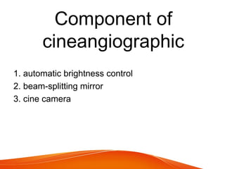
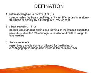
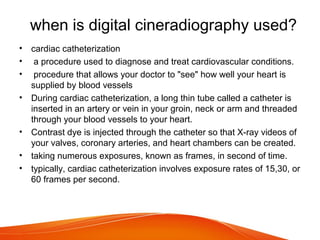
Digital
Cineradiography
Component of
cineangiographic
1. automatic brightness control
2. beam-splitting mirror
3. cine camera
DEFINATION
1. automatic brightness control (ABC) is
compensates the beam quality/quantity for differences in anatomic
thickness or density by adjusting kVp, mA, or both
2. a beam-splitting mirror
permits simultaneous filming and viewing of the images during the
procedure; directs 10% of image to monitor and 90% of image to
cine camera
3. the cine-camera
resembles a movie camera- allowed for the filming of
cineangiographic images but increase the patience dose
when is digital cineradiography used?
• cardiac catheterization
• a procedure used to diagnose and treat cardiovascular conditions.
• procedure that allows your doctor to "see" how well your heart is
supplied by blood vessels
• During cardiac catheterization, a long thin tube called a catheter is
inserted in an artery or vein in your groin, neck or arm and threaded
through your blood vessels to your heart.
• Contrast dye is injected through the catheter so that X-ray videos of
your valves, coronary arteries, and heart chambers can be created.
• taking numerous exposures, known as frames, in second of time.
• typically, cardiac catheterization involves exposure rates of 15,30, or
60 frames per second.

Digital cineradiography

  • 1.
  • 2.
    Component of cineangiographic 1. automaticbrightness control 2. beam-splitting mirror 3. cine camera
  • 3.
    DEFINATION 1. automatic brightnesscontrol (ABC) is compensates the beam quality/quantity for differences in anatomic thickness or density by adjusting kVp, mA, or both 2. a beam-splitting mirror permits simultaneous filming and viewing of the images during the procedure; directs 10% of image to monitor and 90% of image to cine camera 3. the cine-camera resembles a movie camera- allowed for the filming of cineangiographic images but increase the patience dose
  • 4.
    when is digitalcineradiography used? • cardiac catheterization • a procedure used to diagnose and treat cardiovascular conditions. • procedure that allows your doctor to "see" how well your heart is supplied by blood vessels • During cardiac catheterization, a long thin tube called a catheter is inserted in an artery or vein in your groin, neck or arm and threaded through your blood vessels to your heart. • Contrast dye is injected through the catheter so that X-ray videos of your valves, coronary arteries, and heart chambers can be created. • taking numerous exposures, known as frames, in second of time. • typically, cardiac catheterization involves exposure rates of 15,30, or 60 frames per second.