Differentials of neuroendocrine carcinoma
•Download as PPTX, PDF•
1 like•61 views

Alain C.Borczuk, MD

Recommended
More Related Content
What's hot
What's hot (20)
Similar to Differentials of neuroendocrine carcinoma
Similar to Differentials of neuroendocrine carcinoma (20)
More from AbKadir Rifaei Rashid Khairi
Recently uploaded
Recently uploaded (20)
Differentials of neuroendocrine carcinoma
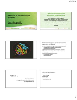
- 1. 3/23/2017 Disclosure of Relevant Differential of Neuroendocrine Carcinoma Alain C. Borczuk,MD Weill Cornell Medicine Problem 1: Small cell carcinoma vs. large cell neuroendocrine carcinoma vs. Combined small cell Financial Relationships USCAP requires that all faculty in a position to influence or control the content of CME disclose any relevant financial relationship WITH COMMERCIAL INTERESTS which they or their spouse/partner have, or have had, within the past 12 months, which relates to the content of this educational activity and creates a conflict of interest. Dr. Alain Borczuk declares he/she has no conflict(s) of interest to disclose. Differential diagnosis of neuroendocrine carcinoma- Problems 1. Small cell carcinoma vs. large cell neuroendocrine carcinoma • Combined small cell or pure large cell neuroendocrine • Organ localized small cell carcinoma 2. Adenocarcinoma or large cell neuroendocrine carcinoma 3. Carcinoid tumors and large cell neuroendocrine carcinoma • Ki-67, mitoses 4. Mimics of neuroendocrine carcinoma • CD56 - perils and problems • Morphologic imitators What is the problem? • Partial sampling • Small samples • Crush artifact • Morphologic overlap 1
- 2. 3/23/2017 Reproducibility in small cell carcinoma diagnosis • Images of 79 tumors, mostly neuroendocrine in 3 tiers - morphology first tier then increasing number of IHC • Moderate agreement (65%) improved to good (78%) with IHC • IHC Panel was variable • Often CK, TTF1 and at least one NE marker Thunnissen E et al J Thorac Oncol. 2017 Feb;12(2):334-346. The Use of ImmunohistochemistryImproves the Diagnosis of Small Cell Lung Cancer and Its Differential Diagnosis. An International ReproducibilityStudy in a Demanding Set of Cases. Small cell carcinoma vs. Large cell neuroendocrine carcinoma - pathology Small cell carcinoma Large cell neuroendocrine • Smaller cells, scant cytoplasm • Larger cells, Visible cytoplasm • Fine “salt and pepper” chromatin • Nucleoli, small (coarse chromatin) • Nuclear molding • Rosettes, palisading (?related to • Crush artifact cytoplasm) • Apoptosis Small cell carcinoma vs. Large cell neuroendocrine carcinoma - clinical Small cell carcinoma Large cell neuroendocrine • Advanced disease • Early stage (50%) • High recurrence rate • Paraneoplastic syndromes rare• Paraneoplastic syndrome - often • Peripheral, early rare • Peripheral, lobulated>spiculated • High PET SUV • High PET SUV • Smokers • Smokers Small cell carcinoma vs. Large cell neuroendocrine carcinoma - IHC Small cell carcinoma Large cell neuroendocrine • Cytokeratin - weak, patchy, dot • Cytokeratin - membranous like • Up to 10% of cases negative • TTF1 - 90% positive • TTF1 - 50% positive (or higher) • Neuroendocrine markers • Neuroendocrine markers • Apoptosis - frequent • Mitoses - frequent (?hard to see) • Necrosis • Mitoses - frequent • Necrosis • Often but not always positive • CD56 most sensitive • Ki-67 high • By definition • Ki-67 high 2
- 3. 3/23/2017 3
- 4. 3/23/2017 Architecture Rosette Palisading Nuclear morphology Problem 1 - Small cell ca vs LCNEC Take home lessons • Mostly morphologic DO NOT FORGET COMBINED SMALL CELL IS SMALL CELL! • Don’t ignore clinical clues (paraneoplastic syndrome, organ localized) LARGE CELL NEUROENDOCRINE AS PART OF • IHC pattern COMBINED SMALL CELL CARCINOMA • Cytokeratin can reveal dual cell population IS COMMON! • If Age <40 or more chest wall than lung (see Problem 4!) 4
- 5. 3/23/2017 What is the problem? Problem 2: Solid type adenocarcinoma vs. large cell neuroendocrine carcinoma Synaptophysin Nuclear Cytoplasm Architecture •IHC definition of neuroendocrine differentiation • “NSCLC with neuroendocrine differentiation”vs. LCNEC • Morphologic overlap •What is the key criterion? Nuclear? Cytoplasm? Architecture? Nuclear Cytoplasm Architecture 5
- 6. 3/23/2017 PROBLEM: Adenocarcinoma or squamous carcinoma with “neuroendocrine differentiation” SYN p40 CD56 Solid AdCa? LCNEC? Clear cell? TTF1 positive Mucin neg Synaptophysin Definition of LCNEC Any NAPSIN - then not LCNEC P40 - squamous Chromogranin - Neuroendocrine Focal NE staining did not exclude Molecular story Do criteria for LCNEC determine molecular results? Many KRAS positive “NSCLC-like” Natasha Rekhtman et al. Clin Cancer Res 2016;22:3618-3629 ©2016 by American Association for Cancer Research AdenoCA or Squamous CA RESULT: NO KRAS positive tumors Rossi et al Overview of key driver mutations and other activating mutations in LCNEC of the lung. KRAS 3 cases (6%) All resected tumors Two of three - one NE marker only One combined with AdCa Tomohiro Miyoshi et al. Clin Cancer Res 2017;23:757-765 ©2017 by American Associationfor Cancer Research 6
- 7. 3/23/2017 Solid adenocarcinoma vs. Large cell neuroendocrine carcinoma Solid adenocarcinoma Large cell neuroendocrine • Nuclear chromatin - • Nuclear chromatin - coarse salt vesicular with nucleoli and pepper and small nucleoli • Ample cytoplasm • Identifiable cytoplasm • TTF1 - >50% positive Or in other words…. •For nucleoli - how big is too big? •For cytoplasm - what is visible versus ample? • TTF1 positive - about 70-80% • Neuroendocrine markers • Neuroendocrine markers • “neuroendocrine differentiation” • By definition • Mitotic rate intermediate • Mitotic rate high • Ki-67 intermediate • Ki-67 high Nucleoli +/-, vesicular Ample cytoplasm Not architecturally NE KRAS positive NO! Solid Adenocarcinoma Did not do neuroendocrine markers (should I have)? Nucleoli - Yes, macro Cytoplasm - present Architecture - no Nucleoli +/- KRAS positive Ample cytoplasm Not architecturally NE KRAS positive 7
- 8. 3/23/2017 Nucleoli - no Cytoplasm - present Palisading - some KRAS positive Did NE markers ? Regretted it MAYBE? Oops…. Nucleoli -yes, variable Cytoplasm- present Architecture - depends on who you ask KRAS positive Problem 2 - Solid adenocarcinoma vs. LCNEC SUMMARY • Adenocarcinoma, solid predominant - • Nuclear features, ample cytoplasm, lack of palisading/rosettes • LCNEC - “molecular small cell-like” • Nuclear features, “less ample” cytoplasm, architecture • High mitotic rate/ apoptosis • “Solid adenocarcinoma with neuroendocrine IHC” vs. LCNEC “adenocarcinoma-like” • Napsin A and lower mitotic rate/Ki-67 = AdCA • KRAS rate seems similar -“Molecular adeno-like” Synatophysin 8
- 9. 3/23/2017 Problem 2 - Solid adenocarcinoma vs. LCNEC Take home message • LCNEC “small cell like” - criteria as in Problem 1 • Molecular is small cell-like • Blurring between some LCNEC “NSCLC-like” and solid adenocarcinoma • Molecular more AdenoCa-like • Criteria and clinical impact need further study What is the problem? • Small samples • Mitotic counting •Including undercalling carcinoids •Ki-67 Problem 3: Atypical carcinoid vs. large cell neuroendocrine carcinoma Atypical carcinoid vs. Large cell neuroendocrine carcinoma Atypical carcinoid Large cell neuroendocrine • Nuclear chromatin - salt and • Nuclear chromatin - coarse, salt pepper and small nucleoli and pepper and small nucleoli • Round nuclei • Irregular nuclear contour • Visible cytoplasm • Visible cytoplasm • Necrosis • Mitotic rate 2-9 in 2mm2 • Ki-67 low/lower • Necrosis • Mitotic rate 10 or greater in 2mm2 • Ki-67 high Mitotic counting •IASLC/WHO classification 2015 • If initial counts are near the cutoff between categories, then the average of 3 sets of fields must be counted. • Impacts lower end more than upper • Upper end - most LCNEC have many more than 10 in 2mm2 Ki67 in pulmonary NE tumors TUMOR KI 67 TC 2.3-4.1 AC 9-17.8 SCLC 65-78 LCNEC 47.5-70 J Thorac Oncol. 2014 Mar;9(3):273-84 9
- 10. 3/23/2017 Natasha Rekhtman et al. Clin Cancer Res 2016;22:3618-3629 ©2016 by American Association for Cancer Research Problem 3 - Atypical carcinoid vs. LCNEC Take home messages • Careful mitotic counting when possible • Ki-67 when small sample • Rare LCNEC may arise from atypical carcinoids What is the problem? • Not considered at sign out, especially when overlapping epidemiology with small cell carcinoma • Presentation in metastatic sites • Small samples • Rare tumors • IHC marker overlap • CD56 • Cytokeratin Problem 4: Mimickers of high grade neuroendocrine carcinomas Mimics of neuroendocrine carcinoma • Primitive neuroectodermal tumor (PNET) • Usually under age 40 • Chest wall more often than lung • Pitfalls - Cytokeratin can be positive; calretinin can be positive; CD56 can be positive • TTF1 negative, WT1 negative • CD99 membranous; FLI1 positive • EWSR1 translocations 10
- 11. 3/23/2017 Mimics of neuroendocrine carcinoma • Pulmonary neuroblastoma • Pediatric tumors often extra-pulmonary • Adult tumors can be pulmonary • Ganglion cells (ganglioneuroblastoma) • Neuropil CD99 Mimics of neuroendocrine carcinoma • Pulmonary paraganglioma • Rare • Depends on definition • Any trabecular pattern, spindle or oncocytic - carcinoid. • Cytokeratin negative • S100 sustentacular cells, even if only focal. 11
- 12. 3/23/2017 CD56 POSITIVE TUMORS Neuroendocrine tumors Non-neuroendocrine tumors Non-neuroendocrine tumors Low High rate (>50%) rate (25%) Merkel cell carcinoma Wilms tumor Papillary thyroid Carcinoma Medullary thyroid CA Rhabdomyosarcoma Mesothelioma Small cell carcinoma Desmoplastic round cell tumor Paraganglioma/Pheochromo Synovial sarcoma PNET (20%) Ovarian/endometrial stromal Mesenchymal chondrosarcoma NK cell tumors AML Myeloma Granular cell tumor Solid pseudopapillary Panel approach for CD56 positive tumor What trigger? • Small cell is unusual (age, location, distribution, non-smoker) • Morphology is variant or non-classical • E.g. no crush or molding, rare apoptosis • CD56 is the only neuroendocrine marker positive Panel approach for CD56 positive tumor What panel? • CD56, Cytokeratin and TTF1 positive combination favors small cell • Rare exceptions • Add WT1, desmin, SMA or actin/HHF35 • Molecular/FISH testing as needed 12
- 13. 3/23/2017 Problem 4 - mimics of neuroendocrine carcinoma Take home messages • High grade pulmonary neuroendocrine tumors - ALERTS! • Age <40, chest wall, no lung tumor • Diagnosis in metastatic site • Non-smoker • Variant histology • Immunohistochemistry panels - be careful when single NE marker is CD56 • Molecular testing/FISH testing Armita Bahrami, Luan D. Truong, and Jae Y. Ro (2008) Undifferentiated Tumor: True Identity by Immunohistochemistry. Archives of Pathology & Laboratory Medicine: March 2008, Vol. 132, No. 3, pp. 326-348 13