Download to read offline
![NERVE SUPPLY
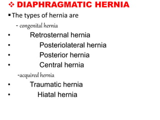
• MOTOR
- The phrenic nerves are the sole motor nerves to
the diaphragm
[ventral rami C3 C4 C5]
• SENSORY
- The phrenic nerves are sensory to the central part,
and the lower six thoracic nerves are sensory to the
peripheral part of diaphragm.](https://image.slidesharecdn.com/diaphragmpr-220308165511/85/Diaphragm-10-320.jpg)
![CLINICAL ANATOMY
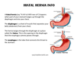
HICCOUGH OR HICCUP- This is due to the
spasmodic contraction of the diaphragm
a. peripheral, due to local irritation of diaphragm
or its nerve
b. central, due to irritation of the hiccough center in
medulla.
SHOULDER TIP PAIN- This is because irritation of
the diaphragm may cause referred pain in the shoulder
because the phrenic nerve and supraclavicular nerves have
the same root values[C3,C4,C5]](https://image.slidesharecdn.com/diaphragmpr-220308165511/85/Diaphragm-14-320.jpg)

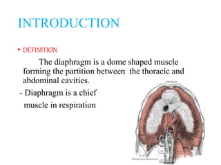
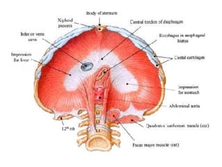
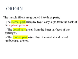
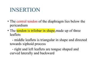
The diaphragm is a dome shaped muscle that forms the partition between the thoracic and abdominal cavities. It has three parts that arise from the sternum, ribs, and lumbar vertebrae. Its central tendon inserts below the pericardium in a trilobar shape. The diaphragm receives motor innervation from the phrenic nerve and sensory innervation from lower thoracic nerves. During respiration, the diaphragm contracts to increase the volume of the thoracic cavity.


