Download to read offline

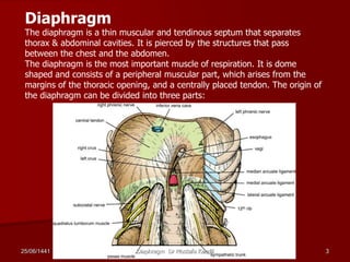
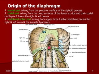
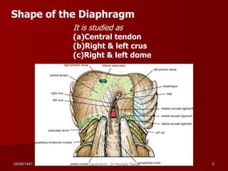
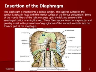
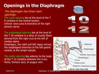
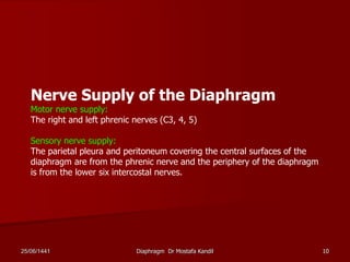

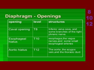
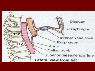
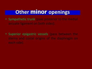
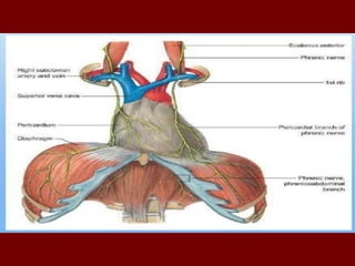


The diaphragm is a thin, dome-shaped muscle that separates the thoracic and abdominal cavities. It has three parts - a central tendon and left and right domes. The diaphragm receives motor innervation from the phrenic nerves and is the primary muscle of respiration, contracting during inhalation to increase the volume of the thoracic cavity. It has several openings that allow structures such as the inferior vena cava, esophagus and aorta to pass between the thorax and abdomen.


![CTEV [ clubfoot] DR ARUN LAL ,DR MOHAMED ASHRAF travancore medical college k...](https://cdn.slidesharecdn.com/ss_thumbnails/ctevclubfootdrarunlaldrmohamedashraftravancoremedicalcollegekollamkeralaindia-260208063247-18fc466c-thumbnail.jpg?width=640&height=640&fit=bounds)


![ONFH[AVN HIP] -TRIPLE REGIME -A NOVAL SURGICAL CONCEPT .pptx](https://cdn.slidesharecdn.com/ss_thumbnails/onfhavnhip2026koaconcalicutdrgokuldevdrmashraf-260210064517-213ec005-thumbnail.jpg?width=640&height=640&fit=bounds)


