SlideShare a Scribd company logo
www.indiandentalacademy.com
INDIAN DENTAL ACADEMY
Leader in continuing dental education
www.indiandentalacademy.com

www.indiandentalacademy.com
Embryology is the study of formation and development of
the embryo. The development of the human embryo from the
time of fertilization through the birth week is an important
period for human appearance .
Every individual spends the first nine months of its life
within the womb.
During this period it develops from one-celled structure to an
organism having billions of cells.
An embryo has no face as such
Webster delimits the face as the front part of the head
comprising the nose,cheeks,jaws,mouth,forehead and the eyes.
www.indiandentalacademy.com
The key primordial begin to gather,and form slight
swelling,depression and thickening that are rapid which
undergo a series of mergers, and enlargements that will
transfer them,forming a cluster of separate masses into a
Face.
Prenatal life may be arbitrarily divided into three periods:
The period of the ovum
(from fertilization to the end of the fourteenth day)
The period of the embryo
(from the fourteenth day to about the fifty-sixth day)
The period of the fetus
(from about the fifty-sixth day until the two hundred and seventieth
day-birth)
www.indiandentalacademy.com
Understanding the development of the structures of the
face requires knowledge of the pharyngeal or branchial
arches. These arches form on either side of the foregut
and correspond to the primitive branchial arches.
The pharyngeal arch consists of a core of mesenchyme
covered externally by ectoderm and covered internally by
endoderm.
Pharyngeal(branchial ) apparatus
The pharyngeal (brachial)arches begin to develop during the
fourth week in utero.
www.indiandentalacademy.com
The pharyngeal apparatus consists of a series of bilaterally
paired arches,pouches(clefts)grooves and membranes
This apparatus gives rise to a significant number of
structure of the head and neck.
The structure of the pharyngeal apparatus are numbered
sequentially.
The first four paired arches make up the lateral wall of the
primordial pharynx,which develop from the foregut.

www.indiandentalacademy.com
The pharyngeal arch apparatus
Cranial
Endoderm

Pharyngeal pouch
Mesenchyme

Pharyngeal
membrane

Pharyngeal cleft
Nerve
Artery
Ectoderm

Caudal

www.indiandentalacademy.com
Weeks 2 through 8 are especially important because
the tissues and organ system are developing from the original
three germ layers.
Formation of Germ layers
At a very early stage in development,the embryo proper
acquires the form of a three-layered disc.,called the
embryonic disc.
The three layers that constitute this embryonic disc are
Endoderm(endo=inside)
Ectoderm(ecto=outside)
Mesoderm(meso=in the middle)
www.indiandentalacademy.com
 

Ectoderm
The ectoderm contributes to the formation of the face is
well around the stomodeum by the fourth week of embryonic
life. It is evident that the ectodermal structures bounding
the stomodeum participate not only in the formation of the
face, but also in the formation of the nasal and oral cavities.
Mesoderm
The mesenchyme that fills the pharyngeal arches is derived
from three sources.. The
Paraxial

Lateral plate

musculature

Neural crest
skeletal

(this musculature is innervated by one nerve that innervates all the
muscles of that particular arch)

www.indiandentalacademy.com
Finally each arch has its own artery.
Folding of the Embryo
Beginning in the fourth week of development the flat
trilaminar embryonic disc folds in two planes to form a
more typical-appearing,cylindric,C-shaped embryo.
Folding in the cranial –caudal plane is mainly a result of
rapid longitudinal growth of the central nervous system.
Growth of the somites account for much of the lateral
folding.

www.indiandentalacademy.com
Folding of the Embryo……
Folding brings the endodermal-lined yolk sac into the
embryo and creates the primordial gut;the
foregut,midgut,and the hindgut.
At the early stages of embryonic development, the
vertebrate face has a common plan. A series of small buds
of tissue called the facial primordial forms around the
stomodeum, which forms the primitive mouth. The facial
primordial are made up mainly of neural crest cells that
have migrated from the cranial crest and settled.
www.indiandentalacademy.com
A summary of the derivatives of the first and second
pharyngeal (ie, branchial)arches is as follows:
·                

Pharyngeal arch I

Arch I-Meckels cartilage
(Mandibular arch)
This pair of arch has distinct maxillary and mandibular
prominence
Its major contribution is in the development of face
o Cranial nerve - Maxillary and Mandibular division of
trigeminal nerve (cranial nerve V)
o  Artery - Maxillary (terminal branch)
www.indiandentalacademy.com
o  Muscles - Muscles of mastication (ie, temporalis,
masseter, pterygoids), mylohyoid, anterior belly of
digastric, tensor tympani, tensor veli palatini
o  Skeleton - Maxillary cartilage (incus, alisphenoid),
mandibular or Meckel cartilage (malleus), arch dermal
mesenchyme (maxilla, zygomatic, squamous portion of
temporal bone, mandible)
www.indiandentalacademy.com
       Pharyngeal arch II (hyoid arch) Reichert’s cartilage
Its major contribution is the development of hyoid bone,the lesser
horn and the superior portion of the body

o  Cranial nerve -Facial nerve VII
o  Artery - Stapedial
o  Muscles - Muscles of facial expression (ie, orbicularis
oculi, orbicularis oris, risorius, buccinator, platysma,
auricularis, frontalis), stapedius muscle, posterior belly of
digastric, stylohyoid muscle
o  Skeleton - Stapes, styloid process, stylohyoid ligament,
lesser cornu of hyoid, upper part of the body of the hyoid
bone
 
www.indiandentalacademy.com
Derivatives of pharyngeal pouches,Grooves &
membranes

Pouch 1-Tympanic cavity,auditory tube,mastoid antrum
Pouch 2-Crypt lining of palatine tonsils
Pouch 3-Inferior parathyroid glands,thymus

Pouch 4-Superior parathyroid glands,Ultimobranchial body
Groove1-External auditory meatus
Groove2 & 4-Cervical sinus
Membrane 1-Tympanic membrane

www.indiandentalacademy.com
Development of the face
Facial development result mainly from enlargement and
movement of the frontonasal prominence and four
prominences from pharyngeal arch1,the paired maxillary
prominences ,and the mandibular prominence.These
structures surround the stomodeum.
The upper jaw develops from 5 main buds of tissue: a
single median frontonasal mass ,the 2 lateral nasal
prominences on both sides,, the 2 maxillary prominences
The lower jaw develops from the paired mandibular
primordia (mandibular prominences).
www.indiandentalacademy.com
Paired maxillary and mandibular prominences are
derivatives of the first pair of branchial or pharyngeal
arches. All these prominences are produced by the
proliferation of the neural crest cells that migrated
into the arches from the neural crest during the
fourth week.
The neural crest cells give rise to the connective
tissue components
Frontal prominence
Nasal placode
Maxillaryprominence

www.indiandentalacademy.com

Mandibular prominence
Arch I
·  

Fourth week of
development

o  Primordia of the face appear at the
cephalic end of the embryo.
o   Two nasal placodes cap the bulbous
frontal prominence.
o   The optic discs appear
posterolateral to the frontal
prominence.
O Three paired branchial arches have
formed.
o The first arches split into maxillary
and mandibular prominences. The hyoid
arches are the second pair.
www.indiandentalacademy.com
o  Between the first arches and frontal prominence, the
buccopharyngeal membrane becomes fenestrated.
Fifth week of
·                     development
o  Nasal pits develop in the
nasal placodes, and the rims
of the placodes differentiate
into medial and lateral nasal
prominences.
o  The lens vesicles
invaginate and close within
the optic discs.
www.indiandentalacademy.com
o  The mesenchyme of the mandibular arch fills in across
the midline.
o   The caudal end of the medial nasal prominences begins
to fuse with the maxillary prominences.
·                     At the beginning of the sixth week of
development
o   The nasals have shifted to a more ventral, central
position.
o    Growing and shifting subectodermal mesenchyme
smooths out the furrows between prominences and
arches, and the second arch becomes more massive.
         
www.indiandentalacademy.com
o   Six auricular hillocks, which will become the pinna of
the ears, form on the mandibular and hyoid arches.
By the end of the sixth week of
·                    
development  
o Medial and lateral nasal
prominences fuse.
o  Maxillary prominences begin the
formation of the upper jaw.
o  The midline approximation of
the medial nasal prominences forms
the nasal septum.
www.indiandentalacademy.com
·                     At the beginning of the seventh week of
development
o The tip of the nose is elevated between the medial nasal
prominences and is visible in profile.
o Eyelids become prominent.
o The pinna of the ear takes shape.
·                 End of the seventh week of development
O The pattern of facial features has taken on a human
appearance. However, facial proportions develop during
the fetal period.
www.indiandentalacademy.com
·   The fusion of the medial nasal prominences is complete,
forming the central axis of the nose and the philtrum of
the lip.
From the beginning of the eighth week of development to
birth, final development of the face occurs slowly and
results mainly from changes in the proportion and relative
positions of the facial components.
  At the end of the 8 week the face has taken on a human
appearance.

www.indiandentalacademy.com
Developmental
Defects
1. Cleft (hare) lip and
cleft jaw: the medial
nasal processes do not
fuse with the maxillary
processes.
2. Cleft palate and/or
uvula: the palatine
shelves fail to fuse with
each other and/or with
the primary palate.
www.indiandentalacademy.com
3.Median cleft of upper lip:
The 2 medial nasal processes
fail to fuse. This is often
accompanied by mental
retardation.
4.Median cleft of lower lip:
Failure of fusion of 2
mandibular arches.

5. Oblique facial cleft: The

nasolacrimal duct remains
exposed due to the lateral
nasal process failing to fuse
with the maxillary process It
can occur along with a cleft
lip.
www.indiandentalacademy.com
Development of Dentition
The development of dentition is an integral part of craniofacial
growth and an Unique structure of body
Tooth is derived from ectoderm & mesoderm
The first indication of tooth formation is seen at about sixth
week of intrauterine life
Life cycle of the tooth
Histo
differentiation

Initiation
Proliferation

www.indiandentalacademy.com

Apposition

Morphodifferentiation
Initiation
The epithelium overlying the convex border of the alveolar
process becomes thickened and projects into the underlying
mesoderm.
This epithelial thickening is called the dental lamina.
Specific cells within the dental lamina have the potential to
form the enamel organ of certain teeth by responding to
those factors that initiate or induce tooth development.
A lack of initiation result in absence of teeth(anodontia)most
frequently the permanent upper laterals,third molars,and lower
second premolars.
Abnormal initiation result in development of supernumerary
teeth

www.indiandentalacademy.com
Proliferation
Enhanced proliferation activity ensures at the point of initiation
and results successively in the Bud,Cap,and Bell stage of the
odontogenic organ.
Proliferative growth causes regular changes in size & proportion of
the growing tooth germ
As the enamel organ grows downwards into the mesenchyme (of
the alveolar process)its lower end assure a cup-shaped
appearance.The cup comes to be occupied by a mass of
mesenchyme called the dental papilla.
The enamel organ and the dental papilla together constitute the
tooth germ.At this stage the developing tooth looks like like a
cap:it is ,therefore ,described as the cap stage of tooth
development.

www.indiandentalacademy.com
Histo differentiation
It succeeds the proliferative stage which undergoes definite
morphologic and functionally changes.
The cells of the enamel organ that line the papilla become
columnar.These are called ameloblasts.
Mesodermal cells of the papilla that are adjacent to the
ameloblasts arrange themselves as a continuous epitheliumlike layer.The cells of this layer are called odontoblasts
Ameloblast lay down enamel on the superficial(outer)surface of
the basement membrane.The odontoblasts lay down dentine
on its deeper surface.As layer after layer of enamel and
dentine are laid down,the layer of ameloblasts and the layer
of odontoblasts move away from each other.

www.indiandentalacademy.com
The ameloblasts and odontoblasts are separated by a
basement membrane.The remaining cells of the papilla
form the pulp of the tooth.The developing tooth now
looks like a bell (Bell stage of tooth development )

Morphodifferentiation & Apposition
Morphodifferentiation is impossible without proliferation.
The basic form and relative size of the tooth is established
by morphodifferentiation.

www.indiandentalacademy.com
In advanced bell stage ,the boundary
between inner enamel epithelium and
odontoblast outline the future dentinoenamel
junction.In addition,the cervical portion of
the enamel organ gives rise to the epithelial
root sheath of Hertwig.
Apposition is the deposition of the matrix of the hard dental
structures.
Appositional growth is characterized by regular and rhythmic
deposition of the extracellular matrix,which is of itself
incapable of further growth.

www.indiandentalacademy.com
Development of Permanent tooth

The Permanent tooth bud appears in the 4th & 5th intrauterine months,
at about the same age at which mineralization of the deciduous teeth
commences.
The dental lamina gives off series of buds, one of which lies on the
medial side of each developing milk tooth.
These buds from enamel organ give rise to the permanent teeth
The permanent molars are formed from bud that arise from the
dental lamina posterior to the region of the last milk tooth
At birth the germs of all the deciduous teeth,and of the
permanent incisors,canine and first molars show considerable
development.
The germs of premolar and of the
permanent second molars are
.
rudimentary.
The germ of third molar is formed
www.indiandentalacademy.com
after birth.
The stage of dentitions
Gum
pads

Primary
dentition

Mixed
dentition

www.indiandentalacademy.com

Permanent
dentition
Gum pad stage:It extends from birth until the eruption of the
first primary tooth in the oral cavity. (The pre-dentition period.)
-This is from birth to six months.
-At this stage, there are no teeth. Clinically, the infant is
edentulous
-Both jaws undergo rapid growth; the growth is in three planes of
space: downward, forward, and laterally (to the side). Forward
growth for the mandible is greater.
Greater the anteroposterior dimension of the gumpads,greater the
possibility of the child developing malocclusion
--occasionally, there is a neonatal tooth present at birth.

www.indiandentalacademy.com
Primary dentition stage:
(first transitional period /Deciduous dentition period. )
It extend from the time of eruption of the primary teeth until
the eruption of the first permanent tooth around 6yerars of
age. –
The dentition of this period manifests several important
characteristics.
1.There is either spacing between the teeth or there is not.
2.There is no sagittal and transversal dimensions alteration,only
vertical changes are seen.
3.The distal surface of the maxillary & mandibular second
primary molars maintain vertical relationship.

www.indiandentalacademy.com
-The tooth buds anticipate the ultimate occlusal pattern.
-Mandibular teeth tend to erupt first.
--Eruption times can be variable.
Spaced dentition is supposed to be good as space in between the
teeth is utilized for adjustment of permanent successors
Primate spaces occur in about 50% of children. They appear in
the deciduous dentition. The spaces appear between the upper
lateral incisor and the upper canine. They also appear between
the lower canine and the deciduous first molar.

www.indiandentalacademy.com
Flush terminal plane
This dentition is completed after the eruption of the second
primary molars,indicating the location for permanent
teeth.Therefore it’s the key factor that influences the future
occlusion of the permanent dentition.
The second primary molar relation is described in terms of
relationship between terminal planes.
In flush terminal plane :The distal surface of upper and lower
teeth are in straight line.It’s a favourable relationship to guide
the permanent molars.

www.indiandentalacademy.com
Mesial step:In mesial step the distal surface of the lower molar
is more mesial to that of the upper.Invariably it is favorable to
guide the permanent molar into a class I relationship.
Distal step:The distal surface of the lower molar is more distal
to that of upper.Its unfavorable as it guides the permanent
molars into distal occlusion.
flush terminal plane

Distal step

Mesial step
ClassIII
ClassI(with late mesial shift)
End-end
ClassIII(normally)
ClassI(desirable)

www.indiandentalacademy.com

ClassII
  Incisor liability:
Mayne has coined the term incisor liability
Permanent incisors are larger than primary incisors,this difference
in size is termed incisal liability.
Eruption of permanent incisors make the
permanent arch circumference wider,normally the interincisal
angle is 150 degree in primary dentition and 120 degree in
permanent dentition
The period of transition determine the successful alignment of
the permanent teeth toward an ideal arch form

www.indiandentalacademy.com
Anterior teeth relation
Over bite:The average over bite in primary dentition is 2mm
Overjet :The average over jet is 1-2mm with a normal range of
2-6mm.
With the excessive wear of the primary canines and molars,the
whole lower dental arch may move anteriorly,the incisors may
assume an edge-to-edge interrelationship by the age of six

Sequence of eruption
Deciduous

Eruption Time

Central
Incisor

6-12 months

Lateral
Incisor

9-13 months

Canine

16-23 months

First Molar

13-19 months

Second
Molar

23-33 months

www.indiandentalacademy.com
Exfoliation of Primary teeth
The replacement is a multifactorial process.
When the permanent tooth begins to erupt,it is apparently guided
by the gubernaculum..
The pathway for the erupting anterior teeth is essentially lingual
to their predecessors resulting in an oblique lingual pattern of
resorption.
Resorption is not a continuous process,but is rather one of
alternating period of destruction and apposition

www.indiandentalacademy.com
Mixed dentition stage:
(second transitional period)
Starts with the eruption of the first permanent tooth,usually
the mandibular central incisor,and is normally completed at the
time the last primary tooth is shed.
During this period ,the vertical dimension of the
face is increased,allowing for a heightening of the alveolar
ridges to accommodate continual root growth of the cuspids and
premolars.

www.indiandentalacademy.com
-If the deciduous dentition is spaced dentition with flush
-terminal plane of second deciduous molars,the eruptive force
-cause a closing of of the existing space causing a decrease in
- arch length-(Early mesial shift)
-

-

When no space exist,the erupting first permanent molars is not
able to close spaces.In these case when the primary molars
exfoliates the permanent molar migrates to use up the “leeway
space”-(Late mesial shift)
www.indiandentalacademy.com
The leeway space.
-The deciduous anteriors--incisors and canines are
narrower than their permanent successors mesiodistally.
-The deciduous molars are wider that their permanent
successors mesiodistally.
-This size difference has clinical significance. The difference is
called the leeway space.
-The leeway space in the lower arch is approximately 3.4 mm.
-The leeway space in the upper arch is approximately 1.8 mm.
In normal development, the leeway space is taken up by the mesial
migration of the first permanent molars.

www.indiandentalacademy.com
Since there are no premolars in the primary dentition, the
primary molars are replaced by permanent premolars. If any
primary teeth are lost before permanent teeth are ready to
replace them, some posterior teeth may drift forward and
cause space to be lost,] This may cause crowding and/or
misplacement once the permanent teeth erupt, which is usually
referred to as maloclussion.

www.indiandentalacademy.com
'Ugly duckling stage.:
' On occasion, the permanent incisors 'spread out' due to
spacing. In the older literature, is called by the 'ugly
duckling stage.' With the eruption of the permanent canines,
the spaces often will close.

This is the norm between ages 7 through 12 years of age, and
usually is not connected with a permanent space between the
teeth. This stage of development needs special comment because
its often mistaken as anwww.indiandentalacademy.com
orthodontic problem.
Permanent dentition
Maxillary / mandibular occlusal relationships are established when
the last of the deciduous teeth are lost.
The adult relationship of the first permanent molars is established
at this time.
The growth of the jaw continues during this period,developing room
for the third molars

www.indiandentalacademy.com
Sequence of eruption
Permanent
Teeth

Eruption
Time

Central
Incisor

6-8 years

Lateral
Incisor

6.5-9 years

Canine

9-12 years

First
Premolar

8-12 years

Second
Premolar

8.5-13
years

First Molar

6-7 years

Second Molar

11-14
years

Third
Molar
(Wisdom
Tooth)

17-25
years

www.indiandentalacademy.com
Physiology of the Stomatognathic
system
• Stomatognathic- of or relating to the jaws and the
mouth :webstar’s medical dictionary
• As a dentist and especially as an orthodontist,it is
essential that we have a sound knowledge of and
understand the importance of the stomatognathic
system
• Until we know what is normal we will not be able to
recognize aberrancy

www.indiandentalacademy.com
Functional Osteology

• Bone is one of the hardest tissues of the human body
and it’s most adaptive and responsive to functional
forces
• Meyer -1864:proposed the trajectorial theory of bone
formation
• 1870:Julius Wolff said that the trabecular
arrangement was due to functional forces and
any change in direction or intensity of the forces would
change the internal architecture & external form of
the bone
• It was expressed by a mechanical mathematical law=
law of orthogonality
www.indiandentalacademy.com
• Tajectories need not pass at
right angles to each other—even
their course may be wavy
• Increased function increases
bone density while decreased
function decreases .
• Muscles have a great influence
over bones and may even change
their shape
• Thus the timed manipulation of
the musculature may be
beneficial to the orthodontist
• It is established that
cartilaginous and membranous
bones react differently to forces
www.indiandentalacademy.com
Beninghoff’s trajectories
He Concluded saying that trajectories can be
demonstrated in the maxillo-facial area
• Lines pass through both the spongy and compact bone
• They obey no individual bone margins
•

Maxilla
Transfers the whole stress to the cranium which
has as thin cortices that are interconnected by
trabeculae
Stress trajectory can be considered as the craniofacial
unit
www.indiandentalacademy.com
3 main pillars of trajectories:
• All arise from the alveolar bones
and end in the base of the skull
The canine pillar
The zygomatic pillar
The pterygoid pillar
• Curve around the sinuses,nasal
and orbital opening
• The supra and infra orbital
ridges and the zygomatic
buttress reinforce these pillars

www.indiandentalacademy.com
Mandible
• Absorbs all the stresses

Thick cortices and more radially arranged
trabecuae
Stress trajectories
• From beneath the teeth-join together
in comman pillar—ends at the condyle

•Mandibular canal and nerve are protected-unloading
of nerve
•Thick cortical layer at the base provides resistance
to bending forces
•Trajectories from the symphysis, gonial angle and
coronoid process join this main pillar
www.indiandentalacademy.com
Myology

Property of muscles
(that are important for help in kinetic activity are)
Elasticity= the ability to return to original length and
shape after contraction or extension
Normally relaxed muscle withstand only a certain
amount of elongation
Contractility=the ability to contract under innervational
impulse.
Strength of contraction depends on
Frequency of stimuli
Number of fibers involved

• The greatest of contraction is seen when the muscle
approximates its resting position

www.indiandentalacademy.com
Other principles of muscle physiology
• All or none law
Intensity of contraction is independent of the
strength of the action potential as long as the
threshold is crossed
Seen only when the muscle is not fatigued
• Muscle tonus
State of slight constant tension and form the basis of
reflex posture
• Resting length
Constant and Permits maintenance of postural
relationship
• Reciprocal innervations and inhibition
Inhibition of contraction may be brought about by
excitation of the antagonist.It is through this that
movements are coordinated
www.indiandentalacademy.com
Buccinator mechanism
There is strong inter relationship between bone and muscle
Muscles are a potent force-at rest or when active
Teeth and supporting tissues are influenced by the contiguous
musculature
The balance between the tongue and the peri-oral musculature
influences the morphology of the dental arches and teeth
The shape and size of the tooth root and the periodontal
spaces also helps in the final adaptation

www.indiandentalacademy.com
• During swallowing and mastication- tongue exerts 2-3
times more force than the peri oral muscles
• Counter acted by the tonal contraction,atmospheric
pressure,peripheral recruitment from the buccal
musculature
• Even though doubts still exist about such a balance of
force-we must admit that aberration of muscles can and
do lead to marked malocclusion
• The continuous band of muscle that encircle the dentition
and attach to the pharyngeal tubercle comprises the
buccinator mechanism

www.indiandentalacademy.com
Tongue
• Opposing the buccinator mechanism is a very powerful
structure- the tongue
• It has multiple functions
• Most developed in a new born
• In infancy extrinsic suspensory muscles attach the
tongue to the various osseous structures and are
responsible for gross movement in the horizontal
direction(suckle-swallow)

www.indiandentalacademy.com
• The tongue has amazingly versatile functional possibilities
by virtue of the fact it is anchored at only one end.
• This freedom permits the tongue to deform the dental arch.
•

It may Leads to proclination of the maxillary
incisors,constriction of buccal segment and may create an
open bite.

• Compensating action of mentalis causes retraction of the
lower incisors
• Abnormal position and size of the tongue is often seen in
the presence of enlarged tonsils and adenoids.

www.indiandentalacademy.com
Muscles of the Tongue

INTRINSIC

Superior longitudinal-Shortens the tongue & makes the dorsum concave
Inferior longitudinal-Shortens the tongue & makes the dorsum convex
Transverse-Makes the tongue narrow & elongated
Vertical-Makes the tongue broad & flattened
EXTRINSIC

Genioglossus-

Protrudes the tongue out of the mouth by pulling the
posterior part forwards

Hyoglossus-depresses the tongue
Styloglossus -pulls it upward & backward
Palatoglossus-brings the palatoglossal arches

together, thus shutting the oral

cavity from the oropharynx
www.indiandentalacademy.com
Temperomandibular Joint

• The TMJ is the ball and socket joint that allows the lower
jaw to swing open and close.  It’s a Compound –movable joint
It articulation with the mandibular condyle and the
inferior surface of the sqamous portion of the temporal
bone
•The condyle: This is the "ball" in the joint.  It is a part of
the mandible (lower jaw), and is covered in a layer of
cartilage which allowswww.indiandentalacademy.com
for smooth motion within the joint
assembly
 
•The glenoid fossa: The fossa is the
"socket", or depression in which the
condyle sits.  It is located in the
temporal bone of the skull. 

The front of the fossa is a more gentle slope of bone called the
articular eminence.  The eminence is also covered with
cartilage.
The articular disc: The articular disc is also called the
meniscus.  It is made of hyaline cartilage. 
The ligament behind the meniscus is called the retrodiscal pad
in deference to its function as a shock absorber
www.indiandentalacademy.com
•The joint capsule is the covering of the TM joint .It
isolates the contents of the joint and allows free movement
of the condyle and articular disk within a small "swimming
pool" of synovial fluid.  The capsule has lots of blood vessels
and nerves as well as connective tissue. 
• It is lined by synovial membrane.

Head of the condyle is tubular or ellipsoidal
Unique feature of the joint is that there
are actually 2 joint spaces- separated by
the disc
Thus the joint is capable of dual function
Lower cavity—rotary/hinge movement
Upper cavity—gliding/translatory movement
www.indiandentalacademy.com
• The rotary movement occurs by itself when opening from
occlusion to physiologic resting after this even gliding
motion takes place
• When opened in his manner the articular disc glides over
the articular eminence
• The condyle rotates against the inferior surface of the
disc.
• Lateral pterygoid helps to move the disc anteriorly

In the lateral shift/bennet movement the condyle on
the working side rotates and moves laterally.
• The condyle on the other side moves forwards and
mesially in an arc
•

• Posterior limit formed by the squamo-tympanic fissure—
medially by the petro-tympanic fissure
www.indiandentalacademy.com
• Due to over closures-the disc is moved forward
and the condyle rides over the posterior border
of the disc. Impinges on the retrodiscal soft
tissues-clicking an discomfort to the patientmisdiagnosed as arthritic changes ,pain may be due
to impingement or due to pterygoid muscle
spasm(mpd)
• The main reason for the mpd is neurogenic or
psychological. The TMJis beautifully engineered to
handle multiple tasks endowed to it
• To maintain it-proper occlusal management an
maintenance of vertical dimensions is essential

www.indiandentalacademy.com
Functional movements of the mandibl
•The mandible is the only movable bone in the head and face.
•It has 13 muscles attached to it
•Due to limitations of morphology and structure of the TMJ
it can be moved only in certain directions
•It has postural stability along with primary movements

www.indiandentalacademy.com
Forced retrusion

Posterior fibres of the
temporalis,masseter,suprahyoid muscles

Opening

lateral pterygoid-main. Supra,infra hyoidgenio,mylo hyoids,digasric>stabilising
Temporalis,masseter,medial pter.>controlled
relaxation

Closure

Temporalis, masseter >main action medial ptery.
>part action
Lateral ptery >cntrolled relaxation

Protrusion

Lateral and medial pter.

Lateral movement

Temporalis + lat. pter
www.indiandentalacademy.com
COMPENSATORY MUSCLE
FUNCTIONS
• While mastication may call for most potent effort
from the associated muscles, the most frequent
demands are made by deglutition, respiration,
speech and posture.
• At times two or more of these functions are
carried on simultaneously.
• where there is malocclusion or abnormal
morphologic relationship certain compensatory or
adaptive muscle functions may arise, either to
restrain the dental malocclusion or to actually
increase the discrepancy.
www.indiandentalacademy.com
Positions of the mandible
•
•
•
•
•
•
•
•

Basic positions are
Postural resting position (Physiologic rest)
Centric relation
Initial contact
Centric occlusion
Most retruded position (terminal hinge position)
Most protruded position
Habitual resting position

• Habitual occlusion position

www.indiandentalacademy.com
POSTURAL RESTING POSITION
It is one of the earliest posture positions to be
developed. Mandible is suspended from the cranial base
by the cradling musculature. Here the jaws are
separated by a constant distance.
Factors influencing the postural position are the
following:
• Body and head posture
• Sleep
• Psychic factors influencing muscle tonus
• Age
• Proprioception from the dentition and muscles
• Occlusal changes such as attrition
• Pain
• Muscle disease and muscle spasm and
• Temporomandibular joint disease.
www.indiandentalacademy.com
CETRIC RELATION
• As far as muscle physiology is concerned, centric
relation may be defined as unstrained, neutral
position of the mandible in which the anterio
superior surfaces of mandibular condyles are in
contact with the concavities of the articular disks
as they approximate the postero inferior third of
their respective articular eminent.
•

This means that the mandible is deviating neither
to the right nor to the left and is neither protruded
no retruded.

•

Such a relation can be the same as the postural
resting position, the point of initial occlusal contact
and centric occlusion.
www.indiandentalacademy.com
INITIAL CONTACT
• As the mandible moves from physiologic rest or the postural
resting position toward occlusion of teeth, if all is normal it
maintains a centric relation position as far as the articular
fossae are concerned.
•

If there is normal occlusion the point of initial contact
produces no change in the function of temporomandibular
joint and all inclined planes are brought together
simultaneously in the maxillary and mandibular teeth.

•

Premature contacts are seen quite frequently, they can
initiate deflections in the mandibular path of closure.

•

This causes traumatic forces to be exerted on the teeth
and severe cases will produce temporomandibular joint
problems
www.indiandentalacademy.com
CENTRIC OCCLUSION
• Centric occlusion is a static position
• It is harmonious with the centric relation
• Teeth brought into contact with unstrained relation
of the condyles
• Few patients can show centric occlusion
• Premature contact,loss of teeth,Overeruption of
teeth,overextension of artificial
restorations,malpositions of individual teeth-all these
mitigate against the establishment of a centric
occlusion

www.indiandentalacademy.com
MOST RETRUDED POSITION (Terminal
Hinge Position)
• It’s a Reproducible retruded mandibular position
with the teeth in occlusion
• It has become a common starting point in occlusal
analysis and rehabilitation
• Some patients can easily retrude a few mm. while
others find it difficult
• Mandible should not be guided or forced beyond the
unstrained position of the mandible-unbiologicalwould compress the tissues.

www.indiandentalacademy.com
MOST PROTRUDED POSITION
• This position is variable from individual to individual
than the retruded position,however,it is
reproducible
• Shows the range of movement of the mandible
• Flaccidity of capsular ligament allows condyle to
over ride the anterior margin of the
eminence,leading to dislocation.

HABITUAL RESTING POSITION

There are certain types of malocclusions that
prevent the patient from achieving a physiologic
resting position.

www.indiandentalacademy.com
• Eg.,in class II div 2 malocclusion with maxillary incisors
inclined lingually there is a tendency to force the
condyles posteriorly and superiorly in the articular
fossae
• The physiologic resting positions can be changed due to
mental disturbances, enlarged adenoids,
temporomandibular joint pathology, psychic trauma,
selective paralysis by poliomyelitis and confirmed
mouth breathing etc.

www.indiandentalacademy.com
HABITUAL OCCLUSAL POSITION
• In normal occlusion the centric occlusion and habitual
occlusion should be the same. But the occlusal
relationship can be changed when there is an
environmental imbalance induced by improper
restoration, tooth loss etc.
• It is vitally important that the dentist make sure that
the habitual occlusal position and the centric occlusal
position are the same and are in harmony with centric
relation and the postural resting position of the

mandible.

www.indiandentalacademy.com
Functions of the system
•
•
•
•

Deglutition
Mastication
Respiration
Speech

www.indiandentalacademy.com
DEGLUTITION

Infantile swallow ( visceral swallow)

Mature swallow ( somatic swallow)

www.indiandentalacademy.com
CHARACTERISTICS OF INFANTILE SWALLOW

The jaws are apart, with the
tongue between the gum pads
The mandible is stabilized by
contraction of the muscles of
the 7th cranial nerve & the
interposed tongue.
The swallow is guided, & to a
great extent controlled by
sensory interchange between
the lips & the tongue.
www.indiandentalacademy.com
TRANSITION PERIOD
At about the 5 to 6th month of age, as the incisors
begin to erupt, certain proprioceptive impulses come into
play & the peripheral portions of the tongue starts to
spread laterally.
An average infant would show a dominant & exclusive
thrusting swallow for the first 6 months of life,a
transitional thrusting & lateral spread of tongue during the
next year & a dominant somatic swallow thereafter.

www.indiandentalacademy.com
CHARACTERISTICS OF
MATURE SWALLOW

The teeth are together

The mandible is stabilized by
contraction of the mandibular
elevators,which are primarily 5th
cranial nerve muscles
The tongue tip is held against the
palate, above & behind the
incisors
There are minimal contractions of
the lips during the mature
swallow.
www.indiandentalacademy.com
DEGLUTITION CYCLE
FLETCHER gave 4 phases
1.Preparatory swallow
2.Oral phase of swallowing
3.Pharyngeal phase of swallowing
4.Esophageal phase of swallowing

www.indiandentalacademy.com
PREPARATORY
SWALLOW
• Starts as soon as
liquids are taken in, or
after the bolus has
been masticated
• The liquid or bolus is
then in a swallowpreparatory position
on the dorsum of the
tongue.
• The oral cavity is
sealed by lip & tongue.
www.indiandentalacademy.com
Oral phase of
swallowing moves
• Soft palate

upward & the tongue
drops downward &
backward
• Larynx & hyoid bone
move upward
• Smooth path for the
bolus as it is pushed
from the oral cavity by
the wave like rippling of
the tongue
• Oral cavity is stabilized
by the muscles of
mastication, &
www.indiandentalacademy.com
maintains the anterior
Pharyngeal phase of
swallowing
• Begins as the bolus passes
through the fauces
• The pharyngeal tube is raised
upward en masse
• Nasopharynx is sealed off by
closure of the soft palate
against the posterior
pharyngeal wall
• Hyoid bone & base of tongue
move forward as the pharynx
& tongue continue their
peristaltic – like movement of
the bolus of the food
www.indiandentalacademy.com
Esophageal phase of
swallowing
• Commences as the food
passes the
cricopharyngeal
sphincter
• While peristaltic
movement carries the
food through the
esophagus, the hyoid
bone , palate & tongue
return to their original
positions
www.indiandentalacademy.com
Mastication
• In the infant food is taken by suckling-unlearned
or automatic reflex
• Rhythmic caving in of the cheeks, bobbing of the
hyoid bone,snake like movement of the tongue,
anterior movement of the tongue and nodding
movement of the head
• Mandible is protruded
• During deglutition rhythmic contraction of the
tongue and facial muscles aids in stabilizing the
mandible

www.indiandentalacademy.com
6 phases outlined by Murphy
• 1)preparatory phase=> food in mouth-tongue
positions it-mandible moves to working side
• 2)food contact=> characterized by a pauseprobably to determine food consistency
• 3)crushing phase=> starts with high velocity and
slows down as food is crushed
• 4)tooth contact=> takes place with slight change in
direction-muscles are already ready for this
• 5)grinding phase=> transgression of the mandible
across the maxillary teeth-most patients chew
unilaterally-cyclic event
• 6)centric occlusion=> mandible back to terminal
position-next cycle begins
www.indiandentalacademy.com
Respiration
Inherent reflex activity
Demand on muscles are more subtle and more difficult to
appreciate
Posture has a significant effect on respiration
Oral musculature is responsible for vital positional
relationships-maintains the airway
All the learned jaw functions are built around and
accommodated to the tongue and mandibular positions which
make clear airway possible
Maintenance of the respiratory spaces and airway is
significant in facial growth
Respiration in the child helps keeping the pharynx patent as
well in the development of speech .
www.indiandentalacademy.com
Speech
Learned activity –depends on maturation of organism
Makes use of muscles that have other functions
WEST lists the “other than speech functions” as follows
1. innate,automatic,vegetative-swallowing,breathing,gagging
2. learned,automatic,vegetative-biting,chewing,sucking
3. learned,automatic,emotional-grimaces,mannerisms
4. innate,automatic,emotional-laugh,smile,sobbing
5. learned,non automatic,discriminatory,voluntary-movement
of with tongue,spreading lips,kissing,blowing
6. learned,automatic practical reactions-whistling,playing
instrument,humming
www.indiandentalacademy.com
Muscles involved
Walls of the torso,the respiratory tract, the pharynx,
the soft palate,the tongue,the lips and face and nasal
passageways are all concerned in the production of
speech.
Simultaneous breathing is necessary to provide vibrations
for sound
Tongue,lips and velopharyngeal closure modify outgoing
stream to produce sound .
Normal structure is necessary for production of sound-in
cleft palate cases its not possible even with muscular
compensation
Speech mechanism acts on the breath streams in a
number of ways,controlling the air mechanism
direction,flow,release,pressure,general and lingual air
path
www.indiandentalacademy.com
With respect to tongue
• The lips and the tongue undergo maturational changes
• Degree of lip protrusions varies the length of the vocal
tract
• With the reduction of suckle swallow activity,more
delicate peripheral lip movements are noted.
• In infancy-suckle reflex is active and extrinsic muscles
are well developed-only later the intrinsic muscles capable
of speech develops

Velopharyngeal valve(closure)
Very important for the dentist-in conditions of cleft
palate-inadequate valving is seen-even in rehabilitated
cases
• Upward and backward movement to contact the post
pharyngeal wall is important for certain sound production
www.indiandentalacademy.com
Speech difficulties related to
Malocclusion

Speech sound

Problem

S/z(sibilants)
T/d

(linguoalveolar stops)

lips

(labiodental fricatives)

Th/sh/ch

gap between incisors,Anterior open bite

difficulty in production

F/v

Related malocclusion

Irregular incisors,lingual position
of maxillary incisors

distortion
distortion

(linguodental fricatives)

www.indiandentalacademy.com

Skeletal classIII
Anterior open bite
Conclusion…
• The study of the intra oral and peri oral structurestheir normal morphology and activity is very
important for us.
• Unless we know what is normal it is difficult to
recognize and correct abnormalities
• Also, the structures are all inter related ::follow
scientific principles and order in their development.
• It becomes important to follow the functions of the
structures which ultimately leads to changes in the
morphology..
www.indiandentalacademy.com
Bibliography
• Orthodontics:Principles and practices-T.M
Graber
• Orthodontics:Graber and Vanarsdal
• Contemporary Orthodontics: William.R.Proffit
• Handbook Of Facial Growth :Donald.H.Enlow

www.indiandentalacademy.com
Leader in continuing dental education

www.indiandentalacademy.com

More Related Content

What's hot

Development of dentition
Development of dentitionDevelopment of dentition
Development of dentition
terranu
 
Development of the dentition
Development of the dentition Development of the dentition
Development of the dentition
Sharanya Majumdar
 
Development Of The Dentition
Development Of The DentitionDevelopment Of The Dentition
Development Of The Dentition
Vaibhav Ambashikar
 
Development of dentition and occlusion (2)
Development of dentition and occlusion (2)Development of dentition and occlusion (2)
Development of dentition and occlusion (2)
Indian dental academy
 
Development of Dentition and Occlusion _Dr. Nabil Al-Zubair
Development of Dentition and Occlusion _Dr. Nabil Al-ZubairDevelopment of Dentition and Occlusion _Dr. Nabil Al-Zubair
Development of Dentition and Occlusion _Dr. Nabil Al-ZubairNabil Al-Zubair
 
development of occlusion
development of occlusiondevelopment of occlusion
development of occlusion
Indian dental academy
 
Development of dentition
Development of dentitionDevelopment of dentition
Development of dentition
Indian dental academy
 
Development of occlusion (2)
Development of occlusion (2)Development of occlusion (2)
Development of occlusion (2)
Indian dental academy
 
Development of dentition & occlusion / /certified fixed orthodontic courses b...
Development of dentition & occlusion / /certified fixed orthodontic courses b...Development of dentition & occlusion / /certified fixed orthodontic courses b...
Development of dentition & occlusion / /certified fixed orthodontic courses b...
Indian dental academy
 
Development of occlusion
Development of occlusionDevelopment of occlusion
Development of occlusion
IAU Dent
 
Development of dentition and occlusion
Development of dentition and occlusionDevelopment of dentition and occlusion
Development of dentition and occlusion
Indian dental academy
 
Development of occlusion 2 /certified fixed orthodontic courses by Indian den...
Development of occlusion 2 /certified fixed orthodontic courses by Indian den...Development of occlusion 2 /certified fixed orthodontic courses by Indian den...
Development of occlusion 2 /certified fixed orthodontic courses by Indian den...
Indian dental academy
 
Modified gum pads,devp of primary dentition
Modified  gum pads,devp of primary dentitionModified  gum pads,devp of primary dentition
Modified gum pads,devp of primary dentition
Indian dental academy
 
Development of occlusion
Development of occlusionDevelopment of occlusion
Development of occlusion
princesoni3954
 
Development of dentiton and occlusion dr ajay srinivas
Development of dentiton and occlusion   dr ajay srinivasDevelopment of dentiton and occlusion   dr ajay srinivas
Development of dentiton and occlusion dr ajay srinivas
Dr. AJAY SRINIVAS
 
Development of dentition & occlusion /certified fixed orthodontic courses by ...
Development of dentition & occlusion /certified fixed orthodontic courses by ...Development of dentition & occlusion /certified fixed orthodontic courses by ...
Development of dentition & occlusion /certified fixed orthodontic courses by ...
Indian dental academy
 
Chronology of dental development and development of occlusion
Chronology  of  dental  development and  development  of  occlusionChronology  of  dental  development and  development  of  occlusion
Chronology of dental development and development of occlusion
shilpathaklotra
 
Development of occlusion
Development of occlusionDevelopment of occlusion
Development of occlusion
roshalmt
 

What's hot (20)

Development of dentition
Development of dentitionDevelopment of dentition
Development of dentition
 
Development of the dentition
Development of the dentition Development of the dentition
Development of the dentition
 
Development Of The Dentition
Development Of The DentitionDevelopment Of The Dentition
Development Of The Dentition
 
Development of dentition and occlusion (2)
Development of dentition and occlusion (2)Development of dentition and occlusion (2)
Development of dentition and occlusion (2)
 
Development of Dentition and Occlusion _Dr. Nabil Al-Zubair
Development of Dentition and Occlusion _Dr. Nabil Al-ZubairDevelopment of Dentition and Occlusion _Dr. Nabil Al-Zubair
Development of Dentition and Occlusion _Dr. Nabil Al-Zubair
 
development of occlusion
development of occlusiondevelopment of occlusion
development of occlusion
 
Development of dentition
Development of dentitionDevelopment of dentition
Development of dentition
 
Development of occlusion (2)
Development of occlusion (2)Development of occlusion (2)
Development of occlusion (2)
 
Development of dentition & occlusion / /certified fixed orthodontic courses b...
Development of dentition & occlusion / /certified fixed orthodontic courses b...Development of dentition & occlusion / /certified fixed orthodontic courses b...
Development of dentition & occlusion / /certified fixed orthodontic courses b...
 
Development of occlusion
Development of occlusionDevelopment of occlusion
Development of occlusion
 
Development of dentition and occlusion
Development of dentition and occlusionDevelopment of dentition and occlusion
Development of dentition and occlusion
 
Development of occlusion 2 /certified fixed orthodontic courses by Indian den...
Development of occlusion 2 /certified fixed orthodontic courses by Indian den...Development of occlusion 2 /certified fixed orthodontic courses by Indian den...
Development of occlusion 2 /certified fixed orthodontic courses by Indian den...
 
Development of dentition
Development of dentitionDevelopment of dentition
Development of dentition
 
Modified gum pads,devp of primary dentition
Modified  gum pads,devp of primary dentitionModified  gum pads,devp of primary dentition
Modified gum pads,devp of primary dentition
 
Development of occlusion
Development of occlusionDevelopment of occlusion
Development of occlusion
 
Development of dentiton and occlusion dr ajay srinivas
Development of dentiton and occlusion   dr ajay srinivasDevelopment of dentiton and occlusion   dr ajay srinivas
Development of dentiton and occlusion dr ajay srinivas
 
Development of dentition & occlusion /certified fixed orthodontic courses by ...
Development of dentition & occlusion /certified fixed orthodontic courses by ...Development of dentition & occlusion /certified fixed orthodontic courses by ...
Development of dentition & occlusion /certified fixed orthodontic courses by ...
 
Chronology of dental development and development of occlusion
Chronology  of  dental  development and  development  of  occlusionChronology  of  dental  development and  development  of  occlusion
Chronology of dental development and development of occlusion
 
Development of occlusion
Development of occlusionDevelopment of occlusion
Development of occlusion
 
clinical features of mixed dentition period
clinical features of mixed dentition periodclinical features of mixed dentition period
clinical features of mixed dentition period
 

Viewers also liked

Smile architect /certified fixed orthodontic courses by Indian dental academy
Smile architect /certified fixed orthodontic courses by Indian dental academy Smile architect /certified fixed orthodontic courses by Indian dental academy
Smile architect /certified fixed orthodontic courses by Indian dental academy
Indian dental academy
 
Hominids
HominidsHominids
Functions of stomatognathic system
Functions of stomatognathic systemFunctions of stomatognathic system
Functions of stomatognathic system
Indian dental academy
 
The Musculature of the Head
The Musculature of the HeadThe Musculature of the Head
The Musculature of the Head
Lheanne Tesoro
 
Physiology of stomatognathic system ppt
Physiology of stomatognathic system pptPhysiology of stomatognathic system ppt
Physiology of stomatognathic system ppt
Indian dental academy
 
Physiology of the stomatognathic system /certified fixed orthodontic courses ...
Physiology of the stomatognathic system /certified fixed orthodontic courses ...Physiology of the stomatognathic system /certified fixed orthodontic courses ...
Physiology of the stomatognathic system /certified fixed orthodontic courses ...
Indian dental academy
 
Development of occlusion
Development  of occlusionDevelopment  of occlusion
Development of occlusion
Dr Pooja Chaturvedi
 
Development & Growth of Maxilla
Development & Growth of Maxilla  Development & Growth of Maxilla
Development & Growth of Maxilla
Menatalla Elhindawy
 
Functions of Stomatognathic System
Functions of Stomatognathic SystemFunctions of Stomatognathic System
Functions of Stomatognathic System
Pam Fabie
 
Stomatognathic System
Stomatognathic SystemStomatognathic System
Stomatognathic System
Maylord Demol
 
Pedia eruption
Pedia eruption Pedia eruption
Pedia eruption
IAU Dent
 
Stomatognathic system
Stomatognathic systemStomatognathic system
Stomatognathic systemEnosh Steward
 
Development of occlusion.
Development of  occlusion.Development of  occlusion.
Development of occlusion.koilonychia
 

Viewers also liked (14)

Development of Occlusion
Development of OcclusionDevelopment of Occlusion
Development of Occlusion
 
Smile architect /certified fixed orthodontic courses by Indian dental academy
Smile architect /certified fixed orthodontic courses by Indian dental academy Smile architect /certified fixed orthodontic courses by Indian dental academy
Smile architect /certified fixed orthodontic courses by Indian dental academy
 
Hominids
HominidsHominids
Hominids
 
Functions of stomatognathic system
Functions of stomatognathic systemFunctions of stomatognathic system
Functions of stomatognathic system
 
The Musculature of the Head
The Musculature of the HeadThe Musculature of the Head
The Musculature of the Head
 
Physiology of stomatognathic system ppt
Physiology of stomatognathic system pptPhysiology of stomatognathic system ppt
Physiology of stomatognathic system ppt
 
Physiology of the stomatognathic system /certified fixed orthodontic courses ...
Physiology of the stomatognathic system /certified fixed orthodontic courses ...Physiology of the stomatognathic system /certified fixed orthodontic courses ...
Physiology of the stomatognathic system /certified fixed orthodontic courses ...
 
Development of occlusion
Development  of occlusionDevelopment  of occlusion
Development of occlusion
 
Development & Growth of Maxilla
Development & Growth of Maxilla  Development & Growth of Maxilla
Development & Growth of Maxilla
 
Functions of Stomatognathic System
Functions of Stomatognathic SystemFunctions of Stomatognathic System
Functions of Stomatognathic System
 
Stomatognathic System
Stomatognathic SystemStomatognathic System
Stomatognathic System
 
Pedia eruption
Pedia eruption Pedia eruption
Pedia eruption
 
Stomatognathic system
Stomatognathic systemStomatognathic system
Stomatognathic system
 
Development of occlusion.
Development of  occlusion.Development of  occlusion.
Development of occlusion.
 

Similar to Development of dentition /certified fixed orthodontic courses by Indian dental academy

Pre Natal Growth & Development
Pre Natal Growth & DevelopmentPre Natal Growth & Development
Pre Natal Growth & Development
Saibel Farishta
 
Embryology part 9
Embryology part 9Embryology part 9
Embryology part 9
Amirrasa Kateb
 
Development of maxilla and mandible/prosthodontic courses
Development of maxilla and mandible/prosthodontic coursesDevelopment of maxilla and mandible/prosthodontic courses
Development of maxilla and mandible/prosthodontic courses
Indian dental academy
 
Development of maxilla and mandible/prosthodontic courses
Development of maxilla and mandible/prosthodontic coursesDevelopment of maxilla and mandible/prosthodontic courses
Development of maxilla and mandible/prosthodontic courses
Indian dental academy
 
Development of face
Development of faceDevelopment of face
Development of face
Ibtisam Odf
 
Growth & development of face/certified fixed orthodontic courses by India...
Growth & development of face/certified fixed orthodontic courses by India...Growth & development of face/certified fixed orthodontic courses by India...
Growth & development of face/certified fixed orthodontic courses by India...
Indian dental academy
 
Growth & development of face/certified fixed orthodontic courses by India...
Growth & development of face/certified fixed orthodontic courses by India...Growth & development of face/certified fixed orthodontic courses by India...
Growth & development of face/certified fixed orthodontic courses by India...
Indian dental academy
 
Craniofacial Outline test 1 .docx
Craniofacial Outline test 1 .docxCraniofacial Outline test 1 .docx
Craniofacial Outline test 1 .docx
nonaaryan3
 
Development of face & Developmental anomalies
Development of face & Developmental anomaliesDevelopment of face & Developmental anomalies
Development of face & Developmental anomalies
Utkal University
 
Development of head and neck final
Development of head and neck finalDevelopment of head and neck final
Development of head and neck finalNurul Mat Rani
 
Development of the Face, Oral Cavity & Pharyngeal arches
Development of the Face, Oral Cavity & Pharyngeal archesDevelopment of the Face, Oral Cavity & Pharyngeal arches
Development of the Face, Oral Cavity & Pharyngeal arches
2015101
 
Development of face and palate
Development of face and palateDevelopment of face and palate
Development of face and palate
Rajvi Nahar
 
Growth and development of facial structures
Growth and development of facial structures Growth and development of facial structures
Growth and development of facial structures
Kunal Ajay Patankar
 
Devlopement of face.pptx
Devlopement of face.pptxDevlopement of face.pptx
Devlopement of face.pptx
PrashantBaviskar10
 
Growth of maxilla & sinus /certified fixed orthodontic courses by Indian den...
Growth of maxilla & sinus  /certified fixed orthodontic courses by Indian den...Growth of maxilla & sinus  /certified fixed orthodontic courses by Indian den...
Growth of maxilla & sinus /certified fixed orthodontic courses by Indian den...
Indian dental academy
 
Prenatal growth /certified fixed orthodontic courses by Indian dental academy
Prenatal growth /certified fixed orthodontic courses by Indian dental academy Prenatal growth /certified fixed orthodontic courses by Indian dental academy
Prenatal growth /certified fixed orthodontic courses by Indian dental academy
Indian dental academy
 
Development of maxilla/ dental crown & bridge courses
Development of maxilla/ dental crown & bridge coursesDevelopment of maxilla/ dental crown & bridge courses
Development of maxilla/ dental crown & bridge courses
Indian dental academy
 
Development of face
Development of faceDevelopment of face
Development of face
Aswini sekar
 
Prenatal growth of maxilla. dr. bilal
Prenatal growth of maxilla. dr. bilalPrenatal growth of maxilla. dr. bilal
Prenatal growth of maxilla. dr. bilal
Bilal Afaq
 
Development of Face Part 1
Development of Face Part 1Development of Face Part 1
Development of Face Part 1
Arun Panwar
 

Similar to Development of dentition /certified fixed orthodontic courses by Indian dental academy (20)

Pre Natal Growth & Development
Pre Natal Growth & DevelopmentPre Natal Growth & Development
Pre Natal Growth & Development
 
Embryology part 9
Embryology part 9Embryology part 9
Embryology part 9
 
Development of maxilla and mandible/prosthodontic courses
Development of maxilla and mandible/prosthodontic coursesDevelopment of maxilla and mandible/prosthodontic courses
Development of maxilla and mandible/prosthodontic courses
 
Development of maxilla and mandible/prosthodontic courses
Development of maxilla and mandible/prosthodontic coursesDevelopment of maxilla and mandible/prosthodontic courses
Development of maxilla and mandible/prosthodontic courses
 
Development of face
Development of faceDevelopment of face
Development of face
 
Growth & development of face/certified fixed orthodontic courses by India...
Growth & development of face/certified fixed orthodontic courses by India...Growth & development of face/certified fixed orthodontic courses by India...
Growth & development of face/certified fixed orthodontic courses by India...
 
Growth & development of face/certified fixed orthodontic courses by India...
Growth & development of face/certified fixed orthodontic courses by India...Growth & development of face/certified fixed orthodontic courses by India...
Growth & development of face/certified fixed orthodontic courses by India...
 
Craniofacial Outline test 1 .docx
Craniofacial Outline test 1 .docxCraniofacial Outline test 1 .docx
Craniofacial Outline test 1 .docx
 
Development of face & Developmental anomalies
Development of face & Developmental anomaliesDevelopment of face & Developmental anomalies
Development of face & Developmental anomalies
 
Development of head and neck final
Development of head and neck finalDevelopment of head and neck final
Development of head and neck final
 
Development of the Face, Oral Cavity & Pharyngeal arches
Development of the Face, Oral Cavity & Pharyngeal archesDevelopment of the Face, Oral Cavity & Pharyngeal arches
Development of the Face, Oral Cavity & Pharyngeal arches
 
Development of face and palate
Development of face and palateDevelopment of face and palate
Development of face and palate
 
Growth and development of facial structures
Growth and development of facial structures Growth and development of facial structures
Growth and development of facial structures
 
Devlopement of face.pptx
Devlopement of face.pptxDevlopement of face.pptx
Devlopement of face.pptx
 
Growth of maxilla & sinus /certified fixed orthodontic courses by Indian den...
Growth of maxilla & sinus  /certified fixed orthodontic courses by Indian den...Growth of maxilla & sinus  /certified fixed orthodontic courses by Indian den...
Growth of maxilla & sinus /certified fixed orthodontic courses by Indian den...
 
Prenatal growth /certified fixed orthodontic courses by Indian dental academy
Prenatal growth /certified fixed orthodontic courses by Indian dental academy Prenatal growth /certified fixed orthodontic courses by Indian dental academy
Prenatal growth /certified fixed orthodontic courses by Indian dental academy
 
Development of maxilla/ dental crown & bridge courses
Development of maxilla/ dental crown & bridge coursesDevelopment of maxilla/ dental crown & bridge courses
Development of maxilla/ dental crown & bridge courses
 
Development of face
Development of faceDevelopment of face
Development of face
 
Prenatal growth of maxilla. dr. bilal
Prenatal growth of maxilla. dr. bilalPrenatal growth of maxilla. dr. bilal
Prenatal growth of maxilla. dr. bilal
 
Development of Face Part 1
Development of Face Part 1Development of Face Part 1
Development of Face Part 1
 

More from Indian dental academy

Indian Dentist - relocate to united kingdom
Indian Dentist - relocate to united kingdomIndian Dentist - relocate to united kingdom
Indian Dentist - relocate to united kingdom
Indian dental academy
 
1ST, 2ND AND 3RD ORDER BENDS IN STANDARD EDGEWISE APPLIANCE SYSTEM /Fixed ort...
1ST, 2ND AND 3RD ORDER BENDS IN STANDARD EDGEWISE APPLIANCE SYSTEM /Fixed ort...1ST, 2ND AND 3RD ORDER BENDS IN STANDARD EDGEWISE APPLIANCE SYSTEM /Fixed ort...
1ST, 2ND AND 3RD ORDER BENDS IN STANDARD EDGEWISE APPLIANCE SYSTEM /Fixed ort...
Indian dental academy
 
Invisalign -invisible aligners course in india
Invisalign -invisible aligners course in india Invisalign -invisible aligners course in india
Invisalign -invisible aligners course in india
Indian dental academy
 
Invisible aligners for your orthodontics pratice
Invisible aligners for your orthodontics praticeInvisible aligners for your orthodontics pratice
Invisible aligners for your orthodontics pratice
Indian dental academy
 
online fixed orthodontics course
online fixed orthodontics courseonline fixed orthodontics course
online fixed orthodontics course
Indian dental academy
 
online orthodontics course
online orthodontics courseonline orthodontics course
online orthodontics course
Indian dental academy
 
Development of muscles of mastication / dental implant courses
Development of muscles of mastication / dental implant coursesDevelopment of muscles of mastication / dental implant courses
Development of muscles of mastication / dental implant courses
Indian dental academy
 
Corticosteriods uses in dentistry/ oral surgery courses  
Corticosteriods uses in dentistry/ oral surgery courses  Corticosteriods uses in dentistry/ oral surgery courses  
Corticosteriods uses in dentistry/ oral surgery courses  
Indian dental academy
 
Cytotoxicity of silicone materials used in maxillofacial prosthesis / dental ...
Cytotoxicity of silicone materials used in maxillofacial prosthesis / dental ...Cytotoxicity of silicone materials used in maxillofacial prosthesis / dental ...
Cytotoxicity of silicone materials used in maxillofacial prosthesis / dental ...
Indian dental academy
 
Diagnosis and treatment planning in completely endntulous arches/dental courses
Diagnosis and treatment planning in completely endntulous arches/dental coursesDiagnosis and treatment planning in completely endntulous arches/dental courses
Diagnosis and treatment planning in completely endntulous arches/dental courses
Indian dental academy
 
Properties of Denture base materials /rotary endodontic courses
Properties of Denture base materials /rotary endodontic coursesProperties of Denture base materials /rotary endodontic courses
Properties of Denture base materials /rotary endodontic courses
Indian dental academy
 
Use of modified tooth forms in complete denture occlusion / dental implant...
Use of modified  tooth forms  in  complete denture occlusion / dental implant...Use of modified  tooth forms  in  complete denture occlusion / dental implant...
Use of modified tooth forms in complete denture occlusion / dental implant...
Indian dental academy
 
Dental luting cements / oral surgery courses  
Dental   luting cements / oral surgery courses  Dental   luting cements / oral surgery courses  
Dental luting cements / oral surgery courses  
Indian dental academy
 
Dental casting alloys/ oral surgery courses  
Dental casting alloys/ oral surgery courses  Dental casting alloys/ oral surgery courses  
Dental casting alloys/ oral surgery courses  
Indian dental academy
 
Dental casting investment materials/endodontic courses
Dental casting investment materials/endodontic coursesDental casting investment materials/endodontic courses
Dental casting investment materials/endodontic courses
Indian dental academy
 
Dental casting waxes/ oral surgery courses  
Dental casting waxes/ oral surgery courses  Dental casting waxes/ oral surgery courses  
Dental casting waxes/ oral surgery courses  
Indian dental academy
 
Dental ceramics/prosthodontic courses
Dental ceramics/prosthodontic coursesDental ceramics/prosthodontic courses
Dental ceramics/prosthodontic courses
Indian dental academy
 
Dental implant/ oral surgery courses  
Dental implant/ oral surgery courses  Dental implant/ oral surgery courses  
Dental implant/ oral surgery courses  
Indian dental academy
 
Dental perspective/cosmetic dentistry courses
Dental perspective/cosmetic dentistry coursesDental perspective/cosmetic dentistry courses
Dental perspective/cosmetic dentistry courses
Indian dental academy
 
Dental tissues and their replacements/ oral surgery courses  
Dental tissues and their replacements/ oral surgery courses  Dental tissues and their replacements/ oral surgery courses  
Dental tissues and their replacements/ oral surgery courses  
Indian dental academy
 

More from Indian dental academy (20)

Indian Dentist - relocate to united kingdom
Indian Dentist - relocate to united kingdomIndian Dentist - relocate to united kingdom
Indian Dentist - relocate to united kingdom
 
1ST, 2ND AND 3RD ORDER BENDS IN STANDARD EDGEWISE APPLIANCE SYSTEM /Fixed ort...
1ST, 2ND AND 3RD ORDER BENDS IN STANDARD EDGEWISE APPLIANCE SYSTEM /Fixed ort...1ST, 2ND AND 3RD ORDER BENDS IN STANDARD EDGEWISE APPLIANCE SYSTEM /Fixed ort...
1ST, 2ND AND 3RD ORDER BENDS IN STANDARD EDGEWISE APPLIANCE SYSTEM /Fixed ort...
 
Invisalign -invisible aligners course in india
Invisalign -invisible aligners course in india Invisalign -invisible aligners course in india
Invisalign -invisible aligners course in india
 
Invisible aligners for your orthodontics pratice
Invisible aligners for your orthodontics praticeInvisible aligners for your orthodontics pratice
Invisible aligners for your orthodontics pratice
 
online fixed orthodontics course
online fixed orthodontics courseonline fixed orthodontics course
online fixed orthodontics course
 
online orthodontics course
online orthodontics courseonline orthodontics course
online orthodontics course
 
Development of muscles of mastication / dental implant courses
Development of muscles of mastication / dental implant coursesDevelopment of muscles of mastication / dental implant courses
Development of muscles of mastication / dental implant courses
 
Corticosteriods uses in dentistry/ oral surgery courses  
Corticosteriods uses in dentistry/ oral surgery courses  Corticosteriods uses in dentistry/ oral surgery courses  
Corticosteriods uses in dentistry/ oral surgery courses  
 
Cytotoxicity of silicone materials used in maxillofacial prosthesis / dental ...
Cytotoxicity of silicone materials used in maxillofacial prosthesis / dental ...Cytotoxicity of silicone materials used in maxillofacial prosthesis / dental ...
Cytotoxicity of silicone materials used in maxillofacial prosthesis / dental ...
 
Diagnosis and treatment planning in completely endntulous arches/dental courses
Diagnosis and treatment planning in completely endntulous arches/dental coursesDiagnosis and treatment planning in completely endntulous arches/dental courses
Diagnosis and treatment planning in completely endntulous arches/dental courses
 
Properties of Denture base materials /rotary endodontic courses
Properties of Denture base materials /rotary endodontic coursesProperties of Denture base materials /rotary endodontic courses
Properties of Denture base materials /rotary endodontic courses
 
Use of modified tooth forms in complete denture occlusion / dental implant...
Use of modified  tooth forms  in  complete denture occlusion / dental implant...Use of modified  tooth forms  in  complete denture occlusion / dental implant...
Use of modified tooth forms in complete denture occlusion / dental implant...
 
Dental luting cements / oral surgery courses  
Dental   luting cements / oral surgery courses  Dental   luting cements / oral surgery courses  
Dental luting cements / oral surgery courses  
 
Dental casting alloys/ oral surgery courses  
Dental casting alloys/ oral surgery courses  Dental casting alloys/ oral surgery courses  
Dental casting alloys/ oral surgery courses  
 
Dental casting investment materials/endodontic courses
Dental casting investment materials/endodontic coursesDental casting investment materials/endodontic courses
Dental casting investment materials/endodontic courses
 
Dental casting waxes/ oral surgery courses  
Dental casting waxes/ oral surgery courses  Dental casting waxes/ oral surgery courses  
Dental casting waxes/ oral surgery courses  
 
Dental ceramics/prosthodontic courses
Dental ceramics/prosthodontic coursesDental ceramics/prosthodontic courses
Dental ceramics/prosthodontic courses
 
Dental implant/ oral surgery courses  
Dental implant/ oral surgery courses  Dental implant/ oral surgery courses  
Dental implant/ oral surgery courses  
 
Dental perspective/cosmetic dentistry courses
Dental perspective/cosmetic dentistry coursesDental perspective/cosmetic dentistry courses
Dental perspective/cosmetic dentistry courses
 
Dental tissues and their replacements/ oral surgery courses  
Dental tissues and their replacements/ oral surgery courses  Dental tissues and their replacements/ oral surgery courses  
Dental tissues and their replacements/ oral surgery courses  
 

Recently uploaded

Chapter 3 - Islamic Banking Products and Services.pptx
Chapter 3 - Islamic Banking Products and Services.pptxChapter 3 - Islamic Banking Products and Services.pptx
Chapter 3 - Islamic Banking Products and Services.pptx
Mohd Adib Abd Muin, Senior Lecturer at Universiti Utara Malaysia
 
The approach at University of Liverpool.pptx
The approach at University of Liverpool.pptxThe approach at University of Liverpool.pptx
The approach at University of Liverpool.pptx
Jisc
 
Additional Benefits for Employee Website.pdf
Additional Benefits for Employee Website.pdfAdditional Benefits for Employee Website.pdf
Additional Benefits for Employee Website.pdf
joachimlavalley1
 
Adversarial Attention Modeling for Multi-dimensional Emotion Regression.pdf
Adversarial Attention Modeling for Multi-dimensional Emotion Regression.pdfAdversarial Attention Modeling for Multi-dimensional Emotion Regression.pdf
Adversarial Attention Modeling for Multi-dimensional Emotion Regression.pdf
Po-Chuan Chen
 
The Challenger.pdf DNHS Official Publication
The Challenger.pdf DNHS Official PublicationThe Challenger.pdf DNHS Official Publication
The Challenger.pdf DNHS Official Publication
Delapenabediema
 
Supporting (UKRI) OA monographs at Salford.pptx
Supporting (UKRI) OA monographs at Salford.pptxSupporting (UKRI) OA monographs at Salford.pptx
Supporting (UKRI) OA monographs at Salford.pptx
Jisc
 
The Roman Empire A Historical Colossus.pdf
The Roman Empire A Historical Colossus.pdfThe Roman Empire A Historical Colossus.pdf
The Roman Empire A Historical Colossus.pdf
kaushalkr1407
 
Acetabularia Information For Class 9 .docx
Acetabularia Information For Class 9  .docxAcetabularia Information For Class 9  .docx
Acetabularia Information For Class 9 .docx
vaibhavrinwa19
 
How to Make a Field invisible in Odoo 17
How to Make a Field invisible in Odoo 17How to Make a Field invisible in Odoo 17
How to Make a Field invisible in Odoo 17
Celine George
 
The French Revolution Class 9 Study Material pdf free download
The French Revolution Class 9 Study Material pdf free downloadThe French Revolution Class 9 Study Material pdf free download
The French Revolution Class 9 Study Material pdf free download
Vivekanand Anglo Vedic Academy
 
BÀI TẬP BỔ TRỢ TIẾNG ANH GLOBAL SUCCESS LỚP 3 - CẢ NĂM (CÓ FILE NGHE VÀ ĐÁP Á...
BÀI TẬP BỔ TRỢ TIẾNG ANH GLOBAL SUCCESS LỚP 3 - CẢ NĂM (CÓ FILE NGHE VÀ ĐÁP Á...BÀI TẬP BỔ TRỢ TIẾNG ANH GLOBAL SUCCESS LỚP 3 - CẢ NĂM (CÓ FILE NGHE VÀ ĐÁP Á...
BÀI TẬP BỔ TRỢ TIẾNG ANH GLOBAL SUCCESS LỚP 3 - CẢ NĂM (CÓ FILE NGHE VÀ ĐÁP Á...
Nguyen Thanh Tu Collection
 
Francesca Gottschalk - How can education support child empowerment.pptx
Francesca Gottschalk - How can education support child empowerment.pptxFrancesca Gottschalk - How can education support child empowerment.pptx
Francesca Gottschalk - How can education support child empowerment.pptx
EduSkills OECD
 
Unit 2- Research Aptitude (UGC NET Paper I).pdf
Unit 2- Research Aptitude (UGC NET Paper I).pdfUnit 2- Research Aptitude (UGC NET Paper I).pdf
Unit 2- Research Aptitude (UGC NET Paper I).pdf
Thiyagu K
 
June 3, 2024 Anti-Semitism Letter Sent to MIT President Kornbluth and MIT Cor...
June 3, 2024 Anti-Semitism Letter Sent to MIT President Kornbluth and MIT Cor...June 3, 2024 Anti-Semitism Letter Sent to MIT President Kornbluth and MIT Cor...
June 3, 2024 Anti-Semitism Letter Sent to MIT President Kornbluth and MIT Cor...
Levi Shapiro
 
special B.ed 2nd year old paper_20240531.pdf
special B.ed 2nd year old paper_20240531.pdfspecial B.ed 2nd year old paper_20240531.pdf
special B.ed 2nd year old paper_20240531.pdf
Special education needs
 
A Strategic Approach: GenAI in Education
A Strategic Approach: GenAI in EducationA Strategic Approach: GenAI in Education
A Strategic Approach: GenAI in Education
Peter Windle
 
Biological Screening of Herbal Drugs in detailed.
Biological Screening of Herbal Drugs in detailed.Biological Screening of Herbal Drugs in detailed.
Biological Screening of Herbal Drugs in detailed.
Ashokrao Mane college of Pharmacy Peth-Vadgaon
 
Polish students' mobility in the Czech Republic
Polish students' mobility in the Czech RepublicPolish students' mobility in the Czech Republic
Polish students' mobility in the Czech Republic
Anna Sz.
 
CLASS 11 CBSE B.St Project AIDS TO TRADE - INSURANCE
CLASS 11 CBSE B.St Project AIDS TO TRADE - INSURANCECLASS 11 CBSE B.St Project AIDS TO TRADE - INSURANCE
CLASS 11 CBSE B.St Project AIDS TO TRADE - INSURANCE
BhavyaRajput3
 
Honest Reviews of Tim Han LMA Course Program.pptx
Honest Reviews of Tim Han LMA Course Program.pptxHonest Reviews of Tim Han LMA Course Program.pptx
Honest Reviews of Tim Han LMA Course Program.pptx
timhan337
 

Recently uploaded (20)

Chapter 3 - Islamic Banking Products and Services.pptx
Chapter 3 - Islamic Banking Products and Services.pptxChapter 3 - Islamic Banking Products and Services.pptx
Chapter 3 - Islamic Banking Products and Services.pptx
 
The approach at University of Liverpool.pptx
The approach at University of Liverpool.pptxThe approach at University of Liverpool.pptx
The approach at University of Liverpool.pptx
 
Additional Benefits for Employee Website.pdf
Additional Benefits for Employee Website.pdfAdditional Benefits for Employee Website.pdf
Additional Benefits for Employee Website.pdf
 
Adversarial Attention Modeling for Multi-dimensional Emotion Regression.pdf
Adversarial Attention Modeling for Multi-dimensional Emotion Regression.pdfAdversarial Attention Modeling for Multi-dimensional Emotion Regression.pdf
Adversarial Attention Modeling for Multi-dimensional Emotion Regression.pdf
 
The Challenger.pdf DNHS Official Publication
The Challenger.pdf DNHS Official PublicationThe Challenger.pdf DNHS Official Publication
The Challenger.pdf DNHS Official Publication
 
Supporting (UKRI) OA monographs at Salford.pptx
Supporting (UKRI) OA monographs at Salford.pptxSupporting (UKRI) OA monographs at Salford.pptx
Supporting (UKRI) OA monographs at Salford.pptx
 
The Roman Empire A Historical Colossus.pdf
The Roman Empire A Historical Colossus.pdfThe Roman Empire A Historical Colossus.pdf
The Roman Empire A Historical Colossus.pdf
 
Acetabularia Information For Class 9 .docx
Acetabularia Information For Class 9  .docxAcetabularia Information For Class 9  .docx
Acetabularia Information For Class 9 .docx
 
How to Make a Field invisible in Odoo 17
How to Make a Field invisible in Odoo 17How to Make a Field invisible in Odoo 17
How to Make a Field invisible in Odoo 17
 
The French Revolution Class 9 Study Material pdf free download
The French Revolution Class 9 Study Material pdf free downloadThe French Revolution Class 9 Study Material pdf free download
The French Revolution Class 9 Study Material pdf free download
 
BÀI TẬP BỔ TRỢ TIẾNG ANH GLOBAL SUCCESS LỚP 3 - CẢ NĂM (CÓ FILE NGHE VÀ ĐÁP Á...
BÀI TẬP BỔ TRỢ TIẾNG ANH GLOBAL SUCCESS LỚP 3 - CẢ NĂM (CÓ FILE NGHE VÀ ĐÁP Á...BÀI TẬP BỔ TRỢ TIẾNG ANH GLOBAL SUCCESS LỚP 3 - CẢ NĂM (CÓ FILE NGHE VÀ ĐÁP Á...
BÀI TẬP BỔ TRỢ TIẾNG ANH GLOBAL SUCCESS LỚP 3 - CẢ NĂM (CÓ FILE NGHE VÀ ĐÁP Á...
 
Francesca Gottschalk - How can education support child empowerment.pptx
Francesca Gottschalk - How can education support child empowerment.pptxFrancesca Gottschalk - How can education support child empowerment.pptx
Francesca Gottschalk - How can education support child empowerment.pptx
 
Unit 2- Research Aptitude (UGC NET Paper I).pdf
Unit 2- Research Aptitude (UGC NET Paper I).pdfUnit 2- Research Aptitude (UGC NET Paper I).pdf
Unit 2- Research Aptitude (UGC NET Paper I).pdf
 
June 3, 2024 Anti-Semitism Letter Sent to MIT President Kornbluth and MIT Cor...
June 3, 2024 Anti-Semitism Letter Sent to MIT President Kornbluth and MIT Cor...June 3, 2024 Anti-Semitism Letter Sent to MIT President Kornbluth and MIT Cor...
June 3, 2024 Anti-Semitism Letter Sent to MIT President Kornbluth and MIT Cor...
 
special B.ed 2nd year old paper_20240531.pdf
special B.ed 2nd year old paper_20240531.pdfspecial B.ed 2nd year old paper_20240531.pdf
special B.ed 2nd year old paper_20240531.pdf
 
A Strategic Approach: GenAI in Education
A Strategic Approach: GenAI in EducationA Strategic Approach: GenAI in Education
A Strategic Approach: GenAI in Education
 
Biological Screening of Herbal Drugs in detailed.
Biological Screening of Herbal Drugs in detailed.Biological Screening of Herbal Drugs in detailed.
Biological Screening of Herbal Drugs in detailed.
 
Polish students' mobility in the Czech Republic
Polish students' mobility in the Czech RepublicPolish students' mobility in the Czech Republic
Polish students' mobility in the Czech Republic
 
CLASS 11 CBSE B.St Project AIDS TO TRADE - INSURANCE
CLASS 11 CBSE B.St Project AIDS TO TRADE - INSURANCECLASS 11 CBSE B.St Project AIDS TO TRADE - INSURANCE
CLASS 11 CBSE B.St Project AIDS TO TRADE - INSURANCE
 
Honest Reviews of Tim Han LMA Course Program.pptx
Honest Reviews of Tim Han LMA Course Program.pptxHonest Reviews of Tim Han LMA Course Program.pptx
Honest Reviews of Tim Han LMA Course Program.pptx
 

Development of dentition /certified fixed orthodontic courses by Indian dental academy

  • 2. INDIAN DENTAL ACADEMY Leader in continuing dental education www.indiandentalacademy.com www.indiandentalacademy.com
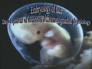
  • 3. Embryology is the study of formation and development of the embryo. The development of the human embryo from the time of fertilization through the birth week is an important period for human appearance . Every individual spends the first nine months of its life within the womb. During this period it develops from one-celled structure to an organism having billions of cells. An embryo has no face as such Webster delimits the face as the front part of the head comprising the nose,cheeks,jaws,mouth,forehead and the eyes. www.indiandentalacademy.com
  • 4. The key primordial begin to gather,and form slight swelling,depression and thickening that are rapid which undergo a series of mergers, and enlargements that will transfer them,forming a cluster of separate masses into a Face. Prenatal life may be arbitrarily divided into three periods: The period of the ovum (from fertilization to the end of the fourteenth day) The period of the embryo (from the fourteenth day to about the fifty-sixth day) The period of the fetus (from about the fifty-sixth day until the two hundred and seventieth day-birth) www.indiandentalacademy.com
  • 5. Understanding the development of the structures of the face requires knowledge of the pharyngeal or branchial arches. These arches form on either side of the foregut and correspond to the primitive branchial arches. The pharyngeal arch consists of a core of mesenchyme covered externally by ectoderm and covered internally by endoderm. Pharyngeal(branchial ) apparatus The pharyngeal (brachial)arches begin to develop during the fourth week in utero. www.indiandentalacademy.com
  • 6. The pharyngeal apparatus consists of a series of bilaterally paired arches,pouches(clefts)grooves and membranes This apparatus gives rise to a significant number of structure of the head and neck. The structure of the pharyngeal apparatus are numbered sequentially. The first four paired arches make up the lateral wall of the primordial pharynx,which develop from the foregut. www.indiandentalacademy.com
  • 7. The pharyngeal arch apparatus Cranial Endoderm Pharyngeal pouch Mesenchyme Pharyngeal membrane Pharyngeal cleft Nerve Artery Ectoderm Caudal www.indiandentalacademy.com
  • 8. Weeks 2 through 8 are especially important because the tissues and organ system are developing from the original three germ layers. Formation of Germ layers At a very early stage in development,the embryo proper acquires the form of a three-layered disc.,called the embryonic disc. The three layers that constitute this embryonic disc are Endoderm(endo=inside) Ectoderm(ecto=outside) Mesoderm(meso=in the middle) www.indiandentalacademy.com
  • 9.   Ectoderm The ectoderm contributes to the formation of the face is well around the stomodeum by the fourth week of embryonic life. It is evident that the ectodermal structures bounding the stomodeum participate not only in the formation of the face, but also in the formation of the nasal and oral cavities. Mesoderm The mesenchyme that fills the pharyngeal arches is derived from three sources.. The Paraxial Lateral plate musculature Neural crest skeletal (this musculature is innervated by one nerve that innervates all the muscles of that particular arch) www.indiandentalacademy.com Finally each arch has its own artery.
  • 10. Folding of the Embryo Beginning in the fourth week of development the flat trilaminar embryonic disc folds in two planes to form a more typical-appearing,cylindric,C-shaped embryo. Folding in the cranial –caudal plane is mainly a result of rapid longitudinal growth of the central nervous system. Growth of the somites account for much of the lateral folding. www.indiandentalacademy.com
  • 11. Folding of the Embryo…… Folding brings the endodermal-lined yolk sac into the embryo and creates the primordial gut;the foregut,midgut,and the hindgut. At the early stages of embryonic development, the vertebrate face has a common plan. A series of small buds of tissue called the facial primordial forms around the stomodeum, which forms the primitive mouth. The facial primordial are made up mainly of neural crest cells that have migrated from the cranial crest and settled. www.indiandentalacademy.com
  • 12. A summary of the derivatives of the first and second pharyngeal (ie, branchial)arches is as follows: ·                 Pharyngeal arch I Arch I-Meckels cartilage (Mandibular arch) This pair of arch has distinct maxillary and mandibular prominence Its major contribution is in the development of face o Cranial nerve - Maxillary and Mandibular division of trigeminal nerve (cranial nerve V) o  Artery - Maxillary (terminal branch) www.indiandentalacademy.com
  • 13. o  Muscles - Muscles of mastication (ie, temporalis, masseter, pterygoids), mylohyoid, anterior belly of digastric, tensor tympani, tensor veli palatini o  Skeleton - Maxillary cartilage (incus, alisphenoid), mandibular or Meckel cartilage (malleus), arch dermal mesenchyme (maxilla, zygomatic, squamous portion of temporal bone, mandible) www.indiandentalacademy.com
  • 14.        Pharyngeal arch II (hyoid arch) Reichert’s cartilage Its major contribution is the development of hyoid bone,the lesser horn and the superior portion of the body o  Cranial nerve -Facial nerve VII o  Artery - Stapedial o  Muscles - Muscles of facial expression (ie, orbicularis oculi, orbicularis oris, risorius, buccinator, platysma, auricularis, frontalis), stapedius muscle, posterior belly of digastric, stylohyoid muscle o  Skeleton - Stapes, styloid process, stylohyoid ligament, lesser cornu of hyoid, upper part of the body of the hyoid bone   www.indiandentalacademy.com
  • 15. Derivatives of pharyngeal pouches,Grooves & membranes Pouch 1-Tympanic cavity,auditory tube,mastoid antrum Pouch 2-Crypt lining of palatine tonsils Pouch 3-Inferior parathyroid glands,thymus Pouch 4-Superior parathyroid glands,Ultimobranchial body Groove1-External auditory meatus Groove2 & 4-Cervical sinus Membrane 1-Tympanic membrane www.indiandentalacademy.com
  • 16. Development of the face Facial development result mainly from enlargement and movement of the frontonasal prominence and four prominences from pharyngeal arch1,the paired maxillary prominences ,and the mandibular prominence.These structures surround the stomodeum. The upper jaw develops from 5 main buds of tissue: a single median frontonasal mass ,the 2 lateral nasal prominences on both sides,, the 2 maxillary prominences The lower jaw develops from the paired mandibular primordia (mandibular prominences). www.indiandentalacademy.com
  • 17. Paired maxillary and mandibular prominences are derivatives of the first pair of branchial or pharyngeal arches. All these prominences are produced by the proliferation of the neural crest cells that migrated into the arches from the neural crest during the fourth week. The neural crest cells give rise to the connective tissue components Frontal prominence Nasal placode Maxillaryprominence www.indiandentalacademy.com Mandibular prominence Arch I
  • 18. ·   Fourth week of development o  Primordia of the face appear at the cephalic end of the embryo. o   Two nasal placodes cap the bulbous frontal prominence. o   The optic discs appear posterolateral to the frontal prominence. O Three paired branchial arches have formed. o The first arches split into maxillary and mandibular prominences. The hyoid arches are the second pair. www.indiandentalacademy.com
  • 19. o  Between the first arches and frontal prominence, the buccopharyngeal membrane becomes fenestrated. Fifth week of ·                     development o  Nasal pits develop in the nasal placodes, and the rims of the placodes differentiate into medial and lateral nasal prominences. o  The lens vesicles invaginate and close within the optic discs. www.indiandentalacademy.com
  • 20. o  The mesenchyme of the mandibular arch fills in across the midline. o   The caudal end of the medial nasal prominences begins to fuse with the maxillary prominences. ·                     At the beginning of the sixth week of development o   The nasals have shifted to a more ventral, central position. o    Growing and shifting subectodermal mesenchyme smooths out the furrows between prominences and arches, and the second arch becomes more massive.           www.indiandentalacademy.com
  • 21. o   Six auricular hillocks, which will become the pinna of the ears, form on the mandibular and hyoid arches. By the end of the sixth week of ·                     development   o Medial and lateral nasal prominences fuse. o  Maxillary prominences begin the formation of the upper jaw. o  The midline approximation of the medial nasal prominences forms the nasal septum. www.indiandentalacademy.com
  • 22. ·                     At the beginning of the seventh week of development o The tip of the nose is elevated between the medial nasal prominences and is visible in profile. o Eyelids become prominent. o The pinna of the ear takes shape. ·                 End of the seventh week of development O The pattern of facial features has taken on a human appearance. However, facial proportions develop during the fetal period. www.indiandentalacademy.com
  • 23. ·   The fusion of the medial nasal prominences is complete, forming the central axis of the nose and the philtrum of the lip. From the beginning of the eighth week of development to birth, final development of the face occurs slowly and results mainly from changes in the proportion and relative positions of the facial components.   At the end of the 8 week the face has taken on a human appearance. www.indiandentalacademy.com
  • 24. Developmental Defects 1. Cleft (hare) lip and cleft jaw: the medial nasal processes do not fuse with the maxillary processes. 2. Cleft palate and/or uvula: the palatine shelves fail to fuse with each other and/or with the primary palate. www.indiandentalacademy.com
  • 25. 3.Median cleft of upper lip: The 2 medial nasal processes fail to fuse. This is often accompanied by mental retardation. 4.Median cleft of lower lip: Failure of fusion of 2 mandibular arches. 5. Oblique facial cleft: The nasolacrimal duct remains exposed due to the lateral nasal process failing to fuse with the maxillary process It can occur along with a cleft lip. www.indiandentalacademy.com
  • 26. Development of Dentition The development of dentition is an integral part of craniofacial growth and an Unique structure of body Tooth is derived from ectoderm & mesoderm The first indication of tooth formation is seen at about sixth week of intrauterine life Life cycle of the tooth Histo differentiation Initiation Proliferation www.indiandentalacademy.com Apposition Morphodifferentiation
  • 27. Initiation The epithelium overlying the convex border of the alveolar process becomes thickened and projects into the underlying mesoderm. This epithelial thickening is called the dental lamina. Specific cells within the dental lamina have the potential to form the enamel organ of certain teeth by responding to those factors that initiate or induce tooth development. A lack of initiation result in absence of teeth(anodontia)most frequently the permanent upper laterals,third molars,and lower second premolars. Abnormal initiation result in development of supernumerary teeth www.indiandentalacademy.com
  • 28. Proliferation Enhanced proliferation activity ensures at the point of initiation and results successively in the Bud,Cap,and Bell stage of the odontogenic organ. Proliferative growth causes regular changes in size & proportion of the growing tooth germ As the enamel organ grows downwards into the mesenchyme (of the alveolar process)its lower end assure a cup-shaped appearance.The cup comes to be occupied by a mass of mesenchyme called the dental papilla. The enamel organ and the dental papilla together constitute the tooth germ.At this stage the developing tooth looks like like a cap:it is ,therefore ,described as the cap stage of tooth development. www.indiandentalacademy.com
  • 29. Histo differentiation It succeeds the proliferative stage which undergoes definite morphologic and functionally changes. The cells of the enamel organ that line the papilla become columnar.These are called ameloblasts. Mesodermal cells of the papilla that are adjacent to the ameloblasts arrange themselves as a continuous epitheliumlike layer.The cells of this layer are called odontoblasts Ameloblast lay down enamel on the superficial(outer)surface of the basement membrane.The odontoblasts lay down dentine on its deeper surface.As layer after layer of enamel and dentine are laid down,the layer of ameloblasts and the layer of odontoblasts move away from each other. www.indiandentalacademy.com
  • 30. The ameloblasts and odontoblasts are separated by a basement membrane.The remaining cells of the papilla form the pulp of the tooth.The developing tooth now looks like a bell (Bell stage of tooth development ) Morphodifferentiation & Apposition Morphodifferentiation is impossible without proliferation. The basic form and relative size of the tooth is established by morphodifferentiation. www.indiandentalacademy.com
  • 31. In advanced bell stage ,the boundary between inner enamel epithelium and odontoblast outline the future dentinoenamel junction.In addition,the cervical portion of the enamel organ gives rise to the epithelial root sheath of Hertwig. Apposition is the deposition of the matrix of the hard dental structures. Appositional growth is characterized by regular and rhythmic deposition of the extracellular matrix,which is of itself incapable of further growth. www.indiandentalacademy.com
  • 32. Development of Permanent tooth The Permanent tooth bud appears in the 4th & 5th intrauterine months, at about the same age at which mineralization of the deciduous teeth commences. The dental lamina gives off series of buds, one of which lies on the medial side of each developing milk tooth. These buds from enamel organ give rise to the permanent teeth The permanent molars are formed from bud that arise from the dental lamina posterior to the region of the last milk tooth At birth the germs of all the deciduous teeth,and of the permanent incisors,canine and first molars show considerable development. The germs of premolar and of the permanent second molars are . rudimentary. The germ of third molar is formed www.indiandentalacademy.com after birth.
  • 33. The stage of dentitions Gum pads Primary dentition Mixed dentition www.indiandentalacademy.com Permanent dentition
  • 34. Gum pad stage:It extends from birth until the eruption of the first primary tooth in the oral cavity. (The pre-dentition period.) -This is from birth to six months. -At this stage, there are no teeth. Clinically, the infant is edentulous -Both jaws undergo rapid growth; the growth is in three planes of space: downward, forward, and laterally (to the side). Forward growth for the mandible is greater. Greater the anteroposterior dimension of the gumpads,greater the possibility of the child developing malocclusion --occasionally, there is a neonatal tooth present at birth. www.indiandentalacademy.com
  • 35. Primary dentition stage: (first transitional period /Deciduous dentition period. ) It extend from the time of eruption of the primary teeth until the eruption of the first permanent tooth around 6yerars of age. – The dentition of this period manifests several important characteristics. 1.There is either spacing between the teeth or there is not. 2.There is no sagittal and transversal dimensions alteration,only vertical changes are seen. 3.The distal surface of the maxillary & mandibular second primary molars maintain vertical relationship. www.indiandentalacademy.com
  • 36. -The tooth buds anticipate the ultimate occlusal pattern. -Mandibular teeth tend to erupt first. --Eruption times can be variable. Spaced dentition is supposed to be good as space in between the teeth is utilized for adjustment of permanent successors Primate spaces occur in about 50% of children. They appear in the deciduous dentition. The spaces appear between the upper lateral incisor and the upper canine. They also appear between the lower canine and the deciduous first molar. www.indiandentalacademy.com
  • 37. Flush terminal plane This dentition is completed after the eruption of the second primary molars,indicating the location for permanent teeth.Therefore it’s the key factor that influences the future occlusion of the permanent dentition. The second primary molar relation is described in terms of relationship between terminal planes. In flush terminal plane :The distal surface of upper and lower teeth are in straight line.It’s a favourable relationship to guide the permanent molars. www.indiandentalacademy.com
  • 38. Mesial step:In mesial step the distal surface of the lower molar is more mesial to that of the upper.Invariably it is favorable to guide the permanent molar into a class I relationship. Distal step:The distal surface of the lower molar is more distal to that of upper.Its unfavorable as it guides the permanent molars into distal occlusion. flush terminal plane Distal step Mesial step ClassIII ClassI(with late mesial shift) End-end ClassIII(normally) ClassI(desirable) www.indiandentalacademy.com ClassII
  • 39.   Incisor liability: Mayne has coined the term incisor liability Permanent incisors are larger than primary incisors,this difference in size is termed incisal liability. Eruption of permanent incisors make the permanent arch circumference wider,normally the interincisal angle is 150 degree in primary dentition and 120 degree in permanent dentition The period of transition determine the successful alignment of the permanent teeth toward an ideal arch form www.indiandentalacademy.com
  • 40. Anterior teeth relation Over bite:The average over bite in primary dentition is 2mm Overjet :The average over jet is 1-2mm with a normal range of 2-6mm. With the excessive wear of the primary canines and molars,the whole lower dental arch may move anteriorly,the incisors may assume an edge-to-edge interrelationship by the age of six Sequence of eruption Deciduous Eruption Time Central Incisor 6-12 months Lateral Incisor 9-13 months Canine 16-23 months First Molar 13-19 months Second Molar 23-33 months www.indiandentalacademy.com
  • 41. Exfoliation of Primary teeth The replacement is a multifactorial process. When the permanent tooth begins to erupt,it is apparently guided by the gubernaculum.. The pathway for the erupting anterior teeth is essentially lingual to their predecessors resulting in an oblique lingual pattern of resorption. Resorption is not a continuous process,but is rather one of alternating period of destruction and apposition www.indiandentalacademy.com
  • 42. Mixed dentition stage: (second transitional period) Starts with the eruption of the first permanent tooth,usually the mandibular central incisor,and is normally completed at the time the last primary tooth is shed. During this period ,the vertical dimension of the face is increased,allowing for a heightening of the alveolar ridges to accommodate continual root growth of the cuspids and premolars. www.indiandentalacademy.com
  • 43. -If the deciduous dentition is spaced dentition with flush -terminal plane of second deciduous molars,the eruptive force -cause a closing of of the existing space causing a decrease in - arch length-(Early mesial shift) - - When no space exist,the erupting first permanent molars is not able to close spaces.In these case when the primary molars exfoliates the permanent molar migrates to use up the “leeway space”-(Late mesial shift) www.indiandentalacademy.com
  • 44. The leeway space. -The deciduous anteriors--incisors and canines are narrower than their permanent successors mesiodistally. -The deciduous molars are wider that their permanent successors mesiodistally. -This size difference has clinical significance. The difference is called the leeway space. -The leeway space in the lower arch is approximately 3.4 mm. -The leeway space in the upper arch is approximately 1.8 mm. In normal development, the leeway space is taken up by the mesial migration of the first permanent molars. www.indiandentalacademy.com
  • 45. Since there are no premolars in the primary dentition, the primary molars are replaced by permanent premolars. If any primary teeth are lost before permanent teeth are ready to replace them, some posterior teeth may drift forward and cause space to be lost,] This may cause crowding and/or misplacement once the permanent teeth erupt, which is usually referred to as maloclussion. www.indiandentalacademy.com
  • 46. 'Ugly duckling stage.: ' On occasion, the permanent incisors 'spread out' due to spacing. In the older literature, is called by the 'ugly duckling stage.' With the eruption of the permanent canines, the spaces often will close. This is the norm between ages 7 through 12 years of age, and usually is not connected with a permanent space between the teeth. This stage of development needs special comment because its often mistaken as anwww.indiandentalacademy.com orthodontic problem.
  • 47. Permanent dentition Maxillary / mandibular occlusal relationships are established when the last of the deciduous teeth are lost. The adult relationship of the first permanent molars is established at this time. The growth of the jaw continues during this period,developing room for the third molars www.indiandentalacademy.com
  • 48. Sequence of eruption Permanent Teeth Eruption Time Central Incisor 6-8 years Lateral Incisor 6.5-9 years Canine 9-12 years First Premolar 8-12 years Second Premolar 8.5-13 years First Molar 6-7 years Second Molar 11-14 years Third Molar (Wisdom Tooth) 17-25 years www.indiandentalacademy.com
  • 49. Physiology of the Stomatognathic system • Stomatognathic- of or relating to the jaws and the mouth :webstar’s medical dictionary • As a dentist and especially as an orthodontist,it is essential that we have a sound knowledge of and understand the importance of the stomatognathic system • Until we know what is normal we will not be able to recognize aberrancy www.indiandentalacademy.com
  • 50. Functional Osteology • Bone is one of the hardest tissues of the human body and it’s most adaptive and responsive to functional forces • Meyer -1864:proposed the trajectorial theory of bone formation • 1870:Julius Wolff said that the trabecular arrangement was due to functional forces and any change in direction or intensity of the forces would change the internal architecture & external form of the bone • It was expressed by a mechanical mathematical law= law of orthogonality www.indiandentalacademy.com
  • 51. • Tajectories need not pass at right angles to each other—even their course may be wavy • Increased function increases bone density while decreased function decreases . • Muscles have a great influence over bones and may even change their shape • Thus the timed manipulation of the musculature may be beneficial to the orthodontist • It is established that cartilaginous and membranous bones react differently to forces www.indiandentalacademy.com
  • 52. Beninghoff’s trajectories He Concluded saying that trajectories can be demonstrated in the maxillo-facial area • Lines pass through both the spongy and compact bone • They obey no individual bone margins • Maxilla Transfers the whole stress to the cranium which has as thin cortices that are interconnected by trabeculae Stress trajectory can be considered as the craniofacial unit www.indiandentalacademy.com
  • 53. 3 main pillars of trajectories: • All arise from the alveolar bones and end in the base of the skull The canine pillar The zygomatic pillar The pterygoid pillar • Curve around the sinuses,nasal and orbital opening • The supra and infra orbital ridges and the zygomatic buttress reinforce these pillars www.indiandentalacademy.com
  • 54. Mandible • Absorbs all the stresses Thick cortices and more radially arranged trabecuae Stress trajectories • From beneath the teeth-join together in comman pillar—ends at the condyle •Mandibular canal and nerve are protected-unloading of nerve •Thick cortical layer at the base provides resistance to bending forces •Trajectories from the symphysis, gonial angle and coronoid process join this main pillar www.indiandentalacademy.com
  • 55. Myology Property of muscles (that are important for help in kinetic activity are) Elasticity= the ability to return to original length and shape after contraction or extension Normally relaxed muscle withstand only a certain amount of elongation Contractility=the ability to contract under innervational impulse. Strength of contraction depends on Frequency of stimuli Number of fibers involved • The greatest of contraction is seen when the muscle approximates its resting position www.indiandentalacademy.com
  • 56. Other principles of muscle physiology • All or none law Intensity of contraction is independent of the strength of the action potential as long as the threshold is crossed Seen only when the muscle is not fatigued • Muscle tonus State of slight constant tension and form the basis of reflex posture • Resting length Constant and Permits maintenance of postural relationship • Reciprocal innervations and inhibition Inhibition of contraction may be brought about by excitation of the antagonist.It is through this that movements are coordinated www.indiandentalacademy.com
  • 57. Buccinator mechanism There is strong inter relationship between bone and muscle Muscles are a potent force-at rest or when active Teeth and supporting tissues are influenced by the contiguous musculature The balance between the tongue and the peri-oral musculature influences the morphology of the dental arches and teeth The shape and size of the tooth root and the periodontal spaces also helps in the final adaptation www.indiandentalacademy.com
  • 58. • During swallowing and mastication- tongue exerts 2-3 times more force than the peri oral muscles • Counter acted by the tonal contraction,atmospheric pressure,peripheral recruitment from the buccal musculature • Even though doubts still exist about such a balance of force-we must admit that aberration of muscles can and do lead to marked malocclusion • The continuous band of muscle that encircle the dentition and attach to the pharyngeal tubercle comprises the buccinator mechanism www.indiandentalacademy.com
  • 59. Tongue • Opposing the buccinator mechanism is a very powerful structure- the tongue • It has multiple functions • Most developed in a new born • In infancy extrinsic suspensory muscles attach the tongue to the various osseous structures and are responsible for gross movement in the horizontal direction(suckle-swallow) www.indiandentalacademy.com
  • 60. • The tongue has amazingly versatile functional possibilities by virtue of the fact it is anchored at only one end. • This freedom permits the tongue to deform the dental arch. • It may Leads to proclination of the maxillary incisors,constriction of buccal segment and may create an open bite. • Compensating action of mentalis causes retraction of the lower incisors • Abnormal position and size of the tongue is often seen in the presence of enlarged tonsils and adenoids. www.indiandentalacademy.com
  • 61. Muscles of the Tongue INTRINSIC Superior longitudinal-Shortens the tongue & makes the dorsum concave Inferior longitudinal-Shortens the tongue & makes the dorsum convex Transverse-Makes the tongue narrow & elongated Vertical-Makes the tongue broad & flattened EXTRINSIC Genioglossus- Protrudes the tongue out of the mouth by pulling the posterior part forwards Hyoglossus-depresses the tongue Styloglossus -pulls it upward & backward Palatoglossus-brings the palatoglossal arches together, thus shutting the oral cavity from the oropharynx www.indiandentalacademy.com
  • 62. Temperomandibular Joint • The TMJ is the ball and socket joint that allows the lower jaw to swing open and close.  It’s a Compound –movable joint It articulation with the mandibular condyle and the inferior surface of the sqamous portion of the temporal bone •The condyle: This is the "ball" in the joint.  It is a part of the mandible (lower jaw), and is covered in a layer of cartilage which allowswww.indiandentalacademy.com for smooth motion within the joint assembly
  • 63.   •The glenoid fossa: The fossa is the "socket", or depression in which the condyle sits.  It is located in the temporal bone of the skull.  The front of the fossa is a more gentle slope of bone called the articular eminence.  The eminence is also covered with cartilage. The articular disc: The articular disc is also called the meniscus.  It is made of hyaline cartilage.  The ligament behind the meniscus is called the retrodiscal pad in deference to its function as a shock absorber www.indiandentalacademy.com
  • 64. •The joint capsule is the covering of the TM joint .It isolates the contents of the joint and allows free movement of the condyle and articular disk within a small "swimming pool" of synovial fluid.  The capsule has lots of blood vessels and nerves as well as connective tissue.  • It is lined by synovial membrane. Head of the condyle is tubular or ellipsoidal Unique feature of the joint is that there are actually 2 joint spaces- separated by the disc Thus the joint is capable of dual function Lower cavity—rotary/hinge movement Upper cavity—gliding/translatory movement www.indiandentalacademy.com
  • 65. • The rotary movement occurs by itself when opening from occlusion to physiologic resting after this even gliding motion takes place • When opened in his manner the articular disc glides over the articular eminence • The condyle rotates against the inferior surface of the disc. • Lateral pterygoid helps to move the disc anteriorly In the lateral shift/bennet movement the condyle on the working side rotates and moves laterally. • The condyle on the other side moves forwards and mesially in an arc • • Posterior limit formed by the squamo-tympanic fissure— medially by the petro-tympanic fissure www.indiandentalacademy.com
  • 66. • Due to over closures-the disc is moved forward and the condyle rides over the posterior border of the disc. Impinges on the retrodiscal soft tissues-clicking an discomfort to the patientmisdiagnosed as arthritic changes ,pain may be due to impingement or due to pterygoid muscle spasm(mpd) • The main reason for the mpd is neurogenic or psychological. The TMJis beautifully engineered to handle multiple tasks endowed to it • To maintain it-proper occlusal management an maintenance of vertical dimensions is essential www.indiandentalacademy.com
  • 67. Functional movements of the mandibl •The mandible is the only movable bone in the head and face. •It has 13 muscles attached to it •Due to limitations of morphology and structure of the TMJ it can be moved only in certain directions •It has postural stability along with primary movements www.indiandentalacademy.com
  • 68. Forced retrusion Posterior fibres of the temporalis,masseter,suprahyoid muscles Opening lateral pterygoid-main. Supra,infra hyoidgenio,mylo hyoids,digasric>stabilising Temporalis,masseter,medial pter.>controlled relaxation Closure Temporalis, masseter >main action medial ptery. >part action Lateral ptery >cntrolled relaxation Protrusion Lateral and medial pter. Lateral movement Temporalis + lat. pter www.indiandentalacademy.com
  • 69. COMPENSATORY MUSCLE FUNCTIONS • While mastication may call for most potent effort from the associated muscles, the most frequent demands are made by deglutition, respiration, speech and posture. • At times two or more of these functions are carried on simultaneously. • where there is malocclusion or abnormal morphologic relationship certain compensatory or adaptive muscle functions may arise, either to restrain the dental malocclusion or to actually increase the discrepancy. www.indiandentalacademy.com
  • 70. Positions of the mandible • • • • • • • • Basic positions are Postural resting position (Physiologic rest) Centric relation Initial contact Centric occlusion Most retruded position (terminal hinge position) Most protruded position Habitual resting position • Habitual occlusion position www.indiandentalacademy.com
  • 71. POSTURAL RESTING POSITION It is one of the earliest posture positions to be developed. Mandible is suspended from the cranial base by the cradling musculature. Here the jaws are separated by a constant distance. Factors influencing the postural position are the following: • Body and head posture • Sleep • Psychic factors influencing muscle tonus • Age • Proprioception from the dentition and muscles • Occlusal changes such as attrition • Pain • Muscle disease and muscle spasm and • Temporomandibular joint disease. www.indiandentalacademy.com
  • 72. CETRIC RELATION • As far as muscle physiology is concerned, centric relation may be defined as unstrained, neutral position of the mandible in which the anterio superior surfaces of mandibular condyles are in contact with the concavities of the articular disks as they approximate the postero inferior third of their respective articular eminent. • This means that the mandible is deviating neither to the right nor to the left and is neither protruded no retruded. • Such a relation can be the same as the postural resting position, the point of initial occlusal contact and centric occlusion. www.indiandentalacademy.com
  • 73. INITIAL CONTACT • As the mandible moves from physiologic rest or the postural resting position toward occlusion of teeth, if all is normal it maintains a centric relation position as far as the articular fossae are concerned. • If there is normal occlusion the point of initial contact produces no change in the function of temporomandibular joint and all inclined planes are brought together simultaneously in the maxillary and mandibular teeth. • Premature contacts are seen quite frequently, they can initiate deflections in the mandibular path of closure. • This causes traumatic forces to be exerted on the teeth and severe cases will produce temporomandibular joint problems www.indiandentalacademy.com
  • 74. CENTRIC OCCLUSION • Centric occlusion is a static position • It is harmonious with the centric relation • Teeth brought into contact with unstrained relation of the condyles • Few patients can show centric occlusion • Premature contact,loss of teeth,Overeruption of teeth,overextension of artificial restorations,malpositions of individual teeth-all these mitigate against the establishment of a centric occlusion www.indiandentalacademy.com
  • 75. MOST RETRUDED POSITION (Terminal Hinge Position) • It’s a Reproducible retruded mandibular position with the teeth in occlusion • It has become a common starting point in occlusal analysis and rehabilitation • Some patients can easily retrude a few mm. while others find it difficult • Mandible should not be guided or forced beyond the unstrained position of the mandible-unbiologicalwould compress the tissues. www.indiandentalacademy.com
  • 76. MOST PROTRUDED POSITION • This position is variable from individual to individual than the retruded position,however,it is reproducible • Shows the range of movement of the mandible • Flaccidity of capsular ligament allows condyle to over ride the anterior margin of the eminence,leading to dislocation. HABITUAL RESTING POSITION There are certain types of malocclusions that prevent the patient from achieving a physiologic resting position. www.indiandentalacademy.com
  • 77. • Eg.,in class II div 2 malocclusion with maxillary incisors inclined lingually there is a tendency to force the condyles posteriorly and superiorly in the articular fossae • The physiologic resting positions can be changed due to mental disturbances, enlarged adenoids, temporomandibular joint pathology, psychic trauma, selective paralysis by poliomyelitis and confirmed mouth breathing etc. www.indiandentalacademy.com
  • 78. HABITUAL OCCLUSAL POSITION • In normal occlusion the centric occlusion and habitual occlusion should be the same. But the occlusal relationship can be changed when there is an environmental imbalance induced by improper restoration, tooth loss etc. • It is vitally important that the dentist make sure that the habitual occlusal position and the centric occlusal position are the same and are in harmony with centric relation and the postural resting position of the mandible. www.indiandentalacademy.com
  • 79. Functions of the system • • • • Deglutition Mastication Respiration Speech www.indiandentalacademy.com
  • 80. DEGLUTITION Infantile swallow ( visceral swallow) Mature swallow ( somatic swallow) www.indiandentalacademy.com
  • 81. CHARACTERISTICS OF INFANTILE SWALLOW The jaws are apart, with the tongue between the gum pads The mandible is stabilized by contraction of the muscles of the 7th cranial nerve & the interposed tongue. The swallow is guided, & to a great extent controlled by sensory interchange between the lips & the tongue. www.indiandentalacademy.com
  • 82. TRANSITION PERIOD At about the 5 to 6th month of age, as the incisors begin to erupt, certain proprioceptive impulses come into play & the peripheral portions of the tongue starts to spread laterally. An average infant would show a dominant & exclusive thrusting swallow for the first 6 months of life,a transitional thrusting & lateral spread of tongue during the next year & a dominant somatic swallow thereafter. www.indiandentalacademy.com
  • 83. CHARACTERISTICS OF MATURE SWALLOW The teeth are together The mandible is stabilized by contraction of the mandibular elevators,which are primarily 5th cranial nerve muscles The tongue tip is held against the palate, above & behind the incisors There are minimal contractions of the lips during the mature swallow. www.indiandentalacademy.com
  • 84. DEGLUTITION CYCLE FLETCHER gave 4 phases 1.Preparatory swallow 2.Oral phase of swallowing 3.Pharyngeal phase of swallowing 4.Esophageal phase of swallowing www.indiandentalacademy.com
  • 85. PREPARATORY SWALLOW • Starts as soon as liquids are taken in, or after the bolus has been masticated • The liquid or bolus is then in a swallowpreparatory position on the dorsum of the tongue. • The oral cavity is sealed by lip & tongue. www.indiandentalacademy.com
  • 86. Oral phase of swallowing moves • Soft palate upward & the tongue drops downward & backward • Larynx & hyoid bone move upward • Smooth path for the bolus as it is pushed from the oral cavity by the wave like rippling of the tongue • Oral cavity is stabilized by the muscles of mastication, & www.indiandentalacademy.com maintains the anterior
  • 87. Pharyngeal phase of swallowing • Begins as the bolus passes through the fauces • The pharyngeal tube is raised upward en masse • Nasopharynx is sealed off by closure of the soft palate against the posterior pharyngeal wall • Hyoid bone & base of tongue move forward as the pharynx & tongue continue their peristaltic – like movement of the bolus of the food www.indiandentalacademy.com
  • 88. Esophageal phase of swallowing • Commences as the food passes the cricopharyngeal sphincter • While peristaltic movement carries the food through the esophagus, the hyoid bone , palate & tongue return to their original positions www.indiandentalacademy.com
  • 89. Mastication • In the infant food is taken by suckling-unlearned or automatic reflex • Rhythmic caving in of the cheeks, bobbing of the hyoid bone,snake like movement of the tongue, anterior movement of the tongue and nodding movement of the head • Mandible is protruded • During deglutition rhythmic contraction of the tongue and facial muscles aids in stabilizing the mandible www.indiandentalacademy.com
  • 90. 6 phases outlined by Murphy • 1)preparatory phase=> food in mouth-tongue positions it-mandible moves to working side • 2)food contact=> characterized by a pauseprobably to determine food consistency • 3)crushing phase=> starts with high velocity and slows down as food is crushed • 4)tooth contact=> takes place with slight change in direction-muscles are already ready for this • 5)grinding phase=> transgression of the mandible across the maxillary teeth-most patients chew unilaterally-cyclic event • 6)centric occlusion=> mandible back to terminal position-next cycle begins www.indiandentalacademy.com
  • 91. Respiration Inherent reflex activity Demand on muscles are more subtle and more difficult to appreciate Posture has a significant effect on respiration Oral musculature is responsible for vital positional relationships-maintains the airway All the learned jaw functions are built around and accommodated to the tongue and mandibular positions which make clear airway possible Maintenance of the respiratory spaces and airway is significant in facial growth Respiration in the child helps keeping the pharynx patent as well in the development of speech . www.indiandentalacademy.com
  • 92. Speech Learned activity –depends on maturation of organism Makes use of muscles that have other functions WEST lists the “other than speech functions” as follows 1. innate,automatic,vegetative-swallowing,breathing,gagging 2. learned,automatic,vegetative-biting,chewing,sucking 3. learned,automatic,emotional-grimaces,mannerisms 4. innate,automatic,emotional-laugh,smile,sobbing 5. learned,non automatic,discriminatory,voluntary-movement of with tongue,spreading lips,kissing,blowing 6. learned,automatic practical reactions-whistling,playing instrument,humming www.indiandentalacademy.com
  • 93. Muscles involved Walls of the torso,the respiratory tract, the pharynx, the soft palate,the tongue,the lips and face and nasal passageways are all concerned in the production of speech. Simultaneous breathing is necessary to provide vibrations for sound Tongue,lips and velopharyngeal closure modify outgoing stream to produce sound . Normal structure is necessary for production of sound-in cleft palate cases its not possible even with muscular compensation Speech mechanism acts on the breath streams in a number of ways,controlling the air mechanism direction,flow,release,pressure,general and lingual air path www.indiandentalacademy.com
  • 94. With respect to tongue • The lips and the tongue undergo maturational changes • Degree of lip protrusions varies the length of the vocal tract • With the reduction of suckle swallow activity,more delicate peripheral lip movements are noted. • In infancy-suckle reflex is active and extrinsic muscles are well developed-only later the intrinsic muscles capable of speech develops Velopharyngeal valve(closure) Very important for the dentist-in conditions of cleft palate-inadequate valving is seen-even in rehabilitated cases • Upward and backward movement to contact the post pharyngeal wall is important for certain sound production www.indiandentalacademy.com
  • 95. Speech difficulties related to Malocclusion Speech sound Problem S/z(sibilants) T/d (linguoalveolar stops) lips (labiodental fricatives) Th/sh/ch gap between incisors,Anterior open bite difficulty in production F/v Related malocclusion Irregular incisors,lingual position of maxillary incisors distortion distortion (linguodental fricatives) www.indiandentalacademy.com Skeletal classIII Anterior open bite
  • 96. Conclusion… • The study of the intra oral and peri oral structurestheir normal morphology and activity is very important for us. • Unless we know what is normal it is difficult to recognize and correct abnormalities • Also, the structures are all inter related ::follow scientific principles and order in their development. • It becomes important to follow the functions of the structures which ultimately leads to changes in the morphology.. www.indiandentalacademy.com
  • 97. Bibliography • Orthodontics:Principles and practices-T.M Graber • Orthodontics:Graber and Vanarsdal • Contemporary Orthodontics: William.R.Proffit • Handbook Of Facial Growth :Donald.H.Enlow www.indiandentalacademy.com
  • 98. www.indiandentalacademy.com Leader in continuing dental education www.indiandentalacademy.com