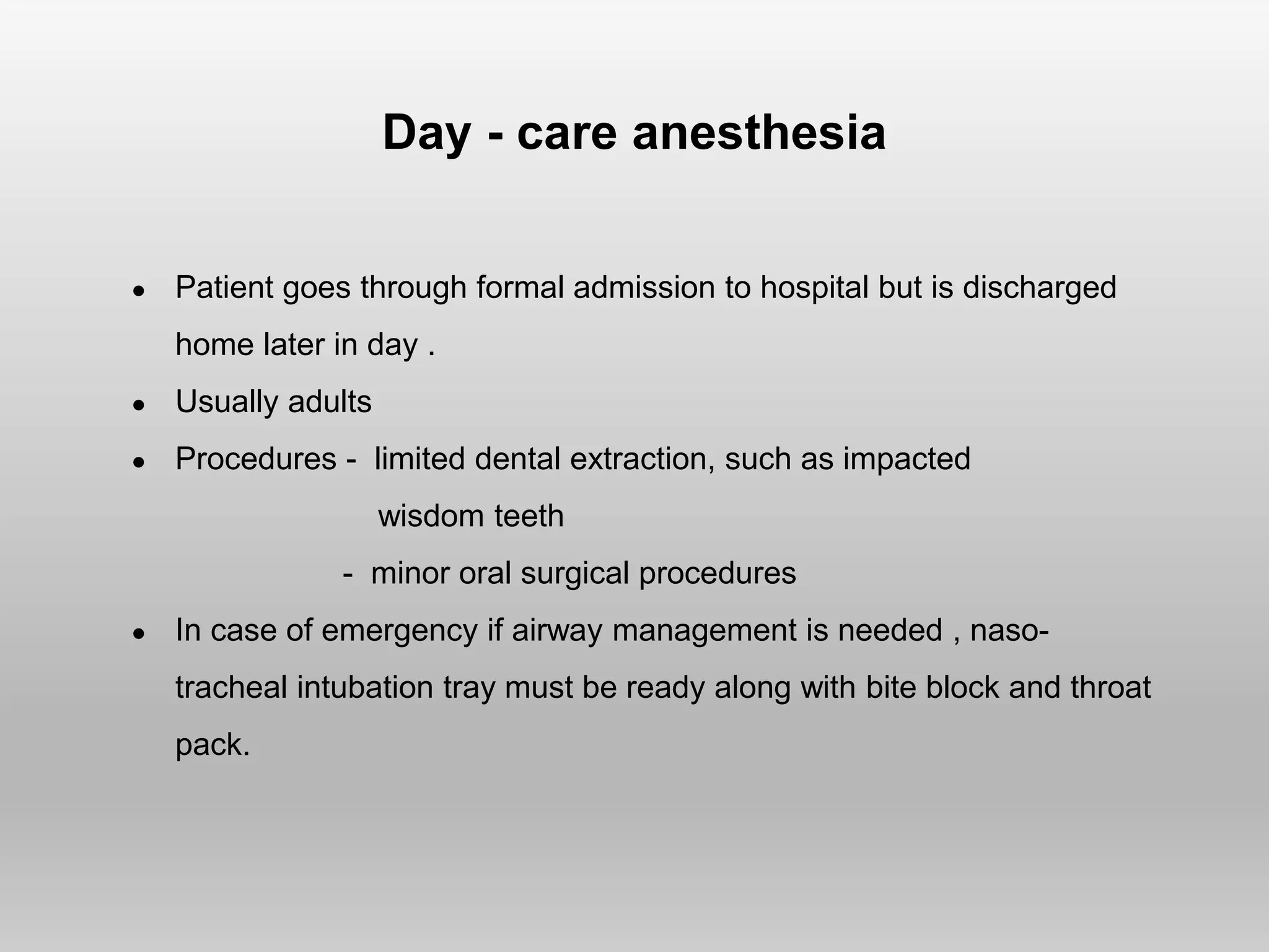
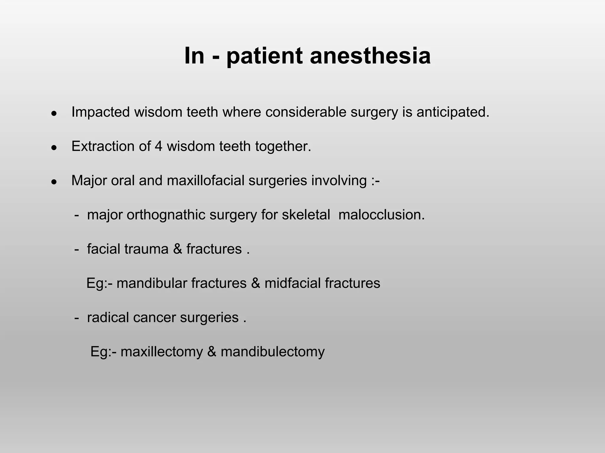
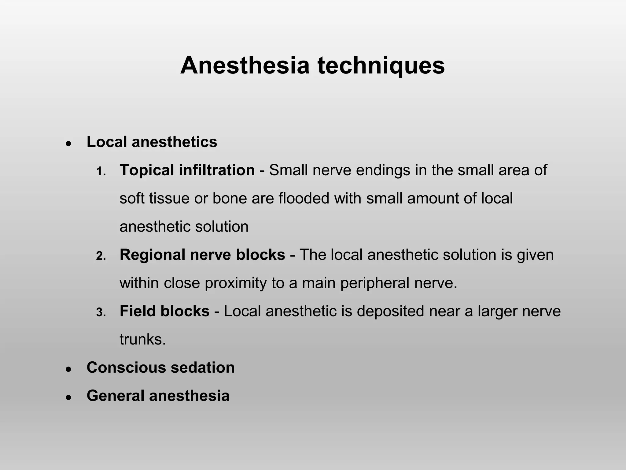
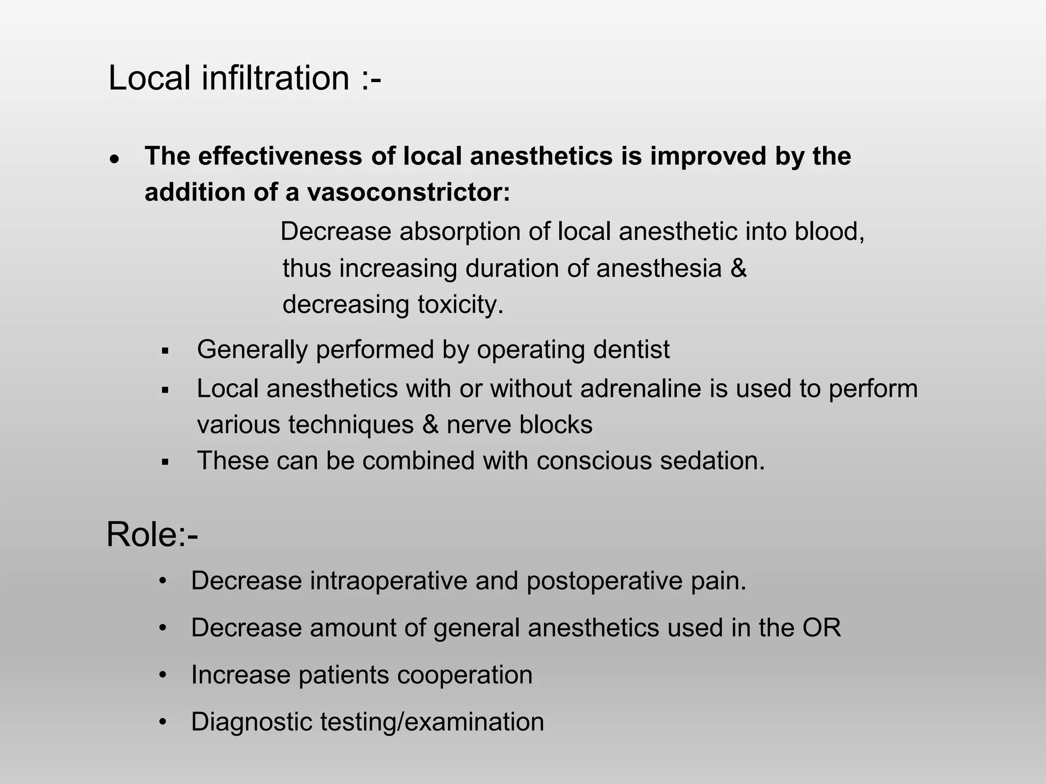
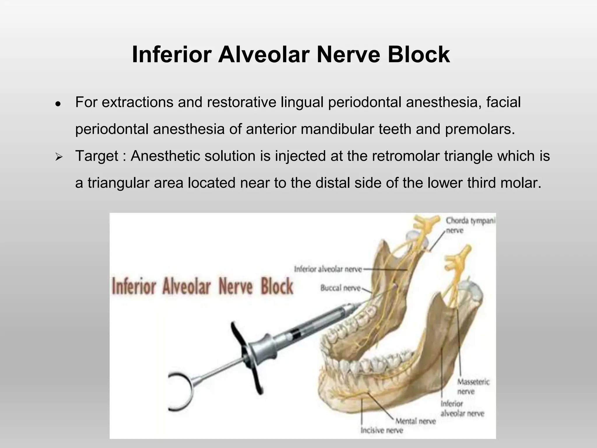
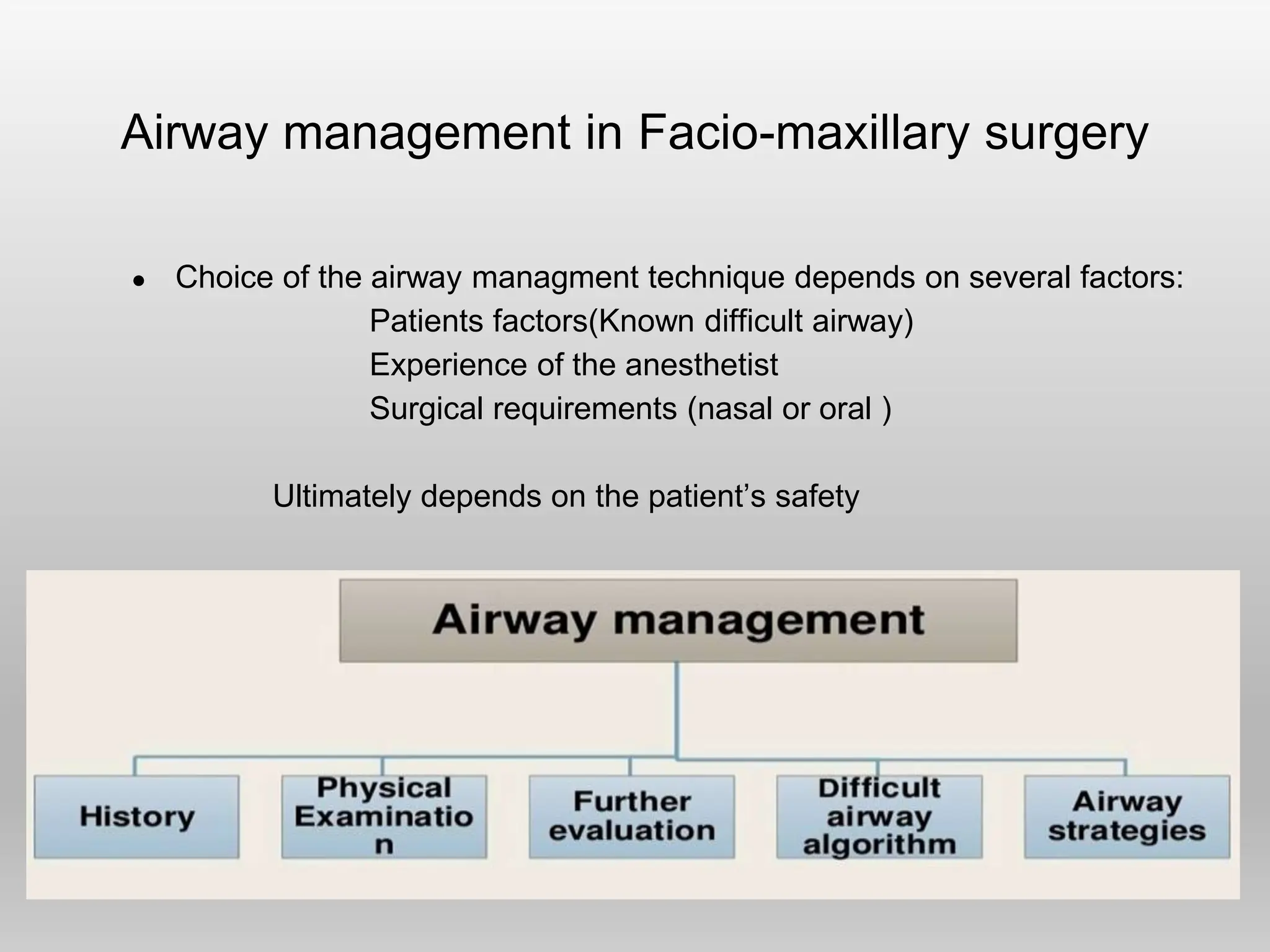
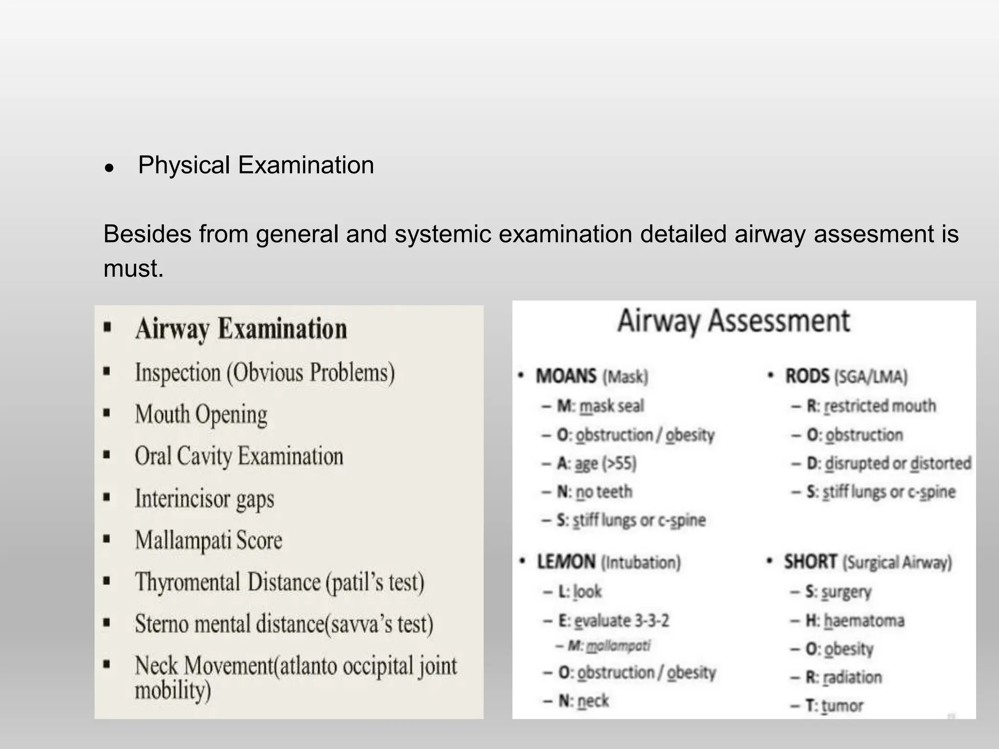
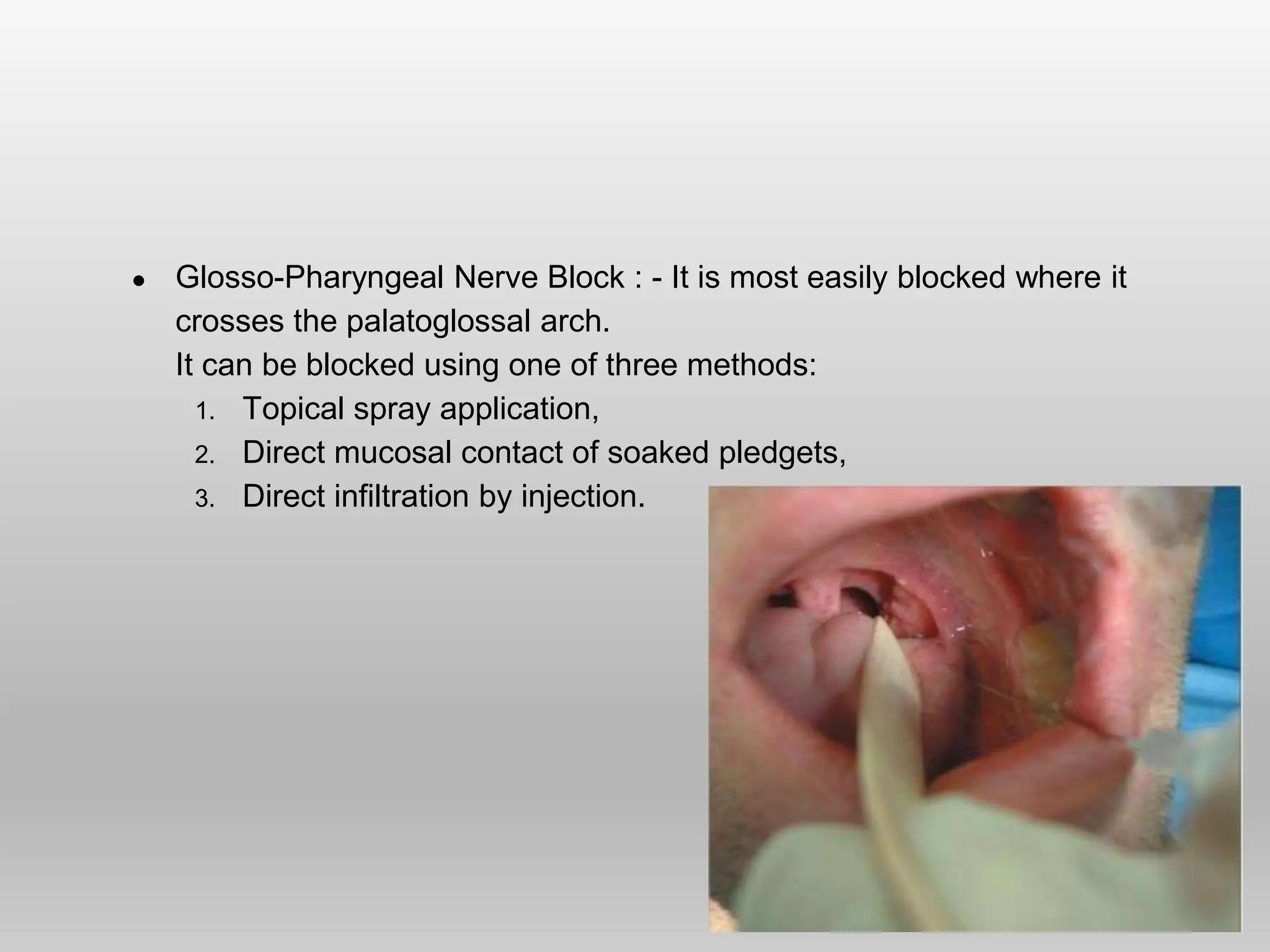
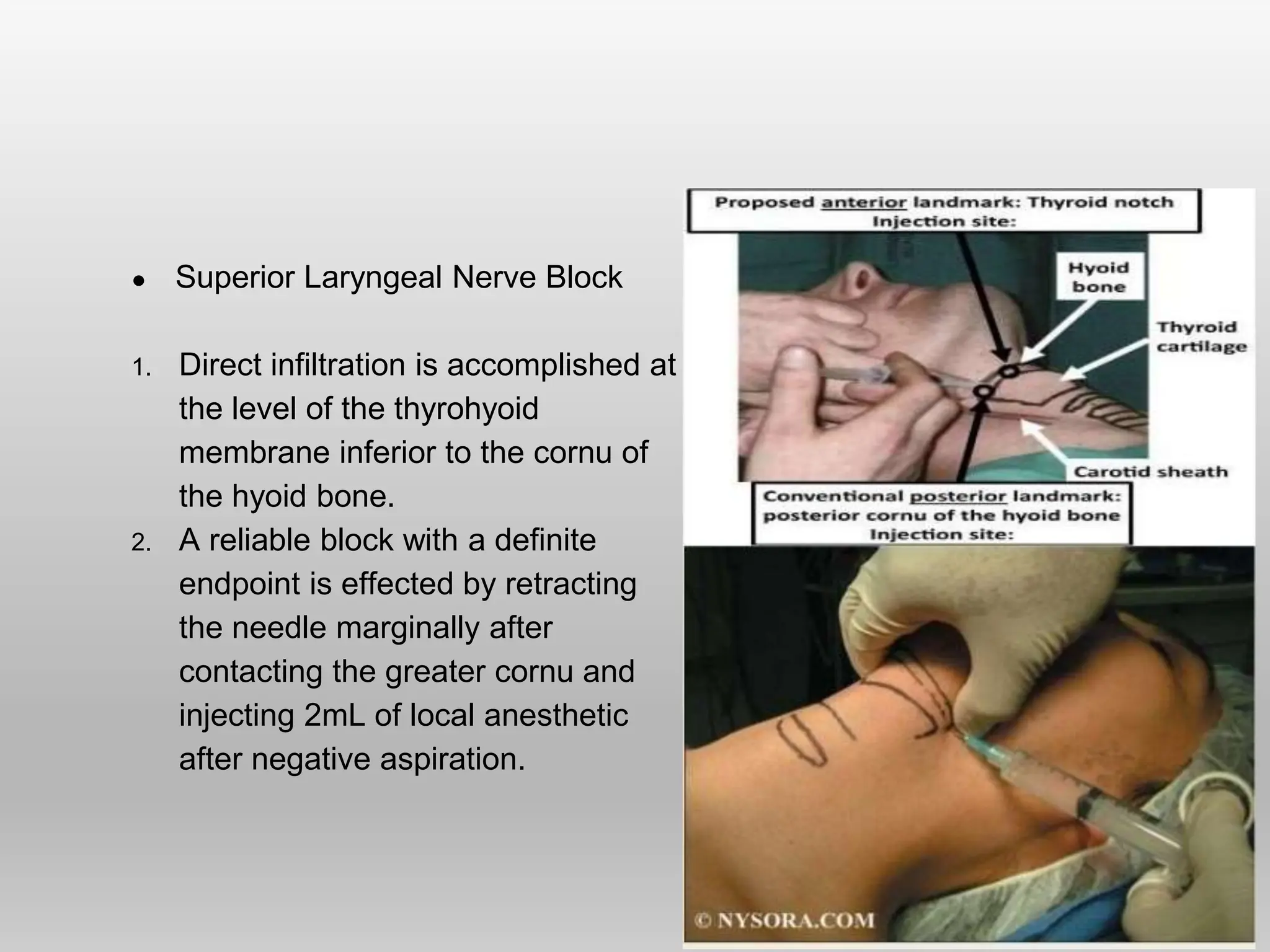
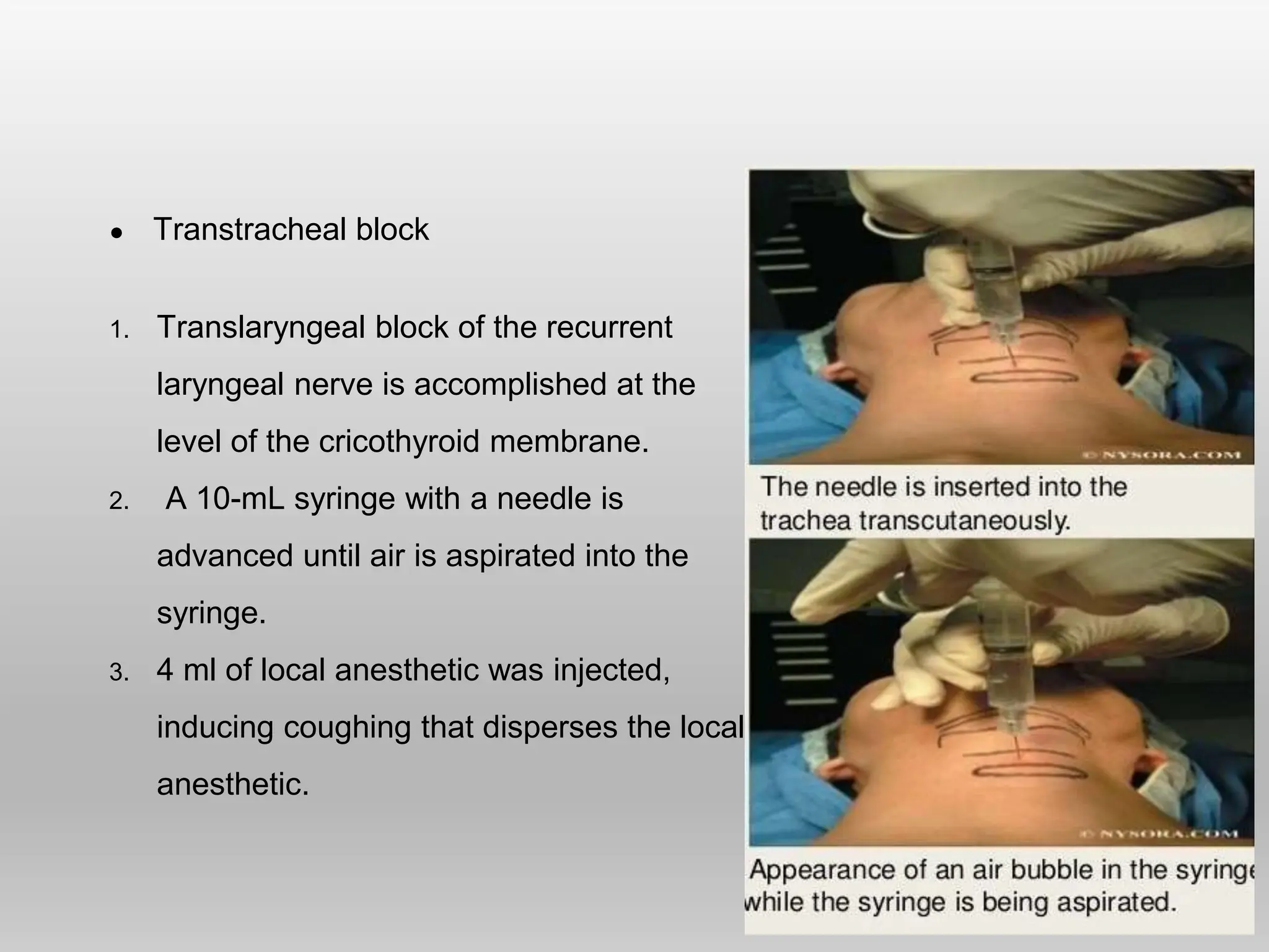
ANESTHESIA FOR
DENTAL PROCEDURES
Presenter : Dr . Jyotsna
Moderator : Dr . Rajeshwar Reddy
Dental anesthesia
Anesthesia started with dentistry
● Horace wells administered himself nitrous oxide – his colleague
extracted his tooth in 1844 .
● In 1846, morton did it again !!
Three types of anesthesia
● Out - patient anesthesia
● Day - care anesthesia
● In - patient anesthesia
Out - patient anesthesia
● Dental chair anesthesia
● Simple extraction of teeth (exodontia) especially in children.
● Conservative dentistry.
● Simple and short duration procedures.
● Incision and drinages.
Indications:-
● Children
● Anxious/ apprehensive patients
● Mentally retarded
● Failure of local anesthesia
Sedation for out patient dentistry: Conscious sedation, is a carefully
controlled technique in which a single intravenous drug or combination of
oxygen and nitrous oxide is used, but allows verbal contact with patients to be
maintained at all the time.
Problems in dental chair
● Venous return decrease
● Venous embolism
● Unprotected airway
● Aspiration of blood or mucus
● Adrenaline in local anesthetic can cause arrhythmias in presence of
halothane.
● Higher incidence of arrhythmias due to stimulation of 5th cranial
nerve.
● Fainting due to cerebral hypoxia.
● Difficulty in initiating CPR once cardiac arrest occurs.
● Foreign body obstruction of the airway by needles or dentures,
necessitating removal by bronchoscopy.
Day - care anesthesia
● Patient goes through formal admission to hospital but is discharged
home later in day .
● Usually adults
● Procedures - limited dental extraction, such as impacted
wisdom teeth
- minor oral surgical procedures
● In case of emergency if airway management is needed , naso-
tracheal intubation tray must be ready along with bite block and throat
pack.
In - patient anesthesia
● Impacted wisdom teeth where considerable surgery is anticipated.
● Extraction of 4 wisdom teeth together.
● Major oral and maxillofacial surgeries involving :-
- major orthognathic surgery for skeletal malocclusion.
- facial trauma & fractures .
Eg:- mandibular fractures & midfacial fractures
- radical cancer surgeries .
Eg:- maxillectomy & mandibulectomy
● Operative procedures require cutting through sensitive structures,
producing extreme discomfort and pain.
● Pain is a result of stimulation of nociceptors that are receptors
preferentially sensitive to a noxious stimulus (Aδ, C fiber afferent
axons)
● Local anesthetics (LA) cause : reversible block sensory nerve
conduction of noxious stimuli from periphery to the CNS.
Anesthesia techniques
● Local anesthetics
1. Topical infiltration - Small nerve endings in the small area of
soft tissue or bone are flooded with small amount of local
anesthetic solution
2. Regional nerve blocks - The local anesthetic solution is given
within close proximity to a main peripheral nerve.
3. Field blocks - Local anesthetic is deposited near a larger nerve
trunks.
● Conscious sedation
● General anesthesia
Local infiltration :-
● The effectiveness of local anesthetics is improved by the
addition of a vasoconstrictor:
Decrease absorption of local anesthetic into blood,
thus increasing duration of anesthesia &
decreasing toxicity.
 Generally performed by operating dentist
 Local anesthetics with or without adrenaline is used to perform
various techniques & nerve blocks
 These can be combined with conscious sedation.
Role:-
• Decrease intraoperative and postoperative pain.
• Decrease amount of general anesthetics used in the OR
• Increase patients cooperation
• Diagnostic testing/examination
Oral nerve blocks
Indications :
Dento-alveolar abscess
Tooth ache, pulpitis, or root impaction
Orofacial laceration repair
Post extraction pain, including dry socket
Dento-alveolar trauma or fractures
Maxillary & mandibular fractures
Maxillary Nerve Anesthesia
● Pulpal anesthesia: is given via the apical foramen to anesthetize the
pulp tissue.
● Periodontal: through the interdental and inter radicular branches
● Palatal: soft and hard tissues of the palatal periodontium (e.g. gingiva,
periodontal ligaments, alveolar bone)
● PSA block: recommended for maxillary molar teeth and associated
buccal tissues in ONE quadrant
● MSA block: recommended for maxillary premolars and associated buccal
tissues
● ASA block: recommended for maxillary canine and the incisors in ONE
quadrant
● Greater palatine block: recommended for palatal tissues distal to the
maxillary canine in ONE quadrant
● Nasopalatine block: recommended for palatal tissues between the right
and left maxillary canines
PSA Block
● Anesthetize the pulps and periodontal ligaments of the maxillary
molars, corresponding buccal alveolar bone and gingival tissue and
posterior portion of the maxillary sinus.
● Technique - between 1st and 2nd molar at a height of insertion of
mucobuccal fold, angle at 45° superiorly and medially.
MSA Block
● Can be indicated for surgery on
maxillary pre-molars and buccal root
of 1st molar.
● Target area: MSA nerve block is
given at the apex of the maxillary 2nd
premolar
- needle inserted into the
mucobuccal fold
ASA Block
● Used in conjunction with an MSA block
● The ASA nerve can cross the midline of the maxilla onto the opposite
side.
● Used in procedures involving the maxillary canines and incisors and
their associated facial tissues
● Anesthetize the pulp tissue + the gingiva, periodontal ligaments and
alveolar bone in that area.
● Target site : ASA nerve block is given at the apex of mucobuccal fold
of maxillary canine.
Greater Palatine Nerve Block
● Anesthetize all palatal mucosa of the side injected
and lingual gingiva posterior to the maxillary
canines and corresponding bone.
● Technique - on the hard palate between the 2nd &
3rd molars approximately 1cm medially.
Nasopalatine Nerve Block
● Anesthetize the anterior maxillary six teeth from
canine one side to canine on other side.
● Technique - Drug injected approximately 1.5 cm
posterior to the alveolar crest between the central
incisors.
Mandibular nerve Blocks
● Infiltration is not as successful as maxillary anesthesia substantial
variability in the anatomy of landmarks when compared to the maxilla
● Pulpal anesthesia
● Periodontal: through the interdental and inter radicular branches.
● Inferior alveolar block: for mandibular teeth + associated lingual tissues
and for the facial tissues anterior to the mandibular 1st molar.
● Buccal block: tissues of the mandibular molars
● Mental block: facial tissues anterior to the mental foramen (mandibular
premolars and anterior teeth)
● Incisive block: for teeth and facial tissue anterior to the mental foramen
● Gow-gates: most of the mandibular nerve for quadrant dentistry.
Inferior Alveolar Nerve Block
● For extractions and restorative lingual periodontal anesthesia, facial
periodontal anesthesia of anterior mandibular teeth and premolars.
 Target : Anesthetic solution is injected at the retromolar triangle which is
a triangular area located near to the distal side of the lower third molar.
Mental & Incisive Nerve Block :
● Nerves anesthetized: Mental & incisive nerves.
● Regions anesthetized: Lower lip, Mucosa anterior to mental foramen,
teeth anterior to second premolar.
Gow-Gates Nerve Block :
● It is an intraoral mandibular nerve block given at neck of condyle &
provides hard & soft tissue anesthesia of mandible upto the midline.
Mandibular nerve & its branches are blocked including its
auriculotemporal subdivision.
Local anesthetics complications
● Tachycardia, hence this should be avoided in patients at risk of
cardiovascular disease particularly when used with a vaso-constrictor.
● Post injection hyper-occlusion, pain and chewing soreness are other
symptoms reported.
● Allergic reaction:- More common with ester based local anesthetics.
● Most allergies are due to preservatives in local anesthetics.
○ Methylparaben
○ Sodium metabisulfite
Local anesthesia complications
 Needle breakage
 Pain on injection
 Burning on injection
 Nerve injury (Persistent anesthesia/parathesia)
 Trismus (Trauma to muscles)
 Hematoma
 Infection
 Edema
 Tissue sloughing
 Facial nerve paralysis (Intraglandular injections)
 LAST
Nerve Injury
● Paresthesia (loss of sensation) commonly involve the tongue and lower lip.
● Hyperesthesia (increased sensitivity to painful stimuli)
● Xerostomia (reduced salivation) - the chorda tympani is traumatized
● Ocular and extraocular symptoms : The passive diffusion of anesthetic
through the orbit leads to ocular and extraocular symptoms:
- paralysis extraocular muscle
- diplopia
- temporary blindness
- Horner´s syndrome (enophthalmos, miosis, palpebral ptosis)
LOCAL ANESTHETIC SYSTEMIC TOXICITY
• Adverse reactions proportional to plasma concentration.
• All system are affected but specially CNS & CVS.
• CNS TOXICITY :- LA produces stimulation followed by CNS depression as
inhibitory neurons are blocked first.
CLINICAL FEATURES:
• CNS excitation – agitation, confusion, twitching, seizures, convulsions
• CNS depression – drowsiness, coma, apnea.
• NON specific CNS- metallic taste, circumoral parathesia, tinnitus, dizziness
• CVS SIGN – initially – hypertension, tachycardia or hypotension or
bradycardia
• CVS hallmark- ventricular ectopic, multiform ventricular tachycardia,
ventricular fibrillation.
• progressive hypotension and bradycardia leading to Asystole and later to
cardiac arrest.
TREATMENT
● Early recognition
● Immediately stop LA administration
● Call for help
● Secure airway & 100% O2 supplement – intubate if required.
● Control seizures – benzodiazepines - inj. Midazolam 0.2mg/kg bolus
repeat after 5 min infusion 2mg/kg/hr.
● Shock – use IV fluid and vasopressin
● Ventricular arrhythmia – Inj amiodarone 150 mg over 10 minutes.
● INTRA-LIPID TREATMENT • Mechanism- increase clearance by
extraction of LA from cardiac tissue.
 Inj. 20% intralipid – 1.5ml/kg over 1 minutes (100ml)
 Infusion @ 0.25ml/kg/min ( 500ml over 30 mins) .Infuse for minimum
30 mins.
Conscious sedation
● A minimally depressed level of consciousness, that retains the patient’s
ability to maintain an airway independently & respond appropriately to
physical stimulation & verbal commands.
Indications:
 In anxious patient in combination with nerve blocks with local anesthesia.
 Complex dental work
 Those patients with movement disorders with physical/mental defects
unlikely allowing safe completion of treatment.
 For patients with severe gag reflex
 To avoid general anesthesia to avoid risks related to GA
● Medical conditions potentially aggravated by stress & affecting the patient’s
ability to cooperate.
Types :
 Inhalational sedation
 Intravenous sedation
 Oral sedation
General anesthesia
● It is required for three main groups of patients:
(i) Patient with learning difficulties
(ii) For extraction of permanent molars more complex or extensive
dental work
(iii) In-patient anesthesia for maxillofacial surgery (Congenital,
cosmetic, Traumatic and Neoplasm)
Conduct of anesthesia for facio-maxillary
surgeries
● Preoperative Assessment
All patients require an appropriate and adequate preoperative
assessment to define:
A. The extent of disease progression, in particular, looking for indicators
of potential airway compromise,
B. Anticipated difficulties in airway management, and
C. Significant comorbidities and selected pathological conditions that
influence airway management e.g cervical instability in trauma ,
D. Associated hemodynamics unstability.
E. Other concerns regarding trauma ,intraoperative blood loss ,post
operative airway problems(airway edema)
Airway management in Facio-maxillary surgery
● Choice of the airway managment technique depends on several factors:
Patients factors(Known difficult airway)
Experience of the anesthetist
Surgical requirements (nasal or oral )
Ultimately depends on the patient’s safety
● Physical Examination
Besides from general and systemic examination detailed airway assesment is
must.
● Airway strategies
 LMA
 Oral intubation
 Nasal intubation
 Retromolar intubation
 Submental intubation
 Retrograde intubation
 Fiberoptic aided intubation
 Trans-tracheal catheter and jet ventilation
 Planned tracheostomy
Nasal intubation
● Nasal intubation with a flexometallic ETT or a preformed nasal (north
facing) preferred
● Nasal patency should be checked
● Nasal passage is well prepared with a vasoconstrictor.
Contraindications
 Midface instability (Le fort II & III)
 Suspected basilar skull fracture
 CSF rhinorrhea
 Coagulopathy
Disadvantages : The presence of nasotracheal tube can interfere with the
surgical reconstruction of naso- orbital - ethmoid (NOE) complex.
● Postoperative ventilation or suctioning are difficult through a nasal tube
owing to its length and contour.
● Bleeding, sinusitis and pharyngeal abscess are occasional complications.
Retromolar intubation
● The retromolar space -space behind the last
erupted upper and lower molar teeth.
● The retromolar tube - stabilized in position by
fixation to 1st or 2nd molar tooth .
Advantages :
● Adequate space for intubation when mouth
opening restricted.
● Can be performed in complete mandibular
occlusion .
● Provides a nobel alternatives to tracheostomy.
Submento-tracheal intubation
● Provides a secure airway, optimal field, allows maxillo-mandibular fixation
while avoiding the drawbacks and complications of nasotracheal
intubation and tracheostomy.
● After induction of general anesthesia - orotracheal intubation is achieved
with an armoured tracheal tube (with a detachable connector).
● 1.5cm skin incision - made in the submental region just medial to the
lower border of mandible.
● Artery forceps - introduced through the submental incision towards the
floor of the mouth.
● Through the given incision the deflated pilot tube cuff along with the
tube is pulled out through the submental incision.
● Connector is reattached and ventilation is checked.
● At the end of the surgery, the tube is pulled back into the oral cavity
and extubated when patient is awake.
● Requires adequate mouth opening for the initial orotracheal
intubation.
Retrograde intubation
● Catheter-over-needle with attached syringe is inserted through
cricothyroid membrane in a cephalad direction.
● Entering the lower airway allows more space for advancing the tube.
● Free air aspiration confirms the location.
● Local anesthesia should be injected through the syringe. The needle
and syringe are removed, and a guide wire is inserted through the
catheter.
● Guide wire is advanced cephalad until it emerges at the oral cavity or
nares.
● Tracheal tube can be threaded over the guide wire, with either through
the Murphy eye or main lumen.
● The tracheal tube is advanced until it reaches the point where the guide
wire enters the airway.
● The guide wire is removed from above and the tracheal tube inserted to
a proper depth.
Fiberoptic Aided intubation
● Indications
 Anticipated difficult intubation
 Unanticipated difficult intubation
 Lower and upper airway obstructions
 Unstable or fixed cervical spine disease
● Contraindications
 Hypoxia
 Significant airway secretions not relieved with anti-sialagogues
and suction.
 Airway bleeding not relieved by suction.
 LA allergy
 Inability to cooperate
● Glosso-Pharyngeal Nerve Block : - It is most easily blocked where it
crosses the palatoglossal arch.
It can be blocked using one of three methods:
1. Topical spray application,
2. Direct mucosal contact of soaked pledgets,
3. Direct infiltration by injection.
● Superior Laryngeal Nerve Block
1. Direct infiltration is accomplished at
the level of the thyrohyoid
membrane inferior to the cornu of
the hyoid bone.
2. A reliable block with a definite
endpoint is effected by retracting
the needle marginally after
contacting the greater cornu and
injecting 2mL of local anesthetic
after negative aspiration.
● Transtracheal block
1. Translaryngeal block of the recurrent
laryngeal nerve is accomplished at the
level of the cricothyroid membrane.
2. A 10-mL syringe with a needle is
advanced until air is aspirated into the
syringe.
3. 4 ml of local anesthetic was injected,
inducing coughing that disperses the local
anesthetic.
● Monitoring and Equipment's
 Minimum ASA monitoring standards must be met.
 Etco2 :to confirm tracheal intubation , to detect disconnection ,detect
Venous air embolism.
 Urine output monitoring: in extensive surgery
 A full range of resuscitation equipment and drugs must be available.
 Difficult airway trolley should be checked and immediately available.
 Invasive lines : Neck dissection, Major prolonged surgery.
Intraoperative concerns and Management
1. Eye Protection : Pads, shields or surgical covers.
2. Remote location in relation to airway :
 Adequate fixation of the airway and circuit connections
 For north-facing tubes or LMAs,- gauze padding on the forehead under
nasal tubes.
 South-facing airways-taped securely away from the surgical site.
 Tube holders to stabilize the circuit.
3. Airway edema : Injection dexamethasone
4. Positioning :
 Once drapes are applied to the patient, head is supported and stabilized
in a horseshoe support or head ring, with a bolster under shoulders to
extend the neck.
 Pressure points protected and vascular access points made accessible.
5. Anti-emetics :
 Prophylaxis of PONV :- 5 HT3 Blockers- Ondansetron
6. Lower airway needs protection against soiling from blood and debris intra
operatively - throat packing is done.
7. Substantial blood loss :
 Extensive blood supply to mid-face (maxillary artery/Pterygoid venous
plexus)
 Methods to decrease : Slight head-up positioning
Controlled hypotension(Hypotensive anesthesia)
Local infiltration with adrenaline
Tranexamic acid .
Emergence and recovery
● Antagonizing neuromuscular blocking agents.
● 100% oxygen administered and any anesthetic agents discontinued.
● Pharyngeal packs should be removed and the pharynx cleared of
secretions and debris by suction.
● The patient should be turned into the lateral position, possibly with a
degree of head- down tilt to encourage the drainage of any blood or
secretions away from the larynx.
● Possibility of post-operative tissue edema - Careful assessment of
tongue and pharynx.
● Cuff leak test.
Postoperative Management
● Supplemental oxygenation .
● Adequate pain control with opioids, NSAIDS, acetaminophen.
● PONV issues treated with antiemetic.
● Airway problems continuously monitored-for airway edema ,hematoma
compressing neck in case of surgery involving neck.
● Airway compromised state-continue intubation and mechanical
ventilation postoperatively.
● Ongoing/extensive bleeding may need to be replaced.
● Fluid and electrolyte supplementation.
● Nutrition supplement.
Thank you

dentalDocument from Aditya.pptx Islam Irish

  • 1.
    ANESTHESIA FOR DENTAL PROCEDURES Presenter: Dr . Jyotsna Moderator : Dr . Rajeshwar Reddy
  • 2.
    Dental anesthesia Anesthesia startedwith dentistry ● Horace wells administered himself nitrous oxide – his colleague extracted his tooth in 1844 . ● In 1846, morton did it again !! Three types of anesthesia ● Out - patient anesthesia ● Day - care anesthesia ● In - patient anesthesia
  • 3.
    Out - patientanesthesia ● Dental chair anesthesia ● Simple extraction of teeth (exodontia) especially in children. ● Conservative dentistry. ● Simple and short duration procedures. ● Incision and drinages. Indications:- ● Children ● Anxious/ apprehensive patients ● Mentally retarded ● Failure of local anesthesia Sedation for out patient dentistry: Conscious sedation, is a carefully controlled technique in which a single intravenous drug or combination of oxygen and nitrous oxide is used, but allows verbal contact with patients to be maintained at all the time.
  • 4.
    Problems in dentalchair ● Venous return decrease ● Venous embolism ● Unprotected airway ● Aspiration of blood or mucus ● Adrenaline in local anesthetic can cause arrhythmias in presence of halothane. ● Higher incidence of arrhythmias due to stimulation of 5th cranial nerve. ● Fainting due to cerebral hypoxia. ● Difficulty in initiating CPR once cardiac arrest occurs. ● Foreign body obstruction of the airway by needles or dentures, necessitating removal by bronchoscopy.
  • 5.
    Day - careanesthesia ● Patient goes through formal admission to hospital but is discharged home later in day . ● Usually adults ● Procedures - limited dental extraction, such as impacted wisdom teeth - minor oral surgical procedures ● In case of emergency if airway management is needed , naso- tracheal intubation tray must be ready along with bite block and throat pack.
  • 6.
    In - patientanesthesia ● Impacted wisdom teeth where considerable surgery is anticipated. ● Extraction of 4 wisdom teeth together. ● Major oral and maxillofacial surgeries involving :- - major orthognathic surgery for skeletal malocclusion. - facial trauma & fractures . Eg:- mandibular fractures & midfacial fractures - radical cancer surgeries . Eg:- maxillectomy & mandibulectomy
  • 7.
    ● Operative proceduresrequire cutting through sensitive structures, producing extreme discomfort and pain. ● Pain is a result of stimulation of nociceptors that are receptors preferentially sensitive to a noxious stimulus (Aδ, C fiber afferent axons) ● Local anesthetics (LA) cause : reversible block sensory nerve conduction of noxious stimuli from periphery to the CNS.
  • 8.
    Anesthesia techniques ● Localanesthetics 1. Topical infiltration - Small nerve endings in the small area of soft tissue or bone are flooded with small amount of local anesthetic solution 2. Regional nerve blocks - The local anesthetic solution is given within close proximity to a main peripheral nerve. 3. Field blocks - Local anesthetic is deposited near a larger nerve trunks. ● Conscious sedation ● General anesthesia
  • 9.
    Local infiltration :- ●The effectiveness of local anesthetics is improved by the addition of a vasoconstrictor: Decrease absorption of local anesthetic into blood, thus increasing duration of anesthesia & decreasing toxicity.  Generally performed by operating dentist  Local anesthetics with or without adrenaline is used to perform various techniques & nerve blocks  These can be combined with conscious sedation. Role:- • Decrease intraoperative and postoperative pain. • Decrease amount of general anesthetics used in the OR • Increase patients cooperation • Diagnostic testing/examination
  • 10.
    Oral nerve blocks Indications: Dento-alveolar abscess Tooth ache, pulpitis, or root impaction Orofacial laceration repair Post extraction pain, including dry socket Dento-alveolar trauma or fractures Maxillary & mandibular fractures
  • 12.
    Maxillary Nerve Anesthesia ●Pulpal anesthesia: is given via the apical foramen to anesthetize the pulp tissue. ● Periodontal: through the interdental and inter radicular branches ● Palatal: soft and hard tissues of the palatal periodontium (e.g. gingiva, periodontal ligaments, alveolar bone) ● PSA block: recommended for maxillary molar teeth and associated buccal tissues in ONE quadrant ● MSA block: recommended for maxillary premolars and associated buccal tissues ● ASA block: recommended for maxillary canine and the incisors in ONE quadrant ● Greater palatine block: recommended for palatal tissues distal to the maxillary canine in ONE quadrant ● Nasopalatine block: recommended for palatal tissues between the right and left maxillary canines
  • 13.
    PSA Block ● Anesthetizethe pulps and periodontal ligaments of the maxillary molars, corresponding buccal alveolar bone and gingival tissue and posterior portion of the maxillary sinus. ● Technique - between 1st and 2nd molar at a height of insertion of mucobuccal fold, angle at 45° superiorly and medially.
  • 14.
    MSA Block ● Canbe indicated for surgery on maxillary pre-molars and buccal root of 1st molar. ● Target area: MSA nerve block is given at the apex of the maxillary 2nd premolar - needle inserted into the mucobuccal fold
  • 15.
    ASA Block ● Usedin conjunction with an MSA block ● The ASA nerve can cross the midline of the maxilla onto the opposite side. ● Used in procedures involving the maxillary canines and incisors and their associated facial tissues ● Anesthetize the pulp tissue + the gingiva, periodontal ligaments and alveolar bone in that area. ● Target site : ASA nerve block is given at the apex of mucobuccal fold of maxillary canine.
  • 16.
    Greater Palatine NerveBlock ● Anesthetize all palatal mucosa of the side injected and lingual gingiva posterior to the maxillary canines and corresponding bone. ● Technique - on the hard palate between the 2nd & 3rd molars approximately 1cm medially. Nasopalatine Nerve Block ● Anesthetize the anterior maxillary six teeth from canine one side to canine on other side. ● Technique - Drug injected approximately 1.5 cm posterior to the alveolar crest between the central incisors.
  • 17.
    Mandibular nerve Blocks ●Infiltration is not as successful as maxillary anesthesia substantial variability in the anatomy of landmarks when compared to the maxilla ● Pulpal anesthesia ● Periodontal: through the interdental and inter radicular branches. ● Inferior alveolar block: for mandibular teeth + associated lingual tissues and for the facial tissues anterior to the mandibular 1st molar. ● Buccal block: tissues of the mandibular molars ● Mental block: facial tissues anterior to the mental foramen (mandibular premolars and anterior teeth) ● Incisive block: for teeth and facial tissue anterior to the mental foramen ● Gow-gates: most of the mandibular nerve for quadrant dentistry.
  • 18.
    Inferior Alveolar NerveBlock ● For extractions and restorative lingual periodontal anesthesia, facial periodontal anesthesia of anterior mandibular teeth and premolars.  Target : Anesthetic solution is injected at the retromolar triangle which is a triangular area located near to the distal side of the lower third molar.
  • 19.
    Mental & IncisiveNerve Block : ● Nerves anesthetized: Mental & incisive nerves. ● Regions anesthetized: Lower lip, Mucosa anterior to mental foramen, teeth anterior to second premolar. Gow-Gates Nerve Block : ● It is an intraoral mandibular nerve block given at neck of condyle & provides hard & soft tissue anesthesia of mandible upto the midline. Mandibular nerve & its branches are blocked including its auriculotemporal subdivision.
  • 20.
    Local anesthetics complications ●Tachycardia, hence this should be avoided in patients at risk of cardiovascular disease particularly when used with a vaso-constrictor. ● Post injection hyper-occlusion, pain and chewing soreness are other symptoms reported. ● Allergic reaction:- More common with ester based local anesthetics. ● Most allergies are due to preservatives in local anesthetics. ○ Methylparaben ○ Sodium metabisulfite
  • 21.
    Local anesthesia complications Needle breakage  Pain on injection  Burning on injection  Nerve injury (Persistent anesthesia/parathesia)  Trismus (Trauma to muscles)  Hematoma  Infection  Edema  Tissue sloughing  Facial nerve paralysis (Intraglandular injections)  LAST
  • 22.
    Nerve Injury ● Paresthesia(loss of sensation) commonly involve the tongue and lower lip. ● Hyperesthesia (increased sensitivity to painful stimuli) ● Xerostomia (reduced salivation) - the chorda tympani is traumatized ● Ocular and extraocular symptoms : The passive diffusion of anesthetic through the orbit leads to ocular and extraocular symptoms: - paralysis extraocular muscle - diplopia - temporary blindness - Horner´s syndrome (enophthalmos, miosis, palpebral ptosis)
  • 23.
    LOCAL ANESTHETIC SYSTEMICTOXICITY • Adverse reactions proportional to plasma concentration. • All system are affected but specially CNS & CVS. • CNS TOXICITY :- LA produces stimulation followed by CNS depression as inhibitory neurons are blocked first. CLINICAL FEATURES: • CNS excitation – agitation, confusion, twitching, seizures, convulsions • CNS depression – drowsiness, coma, apnea. • NON specific CNS- metallic taste, circumoral parathesia, tinnitus, dizziness • CVS SIGN – initially – hypertension, tachycardia or hypotension or bradycardia • CVS hallmark- ventricular ectopic, multiform ventricular tachycardia, ventricular fibrillation. • progressive hypotension and bradycardia leading to Asystole and later to cardiac arrest.
  • 24.
    TREATMENT ● Early recognition ●Immediately stop LA administration ● Call for help ● Secure airway & 100% O2 supplement – intubate if required. ● Control seizures – benzodiazepines - inj. Midazolam 0.2mg/kg bolus repeat after 5 min infusion 2mg/kg/hr. ● Shock – use IV fluid and vasopressin ● Ventricular arrhythmia – Inj amiodarone 150 mg over 10 minutes. ● INTRA-LIPID TREATMENT • Mechanism- increase clearance by extraction of LA from cardiac tissue.  Inj. 20% intralipid – 1.5ml/kg over 1 minutes (100ml)  Infusion @ 0.25ml/kg/min ( 500ml over 30 mins) .Infuse for minimum 30 mins.
  • 25.
    Conscious sedation ● Aminimally depressed level of consciousness, that retains the patient’s ability to maintain an airway independently & respond appropriately to physical stimulation & verbal commands. Indications:  In anxious patient in combination with nerve blocks with local anesthesia.  Complex dental work  Those patients with movement disorders with physical/mental defects unlikely allowing safe completion of treatment.  For patients with severe gag reflex  To avoid general anesthesia to avoid risks related to GA ● Medical conditions potentially aggravated by stress & affecting the patient’s ability to cooperate. Types :  Inhalational sedation  Intravenous sedation  Oral sedation
  • 26.
    General anesthesia ● Itis required for three main groups of patients: (i) Patient with learning difficulties (ii) For extraction of permanent molars more complex or extensive dental work (iii) In-patient anesthesia for maxillofacial surgery (Congenital, cosmetic, Traumatic and Neoplasm)
  • 27.
    Conduct of anesthesiafor facio-maxillary surgeries ● Preoperative Assessment All patients require an appropriate and adequate preoperative assessment to define: A. The extent of disease progression, in particular, looking for indicators of potential airway compromise, B. Anticipated difficulties in airway management, and C. Significant comorbidities and selected pathological conditions that influence airway management e.g cervical instability in trauma , D. Associated hemodynamics unstability. E. Other concerns regarding trauma ,intraoperative blood loss ,post operative airway problems(airway edema)
  • 28.
    Airway management inFacio-maxillary surgery ● Choice of the airway managment technique depends on several factors: Patients factors(Known difficult airway) Experience of the anesthetist Surgical requirements (nasal or oral ) Ultimately depends on the patient’s safety
  • 29.
    ● Physical Examination Besidesfrom general and systemic examination detailed airway assesment is must.
  • 30.
    ● Airway strategies LMA  Oral intubation  Nasal intubation  Retromolar intubation  Submental intubation  Retrograde intubation  Fiberoptic aided intubation  Trans-tracheal catheter and jet ventilation  Planned tracheostomy
  • 31.
    Nasal intubation ● Nasalintubation with a flexometallic ETT or a preformed nasal (north facing) preferred ● Nasal patency should be checked ● Nasal passage is well prepared with a vasoconstrictor. Contraindications  Midface instability (Le fort II & III)  Suspected basilar skull fracture  CSF rhinorrhea  Coagulopathy Disadvantages : The presence of nasotracheal tube can interfere with the surgical reconstruction of naso- orbital - ethmoid (NOE) complex. ● Postoperative ventilation or suctioning are difficult through a nasal tube owing to its length and contour. ● Bleeding, sinusitis and pharyngeal abscess are occasional complications.
  • 32.
    Retromolar intubation ● Theretromolar space -space behind the last erupted upper and lower molar teeth. ● The retromolar tube - stabilized in position by fixation to 1st or 2nd molar tooth . Advantages : ● Adequate space for intubation when mouth opening restricted. ● Can be performed in complete mandibular occlusion . ● Provides a nobel alternatives to tracheostomy.
  • 33.
    Submento-tracheal intubation ● Providesa secure airway, optimal field, allows maxillo-mandibular fixation while avoiding the drawbacks and complications of nasotracheal intubation and tracheostomy. ● After induction of general anesthesia - orotracheal intubation is achieved with an armoured tracheal tube (with a detachable connector). ● 1.5cm skin incision - made in the submental region just medial to the lower border of mandible. ● Artery forceps - introduced through the submental incision towards the floor of the mouth.
  • 34.
    ● Through thegiven incision the deflated pilot tube cuff along with the tube is pulled out through the submental incision. ● Connector is reattached and ventilation is checked. ● At the end of the surgery, the tube is pulled back into the oral cavity and extubated when patient is awake. ● Requires adequate mouth opening for the initial orotracheal intubation.
  • 35.
    Retrograde intubation ● Catheter-over-needlewith attached syringe is inserted through cricothyroid membrane in a cephalad direction. ● Entering the lower airway allows more space for advancing the tube. ● Free air aspiration confirms the location. ● Local anesthesia should be injected through the syringe. The needle and syringe are removed, and a guide wire is inserted through the catheter. ● Guide wire is advanced cephalad until it emerges at the oral cavity or nares. ● Tracheal tube can be threaded over the guide wire, with either through the Murphy eye or main lumen. ● The tracheal tube is advanced until it reaches the point where the guide wire enters the airway. ● The guide wire is removed from above and the tracheal tube inserted to a proper depth.
  • 38.
    Fiberoptic Aided intubation ●Indications  Anticipated difficult intubation  Unanticipated difficult intubation  Lower and upper airway obstructions  Unstable or fixed cervical spine disease ● Contraindications  Hypoxia  Significant airway secretions not relieved with anti-sialagogues and suction.  Airway bleeding not relieved by suction.  LA allergy  Inability to cooperate
  • 39.
    ● Glosso-Pharyngeal NerveBlock : - It is most easily blocked where it crosses the palatoglossal arch. It can be blocked using one of three methods: 1. Topical spray application, 2. Direct mucosal contact of soaked pledgets, 3. Direct infiltration by injection.
  • 40.
    ● Superior LaryngealNerve Block 1. Direct infiltration is accomplished at the level of the thyrohyoid membrane inferior to the cornu of the hyoid bone. 2. A reliable block with a definite endpoint is effected by retracting the needle marginally after contacting the greater cornu and injecting 2mL of local anesthetic after negative aspiration.
  • 41.
    ● Transtracheal block 1.Translaryngeal block of the recurrent laryngeal nerve is accomplished at the level of the cricothyroid membrane. 2. A 10-mL syringe with a needle is advanced until air is aspirated into the syringe. 3. 4 ml of local anesthetic was injected, inducing coughing that disperses the local anesthetic.
  • 42.
    ● Monitoring andEquipment's  Minimum ASA monitoring standards must be met.  Etco2 :to confirm tracheal intubation , to detect disconnection ,detect Venous air embolism.  Urine output monitoring: in extensive surgery  A full range of resuscitation equipment and drugs must be available.  Difficult airway trolley should be checked and immediately available.  Invasive lines : Neck dissection, Major prolonged surgery.
  • 43.
    Intraoperative concerns andManagement 1. Eye Protection : Pads, shields or surgical covers. 2. Remote location in relation to airway :  Adequate fixation of the airway and circuit connections  For north-facing tubes or LMAs,- gauze padding on the forehead under nasal tubes.  South-facing airways-taped securely away from the surgical site.  Tube holders to stabilize the circuit. 3. Airway edema : Injection dexamethasone 4. Positioning :  Once drapes are applied to the patient, head is supported and stabilized in a horseshoe support or head ring, with a bolster under shoulders to extend the neck.  Pressure points protected and vascular access points made accessible.
  • 44.
    5. Anti-emetics : Prophylaxis of PONV :- 5 HT3 Blockers- Ondansetron 6. Lower airway needs protection against soiling from blood and debris intra operatively - throat packing is done. 7. Substantial blood loss :  Extensive blood supply to mid-face (maxillary artery/Pterygoid venous plexus)  Methods to decrease : Slight head-up positioning Controlled hypotension(Hypotensive anesthesia) Local infiltration with adrenaline Tranexamic acid .
  • 45.
    Emergence and recovery ●Antagonizing neuromuscular blocking agents. ● 100% oxygen administered and any anesthetic agents discontinued. ● Pharyngeal packs should be removed and the pharynx cleared of secretions and debris by suction. ● The patient should be turned into the lateral position, possibly with a degree of head- down tilt to encourage the drainage of any blood or secretions away from the larynx. ● Possibility of post-operative tissue edema - Careful assessment of tongue and pharynx. ● Cuff leak test.
  • 46.
    Postoperative Management ● Supplementaloxygenation . ● Adequate pain control with opioids, NSAIDS, acetaminophen. ● PONV issues treated with antiemetic. ● Airway problems continuously monitored-for airway edema ,hematoma compressing neck in case of surgery involving neck. ● Airway compromised state-continue intubation and mechanical ventilation postoperatively. ● Ongoing/extensive bleeding may need to be replaced. ● Fluid and electrolyte supplementation. ● Nutrition supplement.
  • 47.