INTRODUCTION
DEFINITION
EVENTS OF DEVELOPMENT
EMBRYOLOGICAL DEVELOPMENT
DEVELOPMENT OF BRANCHIAL (ARCHES ,
POUCHES AND CLEFTS)
DEVELOPMENT OF FACE
DEVELOPMENTAL ANOMALIES OF FACE
CONCLUSION
REFERENCES
CONTENTS
3.
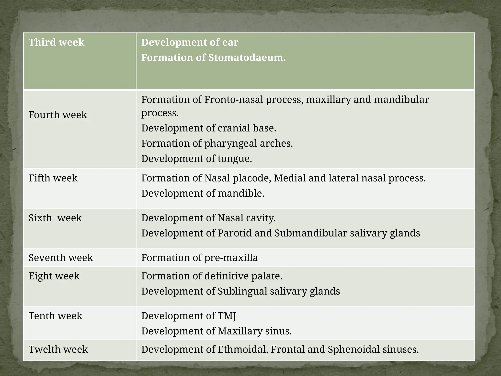
Third week Developmentof ear
Formation of Stomatodaeum.
Fourth week
Formation of Fronto-nasal process, maxillary and mandibular
process.
Development of cranial base.
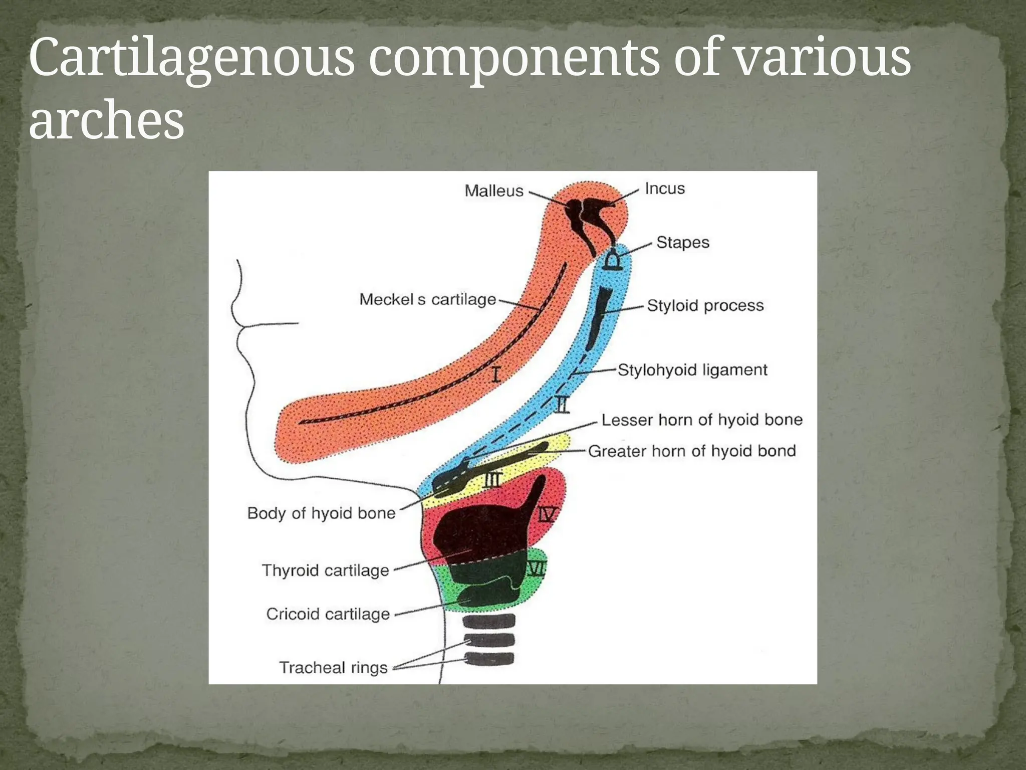
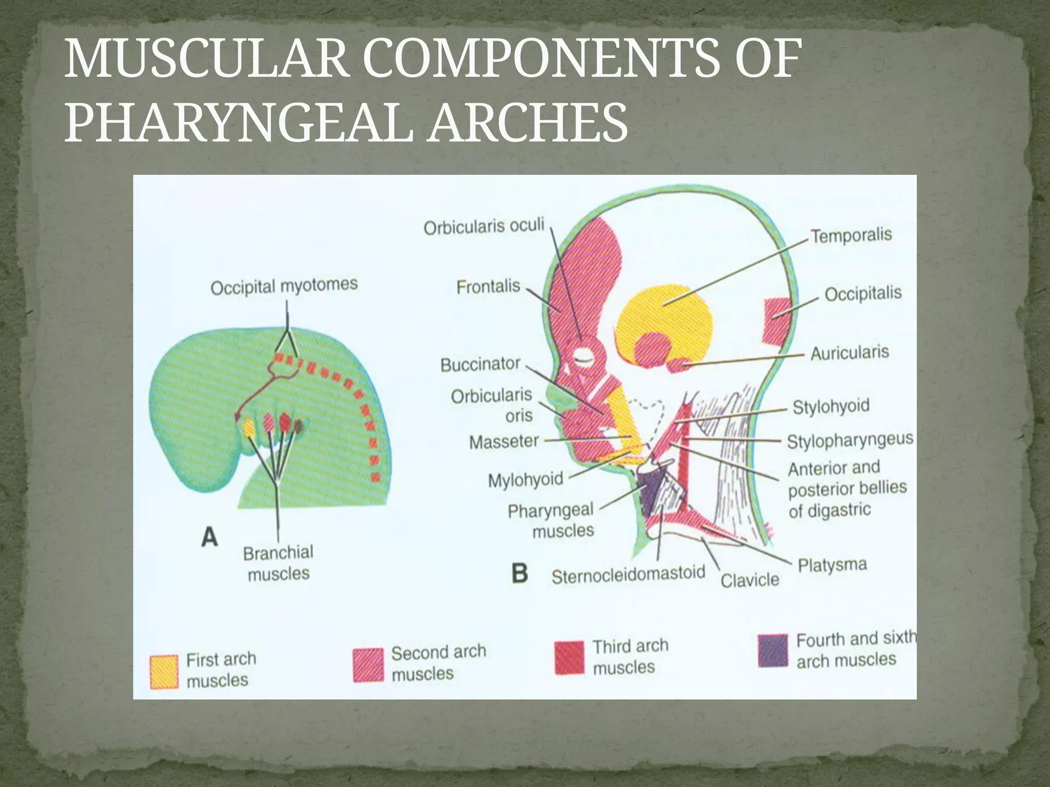
Formation of pharyngeal arches.
Development of tongue.
Fifth week Formation of Nasal placode, Medial and lateral nasal process.
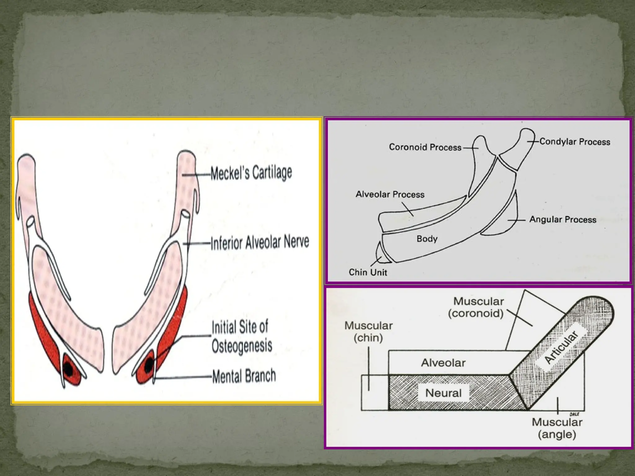
Development of mandible.
Sixth week Development of Nasal cavity.
Development of Parotid and Submandibular salivary glands
Seventh week Formation of pre-maxilla
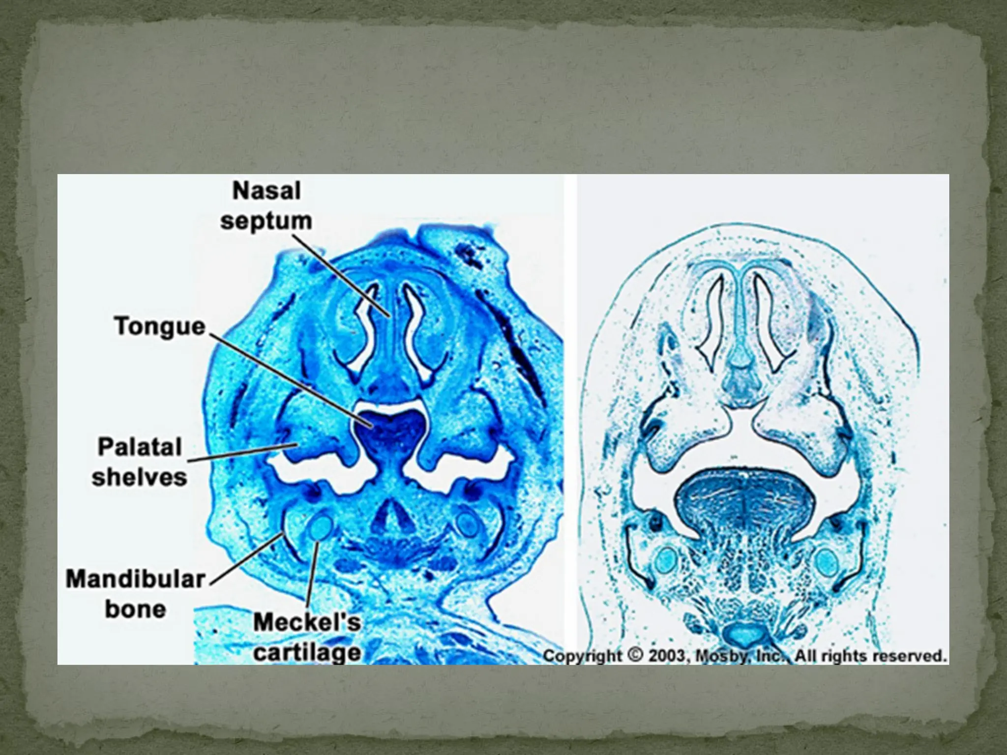
Eight week Formation of definitive palate.
Development of Sublingual salivary glands
Tenth week Development of TMJ
Development of Maxillary sinus.
Twelth week Development of Ethmoidal, Frontal and Sphenoidal sinuses.
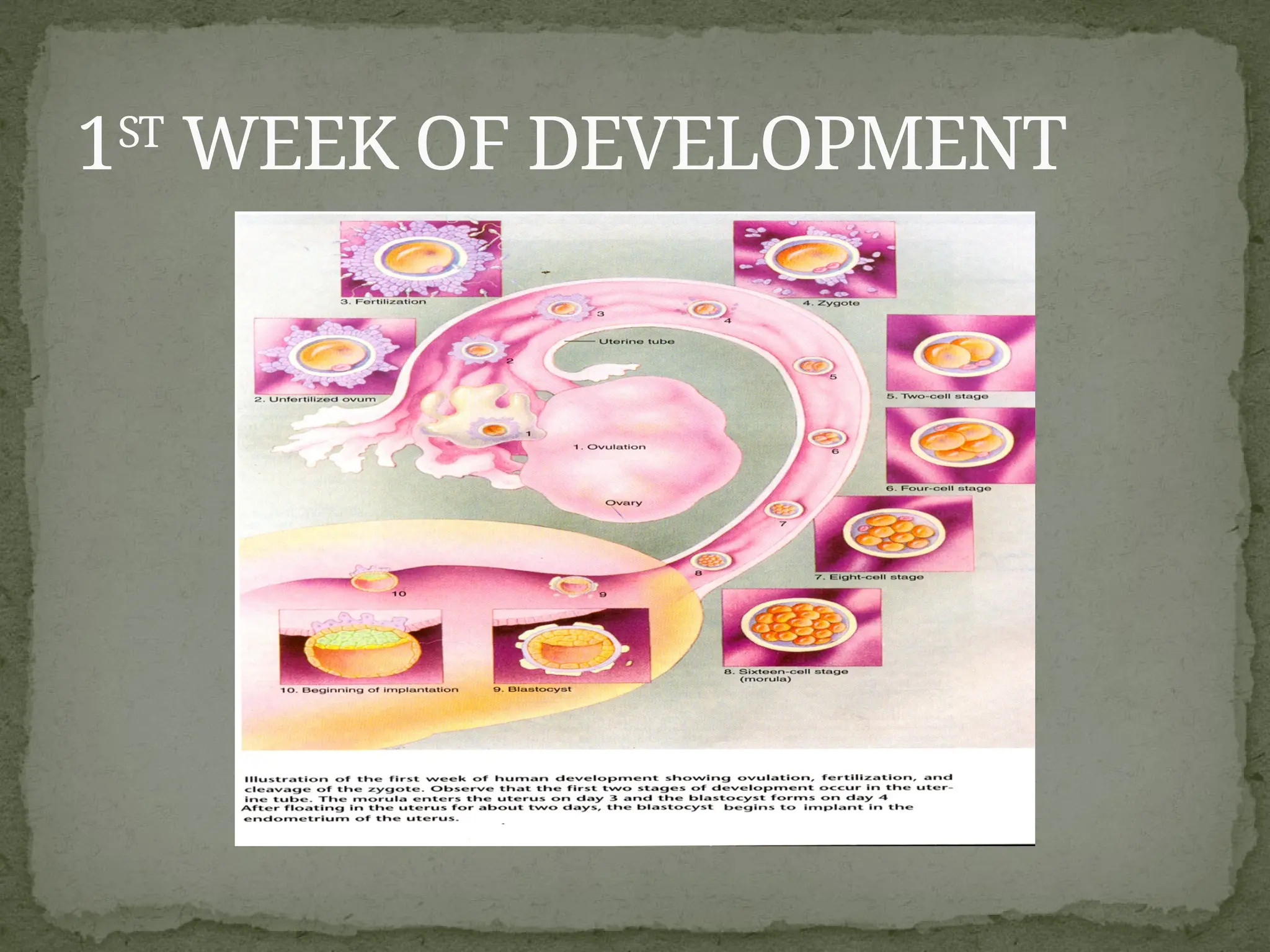
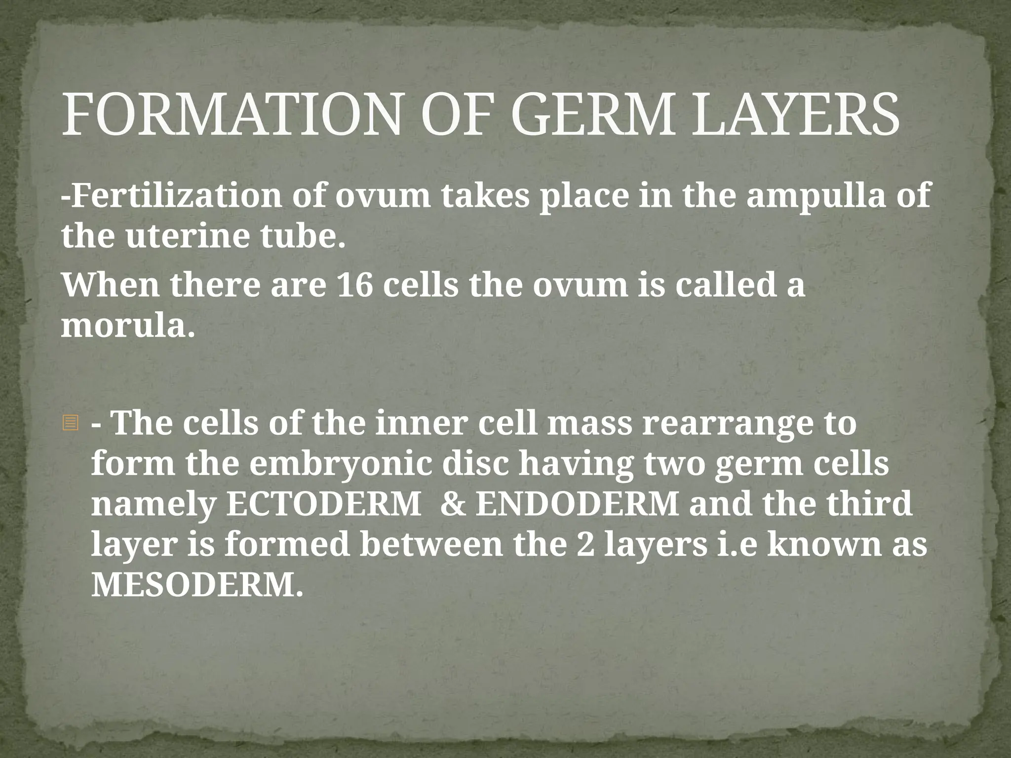
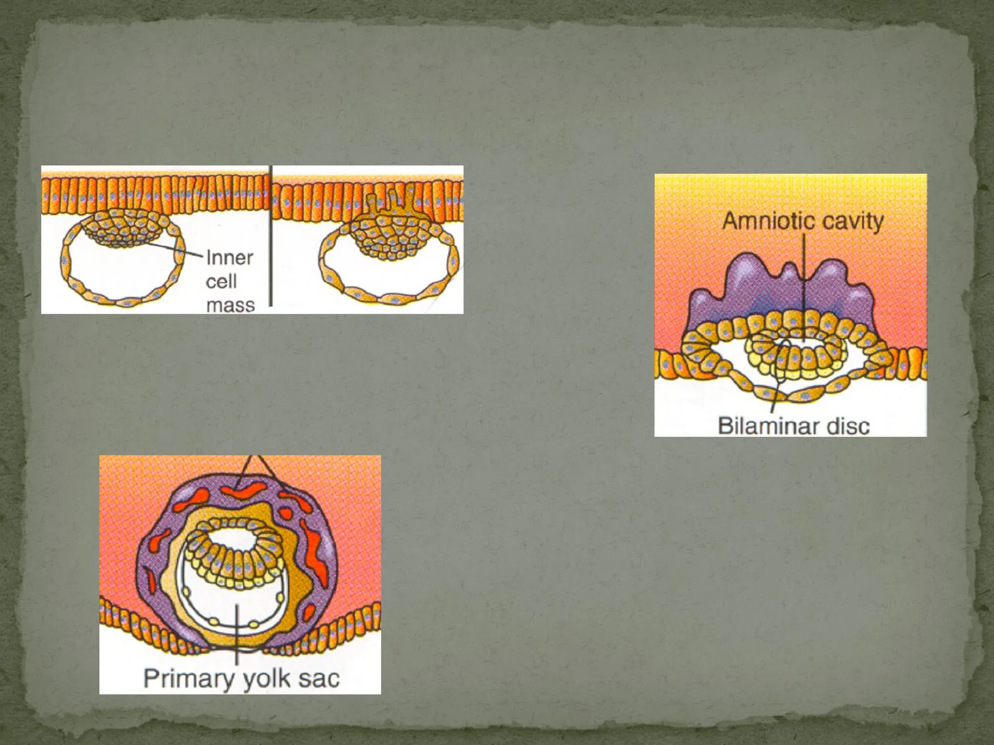
-Fertilization of ovumtakes place in the ampulla of
the uterine tube.
When there are 16 cells the ovum is called a
morula.
- - The cells of the inner cell mass rearrange to
form the embryonic disc having two germ cells
namely ECTODERM & ENDODERM and the third
layer is formed between the 2 layers i.e known as
MESODERM.
FORMATION OF GERM LAYERS
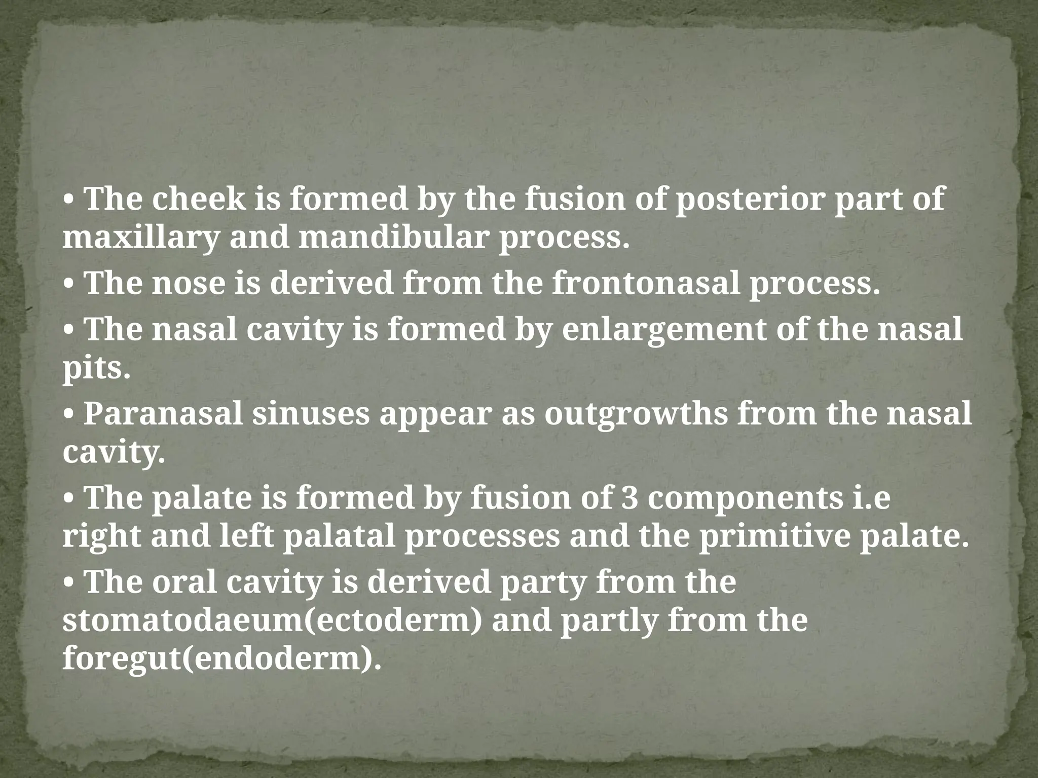
• The cheekis formed by the fusion of posterior part of
maxillary and mandibular process.
• The nose is derived from the frontonasal process.
• The nasal cavity is formed by enlargement of the nasal
pits.
• Paranasal sinuses appear as outgrowths from the nasal
cavity.
• The palate is formed by fusion of 3 components i.e
right and left palatal processes and the primitive palate.
• The oral cavity is derived party from the
stomatodaeum(ectoderm) and partly from the
foregut(endoderm).
14.
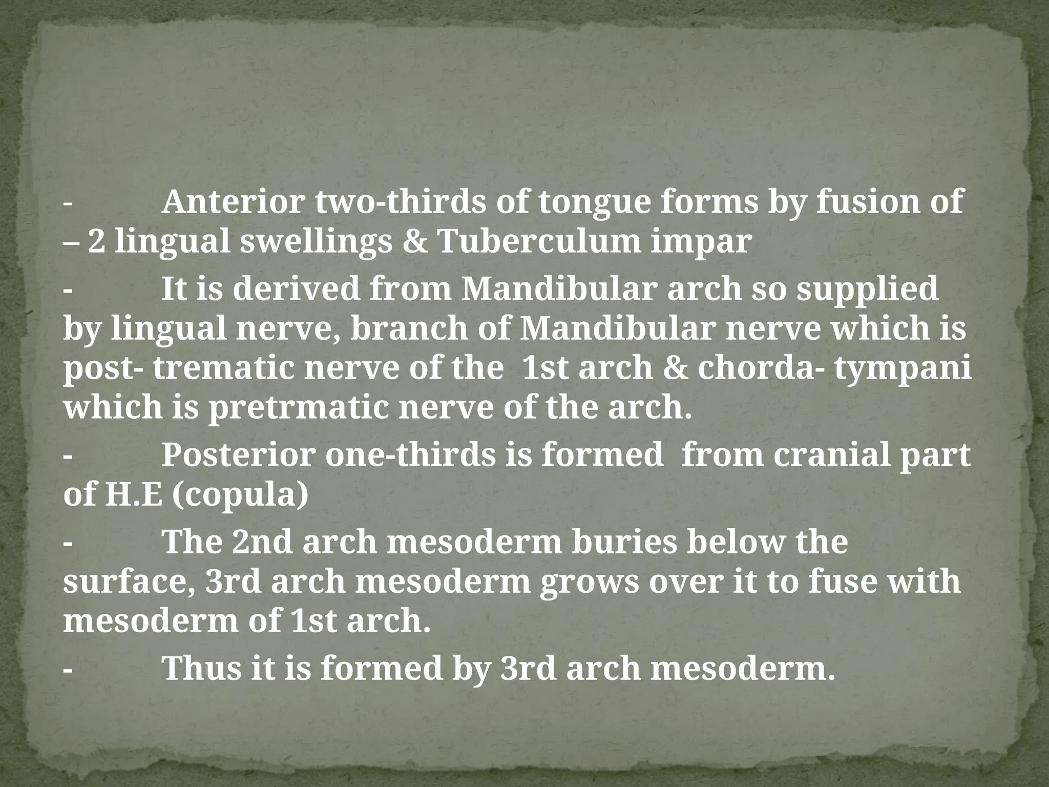
- Anterior two-thirdsof tongue forms by fusion of
– 2 lingual swellings & Tuberculum impar
- It is derived from Mandibular arch so supplied
by lingual nerve, branch of Mandibular nerve which is
post- trematic nerve of the 1st arch & chorda- tympani
which is pretrmatic nerve of the arch.
- Posterior one-thirds is formed from cranial part
of H.E (copula)
- The 2nd arch mesoderm buries below the
surface, 3rd arch mesoderm grows over it to fuse with
mesoderm of 1st arch.
- Thus it is formed by 3rd arch mesoderm.
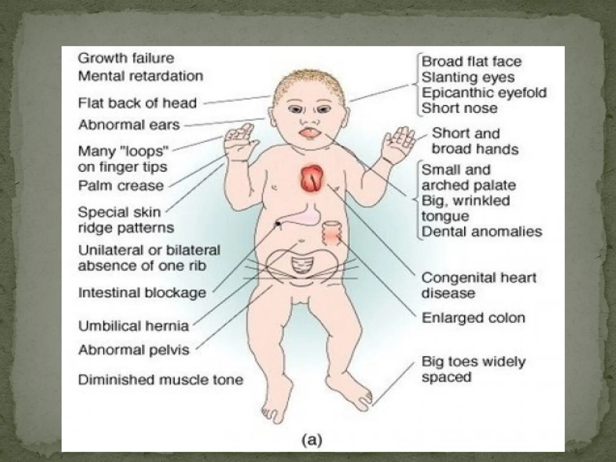
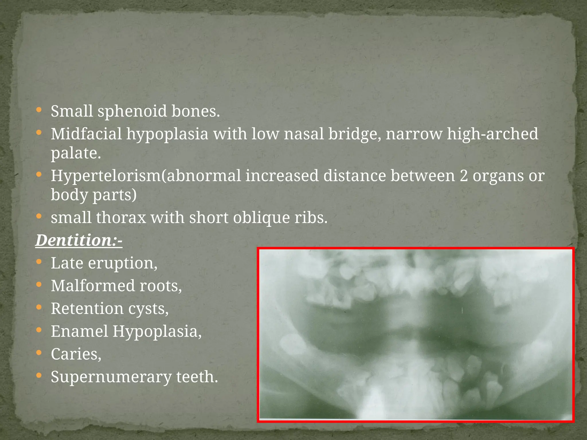
Small sphenoidbones.
Midfacial hypoplasia with low nasal bridge, narrow high-arched
palate.
Hypertelorism(abnormal increased distance between 2 organs or
body parts)
small thorax with short oblique ribs.
Dentition:-
Late eruption,
Malformed roots,
Retention cysts,
Enamel Hypoplasia,
Caries,
Supernumerary teeth.
28.
Atresia (is acondition in which a body orifice or passage
in the body is abnormally closed or absent) of the cavity at
the external nares, at the posterior nasal aperture or in
the cavity
This may be unilateral or bilateral
Congenital defect in the cribriform plate of ethmoid
bone may lead to communication between cranial cavity
and nose
Nasal septum may not be in midline i.e., deflected to
one side
Septum may be absent
Nasal cavity may communicate with the mouth
DEVELOPMENTAL DEFECTS OF
NASAL CAVITIES
29.
Illegal drugs
Marijuana:
prenatal exposureto marijuana leads to infants
reduced weight and size, short term changes in
behaviour e.g. increased startle and a high pitched cry.
Cocaine:
effect of maternal cocaine use - children tend to be
impulsive, highly distractable and difficult to control
and to have problems in language development as
they grow old.
30.
The human faceis a fascinating study of
physiology and psychology. The amount of
information a human face can relay is unending.
Humans are capable of making 10,000 unique
facial expressions! While the face is complicated,
it is also our most useful and most underestimated
tool for communication.
CONCLUSION : -
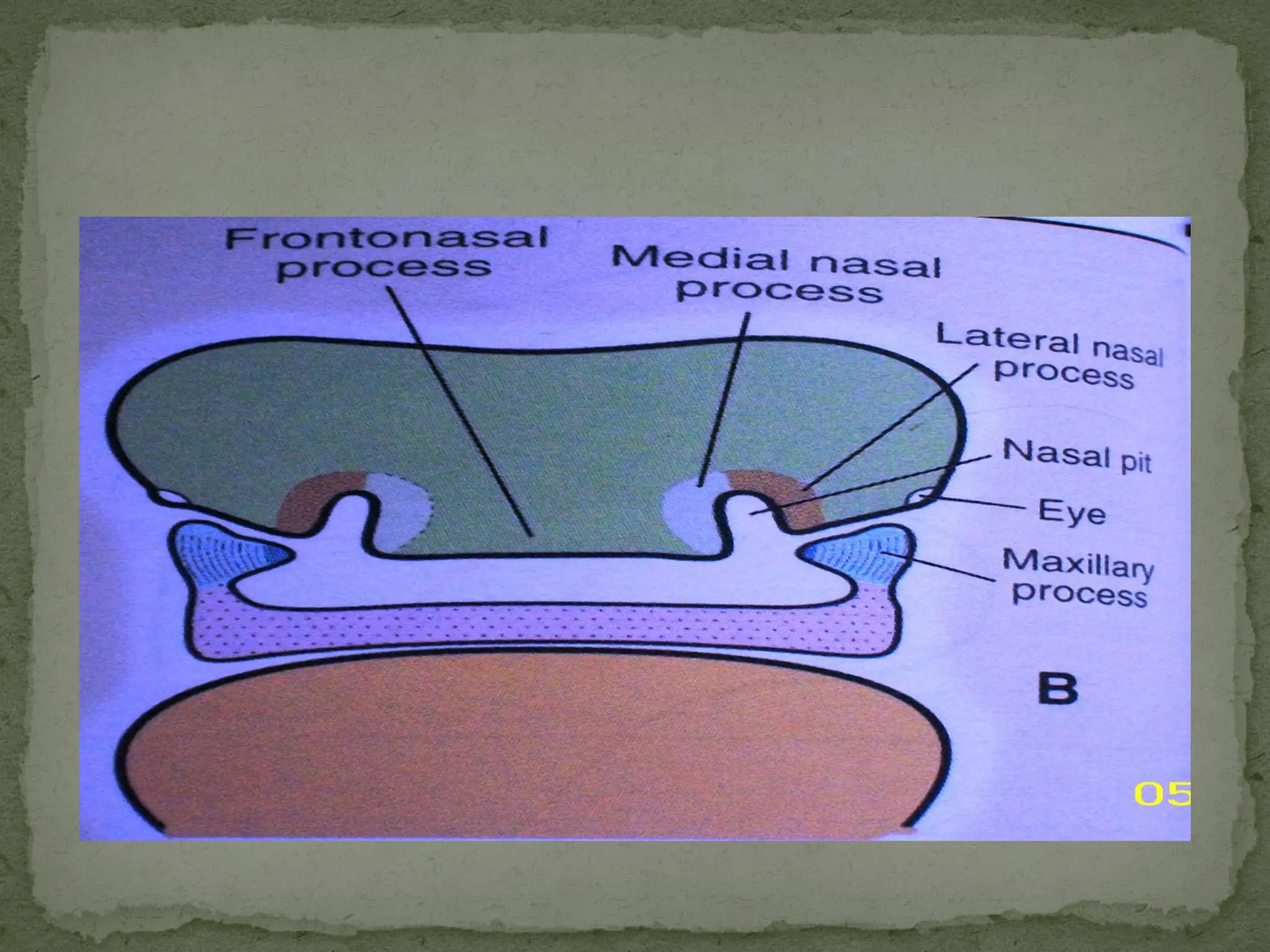
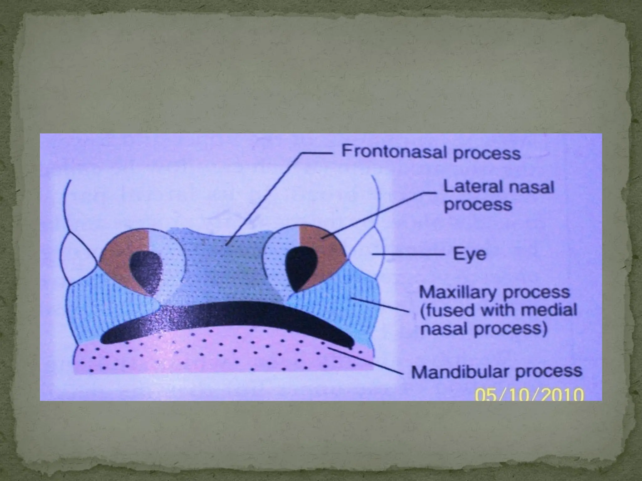
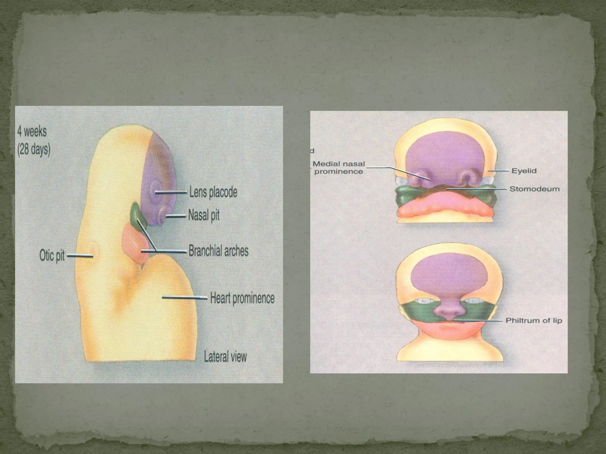
#9 Nasal placodes – formed due to the bilateral localized thickening of the frontonasal process.
Nasal pits – nasal placodes sink below the surface to form nasal pits. The edge of each pit are raised above the surface.
The medial raised edge is called medial nasal process.
lateral edge is called lateral nasal process.
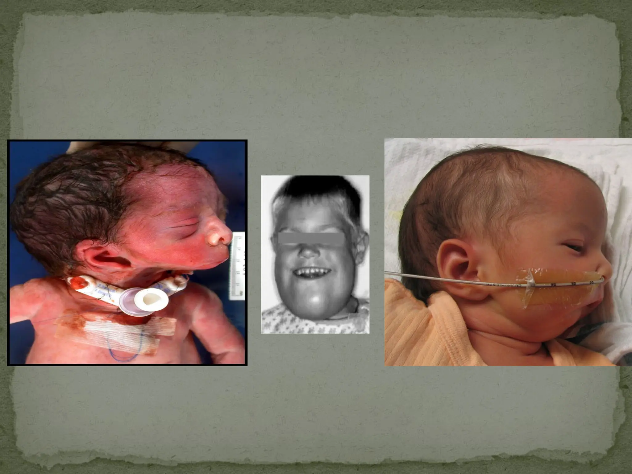
#16 The recognition of craniofacial anomalies in both animals and humans was probably first recorded by the Babylonians. They were the earliest civilization to leave records indicating that malformed infants foretold the future
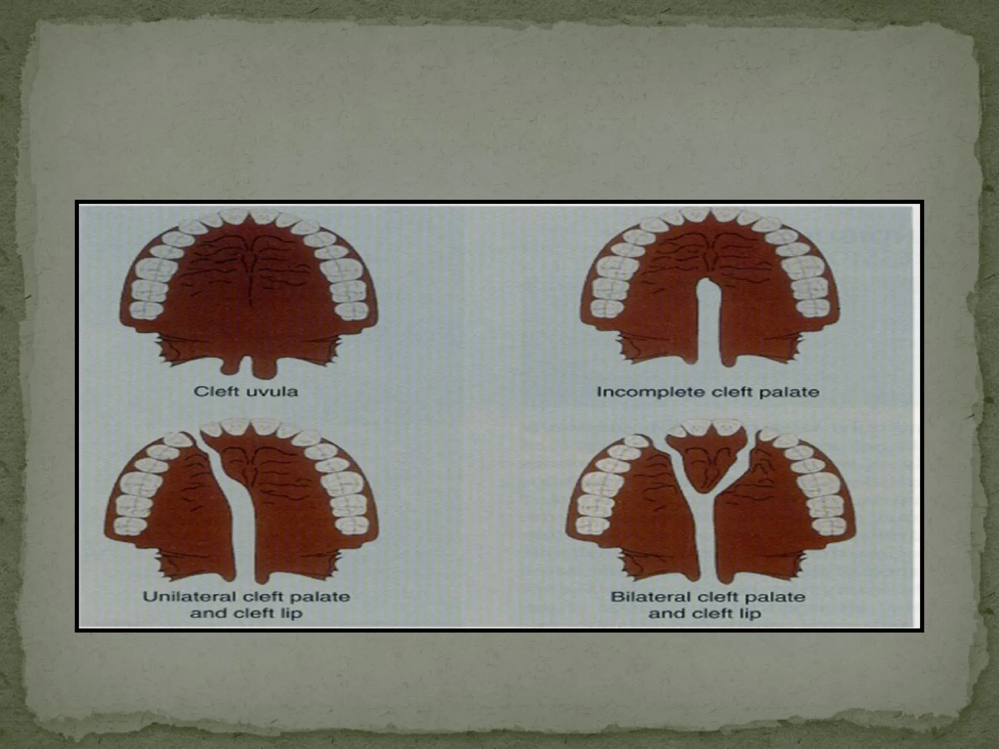
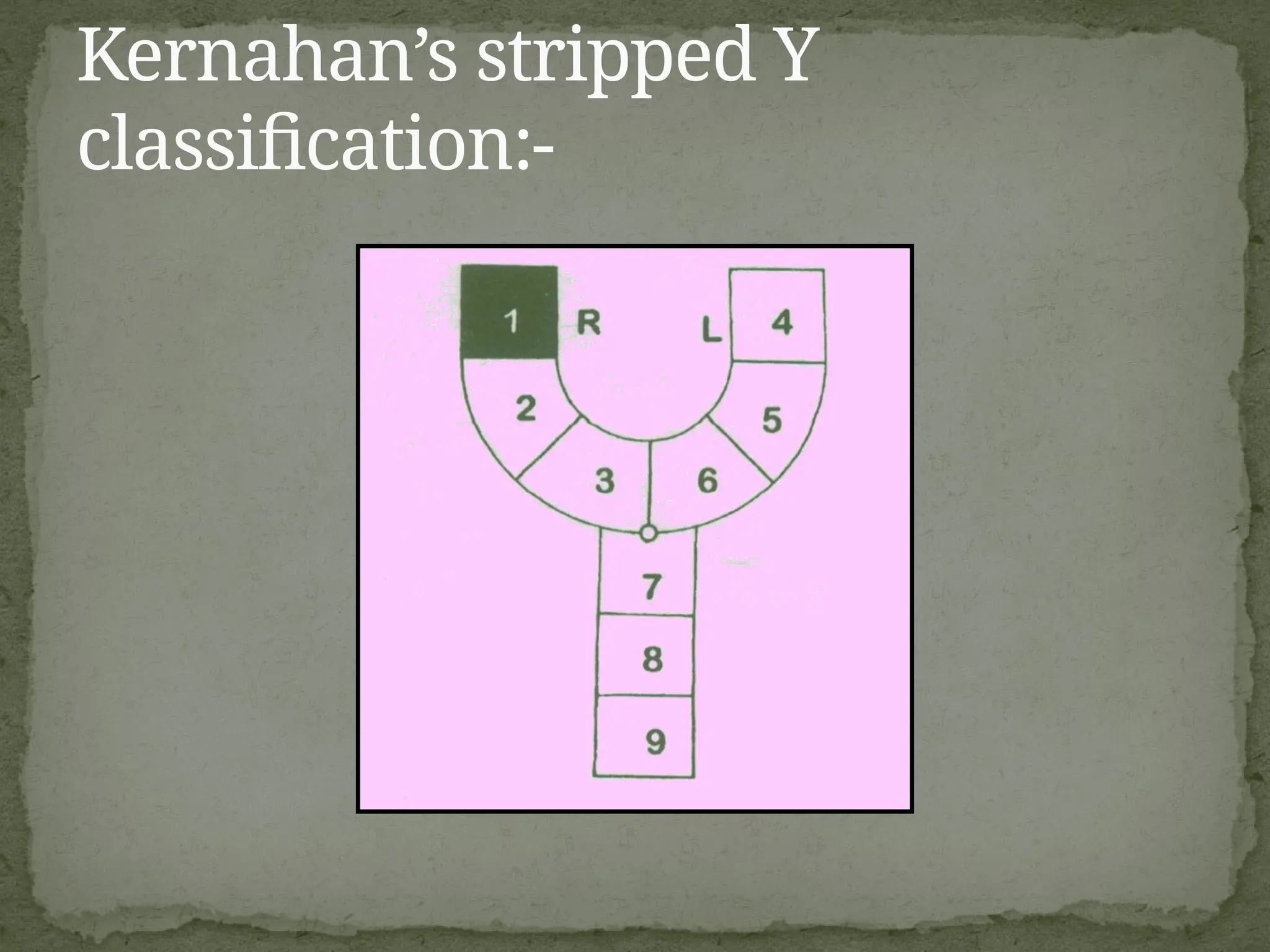
#18 Lip-alveolus-premaxilla-hardpalate-softpalate-submucous cleft
It’s a numerological method but its inadequate n varying complexities
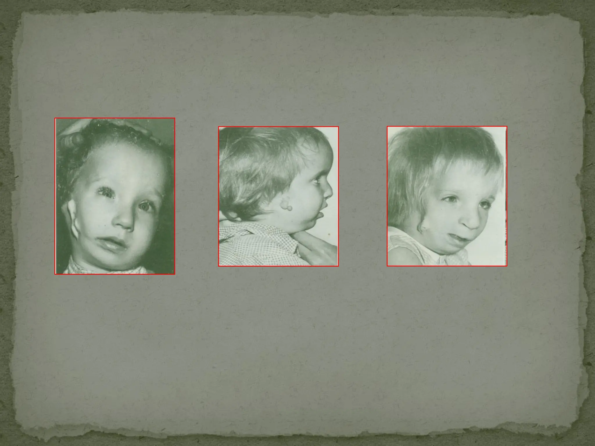
#20 In 1912 crouzon first described a woman and her son with this disorder.
In 1915 he reported a family in which seven of twenty one members affected and thus stressed the genetic aspects of the syndrome.
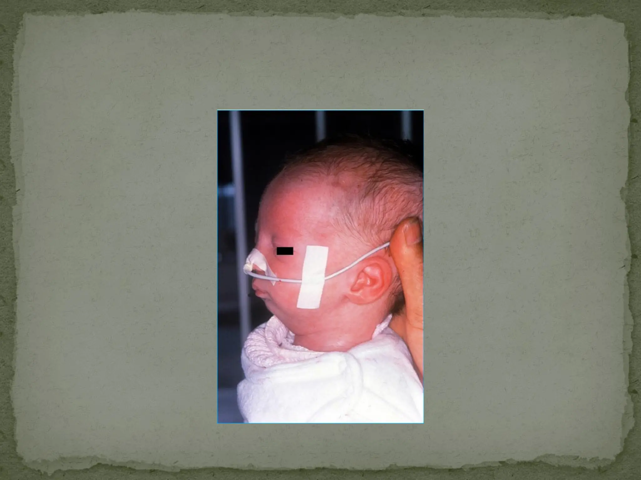
#21 In 1900 Treacher Collins described. congenital deformity of structures derived from first and second branchial arches. some cases - due to teratogens
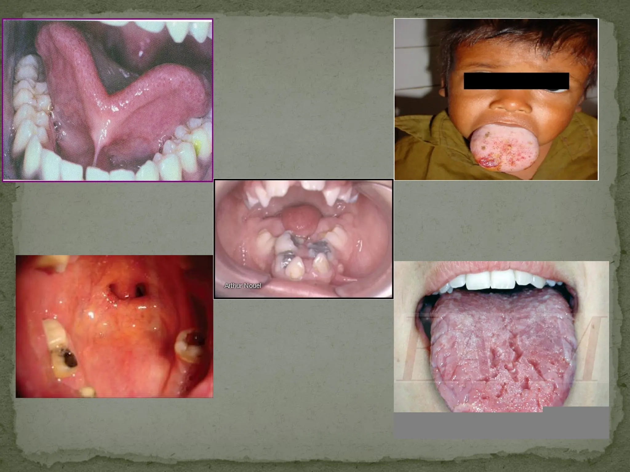
#24 tongue-Red rhomboidal shaped smooth zone may be present on tongue in front of foramen caecum
It is considered to be result of persistence of tuberculum impar.