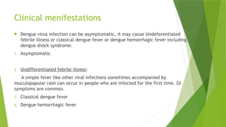
Dengue fever
• DR TOQEER HUSSAIN
• PGR MEDICINE
• SKBZ CMH MZD
 Dengue is a self-limiting Mosquito transmitting disease characterized by Fever,
headache, arthalgias, myalgias, rash, nausea and vomiting.
 Dengue virus is member of genus flavi virus. It has four serotypes DENV1, DENV2,
DENV3, DENV4.
 Dengue viruses are transmitted by bite of female aedes aegypti.
 All the ages and both sexes are equally susceptible to dengue virus.
 Carries: humans
 Incubation period in human: 4-10 days
 Infected person can transmit the infection to mosquito 1 day before the onset of
febrile phase and remain infectious for next 6-7 days.
 Incubation period in mosquitos:8-12 days then remain infected throughout their
life.
Epidemiology
According to WHO fact sheet.
 Endemic in 128 countries
 First confirmed dengue breakout in Pakistan – 1994
 Since 2010 – 16580 confirmed cases, 257 deaths in lahore only and 5000
cases and 60 deaths from rest of the country.
 3.9 billion population is at risk of dengue fever
 390 million cases annually
 100 million cases present with clinical manifestations
 5 milliom cases of dengue hemorrhagic fever
 25000 deaths worldwide annually
Clinical menifestations
 Dengue virus infection can be asymptomatic, it may cause Undeferentiated
febrile illness or classical dengue fever or dengue hemorrhagic fever including
dengue shock syndrome.
1. Asymptomatic
2. Undifferentiated febrile illness:
A simple fever like other viral infections sometimes accompanied by
muculopapular rash can occur in people who are infected for the first time. GI
symptoms are common.
3. Classical dengue fever
4. Dengue hemorrhagic fever
Classical dengue fever
 The onset of CDF in sudden with high grade fever and chills, headache,
arthalgias, myalgias and joint pains restricting movements.
 Within 24 hours retro orbital pain and photophobia develops
 Fever is usually between 102- 103 F.
 Other symptoms include lethargy, anorexia, constipation, abdominal
pain and tenderness, pain in inguinal region, altered sensorium and
general depression.
 Rash in dengue fever is a maculopapular or petechial rash over the
face, thorax, and flexor surfaces, with islands of skin sparing. The
rash typically begins on day 3 and persists 2-3 days.
Petechial rash Muculopapular rash
Dengue hemorrhagic fever
 It has three phases.
 Febrile phase, Critical phase and Recovery phase.
 Febrile phase:
During first few days it usually has same presentation like classical dengue fever.
 Critical phase:
 When temperature drops to 99F or 100F on day 3-7 disease. Increase in
capillary permeability occurs parallel to the increasing hematocrit. Progressive
Leukopenia followed by decreased platelet count usually precedes plasma leakage.
 Pleural effusion on the right side and ascites maybe clinically detectable.
 The period of plasma leakage usually lasts for 1-2 days.
 Shock is often preceded by warning sign like abdominal pain and
tenderness, persistent vomiting, fluid accumulation, mucosal
bleeding, lethargy, restlessness, hepatomegaly more than 2 cm and
oliguria.
 With prolonged shock, consistent organ hypoperfusion leads to organ
impairment, acidosis DIC which then leads to severe bleeding.
 Hepatitis, encephalitis, myocarditis may develop.
 RECOVERY PHASE:
 If patient survives 24-48 hours of critical phase a gradual reabsorption
of fluids takes place following 3 – 4 days.
 Appetite returns, GI symptoms improve, patient stabilizes
hemodynamically and urine output becomes normal.
 During critical or recovery phase excessive fluid administration is
associated with massive ascites, pleural effusion, pulmonary oedema
and congestive cardiac failure.
Criteria for clinical diagnosis
 CLASSICAL DENGUE FEVER :-
 Probable diagnosis
Acute febrile illness of 2-7 days with two or more of following features
 Headache
 Retroorbital pain
 Myalgias
 Arthalgias
 Rash
 Hemorrhages
 Leukopenia (WBC < 5000 cells/mm3)
 Thrombocytopenia (<150000 cells/mm3)
 Rising hematocrit.
And one of the following:
 IgG titre > 1280 with hemoaggltination test
 Occourance in same location and same time as confirmed cases of
dengue fever.
 Confirmed diagnosis:
Probable case with one of the following-
1. Isolation of dengue virus from serum, CSF, autopsy.
2. Fourfold or greater increase in S/IgG or increase in IgM ab specific to
dengue virus.
3. Detection of dengue antigen in tissue, serum or CSF by ELISA or dot
blot assay directed against non structural protein-1 (NS-1).
4. Detection of dengue virus genomic sequences by reverse transcription
PCR.
 Dengue hemorrhagic fever
 all should be present:
1. Acute onset fever of 2-7 days
2. Positive tourniquet test, petechiae, ecchymoses, purpura or bleeding
from mucosa, GI tract, injection sites.
3. Platelet count < 100000cells/mm3
4. Rising hematocrit > 20% from the baseline. OR Pleural effusion,
ascites or
5. hypoproteinemia/hypoabluminemia.
 Dengue Shock Syndrome
criteria of DHF plus signs of shock including:
 Tachycardia, cold Extremities, delayed capillary refill, weak pulse,
lethargy.
 Pulse pressure < 20mmHg with increased diastolic pressure.
 Hypotension by age (systolic BP <80 mmHg for aged <5 years and 80-
90mmHg for older children and adults.
Tourniquet test
It is performed bs inflating BP cuff to a mid-point between systolic and
diastolic pressure for 5 minutes.
 Test is considered positive when 10 or more petechiae per square inch
appears above forearm.
 In DHF, test usually gives a definite positive result with 20 or more
petechiae.
 This test maybe negative during phase of profound shock. If negative
then repeat test.
Management of dengue fever
Investigations
 All the baseline should be done including CBC, LFTs, RFTS,
S/Electrolytes .
 Coagulation profile
 PT and APTT can be elavated.
 BUN is elevated in shock
 Hyponatremia is most common in DHF and DSS.
 Stool for occult blood
 Chest X-ray and ultrasound.
 Cardiac enzymes and ECG.
investigations
 Full Blood Count (FBC)
 1. White blood cell count (WBC):
In the beginning of the febrile phase WBC is usually normal but will decrease rapidly as
the disease progresses.13 WBC may show relative lymphocytosis.
 2. Hematocrit (HCT):
 Hemo concentration as depicted by rising HCT is a marker of plasma leakage in dengue
infection and helps to differentiate between DF and DHF –
 A significant blood loss and early fluid replacement may mask the trend.
 It is of paramount importance to get a baseline HCT in the early febrile phase of
disease. It would be easier later on to recognize start of plasma leak – detection of
rising HCT level.
investigation
 3. Thrombocytopenia:
 Thrombocytopenia is perhaps the most common laboratory
investigation in dengue infection.
 In the early febrile phase, platelet count is usually within normal
range but it will decrease rapidly as the disease progresses to the late
febrile phase or at defervescence and it may continue to remain low
for the first few days of recovery.
 4. Liver Function Test
 Abnormal LFTs in the form of elevated transaminases is common and
is characterized by greater elevation of the AST as compared to the
ALT. The frequency and degree of elevation of the liver enzymes are
higher with DHF compared to DF.
Suggestions
 Leucopenia followed by progressive thrombocytopenia is suggestive of
dengue infection.
 A rising HCT with accompanying progressive thrombocytopenia is
suggestive of DHF.
 There is no local data available on the normal range of HCT in adults.
In the absence of a baseline HCT level, a HCT value of >40% in female
adults and >46% in male adults should raise the suspicion of plasma
leakage.
Recommendations
The baseline HCT and WCC should be established as early as possible in
all patients with suspected dengue.
Serial FBC and HCT must be monitored as the disease progresses.
Dengue Serology Tests
 Hemagglutination Inhibition Test
HI test is considered a gold standard for the serological diagnosis of dengue.
 Dengue IgM test:
Dengue-IgM capture enzyme-linked immunosorbent assay (ELISA) is the most widely used
serological test, to diagnose dengue. The titer of IgM tends to be significantly higher in
primary infections (10) as compared to secondary (20) infections.
 Indirect IgG ELISA test:
Both primary & secondary dengue infection, dengue IgG becomes detectable in 100% of
patients after day seven of onset of fever. A paired dengue IgG is, therefore, a recommended
test to see the seroconversion; if dengue IgM stays negative after day seven and the disease is
still suspected clinically
Recommendations
 Seroconversion for dengue IgM in a paired sample is conclusive evidence of dengue
fever. Therefore, dengue IgM should be taken as soon as the disease is suspected.
 Dengue IgM is usually becomes positive after day 5-7 of illness. Therefore, a
negative IgM taken before day 5-7 of illness does not exclude dengue infection.
 If dengue IgM is negative before day seven, a repeat sample must be taken in
recovery phase.
 If dengue IgM is still negative after day seven with negative IgG test reported at
less than seven days, a fourfold rise in reciprocal IgG antibody titre between acute
and convalescent sera is needed for diagnostic confirmation.
Polymerase Chain Reaction– (DENV-RNA
by RT-PCR)
In the early phase (< 5 days of illness), molecular tests such as the
reverse transcriptase – polymerase chain reaction (RT- PCR) can be very
useful for the diagnosis of dengue infection.
It was shown to have a sensitivity of 100% in the first 5 days of disease,
but is reduced to about 70% by the day six.
An additional advantage of RT-PCR is the ability to determine dengue
serotypes
Non-Structural Protein-1 (NS1 Antigen)
 The sensitivity of NS1 antigen detection starts to drop off from the
day 4-5 of illness
 and is usually undetectable in the convalescence phase. It is
recommended to
 carry out Dengue NS1 testing by ELISA technique. Simple rapid tests
such as strip
 assays (Immune chromatography technique ECT) have significantly
reduced sensitivity
 and specificity and must be interpreted in clinical context.
Admission criteria
 Presence of any warning sign.
 Bleeding from any site independent of platelet count.
 Signs of organ impairment(renal, hepatic etc.)
 co-existing conditions such as peptic ulcer, hemolytic anemias,
obesity.
 Sign and symptoms of hypotension
 Living very far from health facility
 Increased hematocrit
 Pleural effusion, ascites.
Depending on the severity of disease patient can be:
 Sent home (group A)
 Treated in hospital (group B)
 Given emergency treatment and urgently referred to tertiary care hospital.
(Group C)
Group A
Patients who don’t have warnings signs and who are able to
 Tolerate adequate amount of oral fluids
 Pass urine atleast every 6 hours.
 Send home with instructions (group A)
 ORS intake.
 Paracetamol for high grade fever and tapid sponging.
 Avoid NSAIDS as these may aggravate bleeding.
 Aspirin can cause Reye’s syndrome in children.
Group B
 Patients with warning signs.
 Having co-existing conditions such as renal failure, hemolytic
anemias.
 Living far from hospital with no appropriate traveling facility.
The management of group B is through IV fluids.
Monitoring
 Vitals monitoring (1- 4 hrly)
 Monitoring of peripheral perfusion 1-4 houly.
 Urine output (4 – 6 hourly)
 Hematocrit before and after fluid replacement then 6- 12 hourly.
 Blood glucose levels
 Other organ functions such as liver, kidney, heart.
Group c
 It includes patients with severe plasma leakage leading to dengue shock or fluid
accumulation with respiratory distress.
 Severe hemorrhage
 Severe organ impairment.
Patients in group C should be urgently referred to tertiary care hospital.
Fluid management in shock
 Choice of intravenous fluid is
 Crystalloids( ringer lactate and 0.9% NaCl) have beem shown to be
the safe and effective as colloids in reducing the recurrence of shock
and mortality.
 Crystalloids should be used as first line to fluid resuscitation In
compensated dengue shock.
 Colloids have been shown to restore cardiac index and reduce the
levels of hematocrit faster. Colloids can be fluid of choice if blood
pressure has to be restored urgently i. e in those with pulse pressue
less than 10 mmHg.
Management of Bleeding in Dengue
 Mucosal bleed may occur in any patient with dengue fever but if
patient remains stable fluid resuscitation it should ne considered as
minor.
 In patients with profound Thrombocytopenia
1. Ensure bedrest and protection from any trauma to reduce the risk of
bleed.
2. Don’t give intramuscular injections to avoid hematoma.
 Severe bleeding can be recognized bs decrease in HCT after fluid
resuscitation together with hemodynamically unstable status.
 In severe bleed give 5 – 10 ml/kg of packed red cells or 10 – 20 ml/kg
of fresh whole blood at an appropriate rate and observe clinical
response.
 Consider repeating transfusion if there is further blood loss or there is
no increase on hematocrit after transfusion
 Transfuse platelet concentrates when massive bleed cannot be
managed with fresh whole blood.
Indications of platelet transfusion
 Prophylactic platelet may be given at level of platelets <10000
cells/mm3.
 Prolonged shock with coagulopathy.
 In case of massive systemic bleed platelet along with RCC are needed
to be transfused.
Management of Upper Gastrointestinal
Bleeding
 most of the GIT bleed will improve after 48-72 hours of the defervescence.
 Any bleed that persists beyond this time will require further investigation.
 Recommendations
 Endoscopy and endoscopic injection therapy in upper GIT hemorrhage
should be avoided.
 Blood transfusion with whole blood or packed cell (as fresh as is available,
preferably less than one week old) is indicated in significant bleeding.
Management of Cardiac Complications
in Dengue Infection
 Cardiac complications of dengue should be suspected in those with fluid
refractory shock or haemodynamic compromise disproportionate to
capillary leakage/HCT increase
 it is recommended to do ECG, cardiac biomarkers and echocardiography.
 Common ECG changes reported are: Sinus bradycardia
 
Atrioventricular block Atrial fibrillation T-wave and ST-segment
 
abnormalities
 Echocardiography
 There is evidence of significantly low ejection fraction during the critical
stage of severe dengue infection
DENGUE FEVER IN PREGNANCY
 the signs and symptoms may be confused with other complications of
pregnancy such as toxemia, Hemolysis, Elevated Liver Enzymes, Low Platelets
(HELLP) syndrome.There are some reports of an increased incidence of
prematurity, in-utero death and abruptio placenta in these women.
 Elevation of HCT in dengue is masked by hemodilution due to increase in
plasma volume especially in the 2nd and 3rd trimester. Serial HCT
measurement is crucial for disease monitoring in pregnancy.
 The detection of third space fluid accumulation is difficult due to the
presence of gravid uterus.
 Baseline blood pressure is often lower and pulse pressure wider
 Baseline heart rate may be higher
Management of infected pregnant
patients close to delivery:
Risk of bleeding is at its highest during the period of plasma leakage
(critical phase). If possible, avoid Lower Segment Caesarean Section
(LSCS) or induction of labor during critical phase (plasma leakage).96
Procedures/maneuvers that may provoke or augment labor should be
avoided during this critical phase.
Care for the mother should be provided in a multidisciplinary way in
an area of the hospital where there are trained personnel available to
handle labor and its complications.
The baby should be observed for vertical transmission of dengue after
delivery
ANTIHYPERTENSIVE TREATMENT IN
DENGUE FEVER:
 Diuretics should be stopped as soon as the probable diagnosis of
dengue is made.
 Other anti-hypertensive treatment may be continued if the Pulse
Pressure stays above 30 mm of Hg.
 In case the Pulse Pressure drops, ≤30 mm Hg, stop all anti-
hypertensive treatment and introduce monitoring
DENGUE FEVER IN ANTITHROMBOTIC
THERAPY
 As a general rule acetyl salicylic acid should be avoided in patients
with DF
 because of worsening of complications from thrombocytopenia and
bleeding –
 unless patient is at a high risk of thromboembolism.
 High risk patients - need of anti-coagulation obligatory:
 For patients who are at a high risk of thrombo-embolic event and are
already using clopidogrel and asprin these drugs should be continued.
DENGUE FEVER IN ANTITHROMBOTIC
THERAPY
 High Risk patients needing obligatory anti-coagulation:
  In patients using warfarin, the current recommendation is to withhold it and perform
serial platelet and coagulation monitoring.
  As soon as the INR is below the therapeutic range introduce heparin to keep INR within
the desired range - continue serial platelet and coagulation monitoring.
 Even in these High Risk patients –
 All anti- coagulants should be withheld if:
 Platelet count falls below 50,000/mm3
 There is clinical or laboratory evidence of bleeding
 Patient is in shock.
recommendation
 Current recommendation for the low risk group is to withhold asprin
and consider
 withholding clopidogrel and warfarin for one week.
Criteria for discharge
All of the following should be present
1. No fever for almost 48 hours
2. Hemodynamically stable, tolerating orally, passing adequate amounts
of urine.
3. Minimun 48 – 72 hours have been passed after recovery from shock.
4. Increasing trend of platelet count.
5. Stable hematocrit without IV fluids.
Preventive measures
Thank you and beware of dengue

Dengue.pptx.,.................,..........

  • 1.
    Dengue fever • DRTOQEER HUSSAIN • PGR MEDICINE • SKBZ CMH MZD
  • 2.
     Dengue isa self-limiting Mosquito transmitting disease characterized by Fever, headache, arthalgias, myalgias, rash, nausea and vomiting.  Dengue virus is member of genus flavi virus. It has four serotypes DENV1, DENV2, DENV3, DENV4.  Dengue viruses are transmitted by bite of female aedes aegypti.  All the ages and both sexes are equally susceptible to dengue virus.  Carries: humans  Incubation period in human: 4-10 days  Infected person can transmit the infection to mosquito 1 day before the onset of febrile phase and remain infectious for next 6-7 days.  Incubation period in mosquitos:8-12 days then remain infected throughout their life.
  • 3.
    Epidemiology According to WHOfact sheet.  Endemic in 128 countries  First confirmed dengue breakout in Pakistan – 1994  Since 2010 – 16580 confirmed cases, 257 deaths in lahore only and 5000 cases and 60 deaths from rest of the country.  3.9 billion population is at risk of dengue fever  390 million cases annually  100 million cases present with clinical manifestations  5 milliom cases of dengue hemorrhagic fever  25000 deaths worldwide annually
  • 4.
    Clinical menifestations  Denguevirus infection can be asymptomatic, it may cause Undeferentiated febrile illness or classical dengue fever or dengue hemorrhagic fever including dengue shock syndrome. 1. Asymptomatic 2. Undifferentiated febrile illness: A simple fever like other viral infections sometimes accompanied by muculopapular rash can occur in people who are infected for the first time. GI symptoms are common. 3. Classical dengue fever 4. Dengue hemorrhagic fever
  • 5.
    Classical dengue fever The onset of CDF in sudden with high grade fever and chills, headache, arthalgias, myalgias and joint pains restricting movements.  Within 24 hours retro orbital pain and photophobia develops  Fever is usually between 102- 103 F.  Other symptoms include lethargy, anorexia, constipation, abdominal pain and tenderness, pain in inguinal region, altered sensorium and general depression.  Rash in dengue fever is a maculopapular or petechial rash over the face, thorax, and flexor surfaces, with islands of skin sparing. The rash typically begins on day 3 and persists 2-3 days.
  • 6.
  • 7.
    Dengue hemorrhagic fever It has three phases.  Febrile phase, Critical phase and Recovery phase.  Febrile phase: During first few days it usually has same presentation like classical dengue fever.  Critical phase:  When temperature drops to 99F or 100F on day 3-7 disease. Increase in capillary permeability occurs parallel to the increasing hematocrit. Progressive Leukopenia followed by decreased platelet count usually precedes plasma leakage.  Pleural effusion on the right side and ascites maybe clinically detectable.  The period of plasma leakage usually lasts for 1-2 days.
  • 11.
     Shock isoften preceded by warning sign like abdominal pain and tenderness, persistent vomiting, fluid accumulation, mucosal bleeding, lethargy, restlessness, hepatomegaly more than 2 cm and oliguria.  With prolonged shock, consistent organ hypoperfusion leads to organ impairment, acidosis DIC which then leads to severe bleeding.  Hepatitis, encephalitis, myocarditis may develop.
  • 12.
     RECOVERY PHASE: If patient survives 24-48 hours of critical phase a gradual reabsorption of fluids takes place following 3 – 4 days.  Appetite returns, GI symptoms improve, patient stabilizes hemodynamically and urine output becomes normal.  During critical or recovery phase excessive fluid administration is associated with massive ascites, pleural effusion, pulmonary oedema and congestive cardiac failure.
  • 13.
    Criteria for clinicaldiagnosis  CLASSICAL DENGUE FEVER :-  Probable diagnosis Acute febrile illness of 2-7 days with two or more of following features  Headache  Retroorbital pain  Myalgias  Arthalgias  Rash  Hemorrhages
  • 14.
     Leukopenia (WBC< 5000 cells/mm3)  Thrombocytopenia (<150000 cells/mm3)  Rising hematocrit. And one of the following:  IgG titre > 1280 with hemoaggltination test  Occourance in same location and same time as confirmed cases of dengue fever.
  • 15.
     Confirmed diagnosis: Probablecase with one of the following- 1. Isolation of dengue virus from serum, CSF, autopsy. 2. Fourfold or greater increase in S/IgG or increase in IgM ab specific to dengue virus. 3. Detection of dengue antigen in tissue, serum or CSF by ELISA or dot blot assay directed against non structural protein-1 (NS-1). 4. Detection of dengue virus genomic sequences by reverse transcription PCR.
  • 16.
     Dengue hemorrhagicfever  all should be present: 1. Acute onset fever of 2-7 days 2. Positive tourniquet test, petechiae, ecchymoses, purpura or bleeding from mucosa, GI tract, injection sites. 3. Platelet count < 100000cells/mm3 4. Rising hematocrit > 20% from the baseline. OR Pleural effusion, ascites or 5. hypoproteinemia/hypoabluminemia.
  • 18.
     Dengue ShockSyndrome criteria of DHF plus signs of shock including:  Tachycardia, cold Extremities, delayed capillary refill, weak pulse, lethargy.  Pulse pressure < 20mmHg with increased diastolic pressure.  Hypotension by age (systolic BP <80 mmHg for aged <5 years and 80- 90mmHg for older children and adults.
  • 19.
    Tourniquet test It isperformed bs inflating BP cuff to a mid-point between systolic and diastolic pressure for 5 minutes.  Test is considered positive when 10 or more petechiae per square inch appears above forearm.  In DHF, test usually gives a definite positive result with 20 or more petechiae.  This test maybe negative during phase of profound shock. If negative then repeat test.
  • 21.
  • 26.
    Investigations  All thebaseline should be done including CBC, LFTs, RFTS, S/Electrolytes .  Coagulation profile  PT and APTT can be elavated.  BUN is elevated in shock  Hyponatremia is most common in DHF and DSS.  Stool for occult blood  Chest X-ray and ultrasound.  Cardiac enzymes and ECG.
  • 27.
    investigations  Full BloodCount (FBC)  1. White blood cell count (WBC): In the beginning of the febrile phase WBC is usually normal but will decrease rapidly as the disease progresses.13 WBC may show relative lymphocytosis.  2. Hematocrit (HCT):  Hemo concentration as depicted by rising HCT is a marker of plasma leakage in dengue infection and helps to differentiate between DF and DHF –  A significant blood loss and early fluid replacement may mask the trend.  It is of paramount importance to get a baseline HCT in the early febrile phase of disease. It would be easier later on to recognize start of plasma leak – detection of rising HCT level.
  • 28.
    investigation  3. Thrombocytopenia: Thrombocytopenia is perhaps the most common laboratory investigation in dengue infection.  In the early febrile phase, platelet count is usually within normal range but it will decrease rapidly as the disease progresses to the late febrile phase or at defervescence and it may continue to remain low for the first few days of recovery.  4. Liver Function Test  Abnormal LFTs in the form of elevated transaminases is common and is characterized by greater elevation of the AST as compared to the ALT. The frequency and degree of elevation of the liver enzymes are higher with DHF compared to DF.
  • 29.
    Suggestions  Leucopenia followedby progressive thrombocytopenia is suggestive of dengue infection.  A rising HCT with accompanying progressive thrombocytopenia is suggestive of DHF.  There is no local data available on the normal range of HCT in adults. In the absence of a baseline HCT level, a HCT value of >40% in female adults and >46% in male adults should raise the suspicion of plasma leakage.
  • 30.
    Recommendations The baseline HCTand WCC should be established as early as possible in all patients with suspected dengue. Serial FBC and HCT must be monitored as the disease progresses.
  • 31.
    Dengue Serology Tests Hemagglutination Inhibition Test HI test is considered a gold standard for the serological diagnosis of dengue.  Dengue IgM test: Dengue-IgM capture enzyme-linked immunosorbent assay (ELISA) is the most widely used serological test, to diagnose dengue. The titer of IgM tends to be significantly higher in primary infections (10) as compared to secondary (20) infections.  Indirect IgG ELISA test: Both primary & secondary dengue infection, dengue IgG becomes detectable in 100% of patients after day seven of onset of fever. A paired dengue IgG is, therefore, a recommended test to see the seroconversion; if dengue IgM stays negative after day seven and the disease is still suspected clinically
  • 32.
    Recommendations  Seroconversion fordengue IgM in a paired sample is conclusive evidence of dengue fever. Therefore, dengue IgM should be taken as soon as the disease is suspected.  Dengue IgM is usually becomes positive after day 5-7 of illness. Therefore, a negative IgM taken before day 5-7 of illness does not exclude dengue infection.  If dengue IgM is negative before day seven, a repeat sample must be taken in recovery phase.  If dengue IgM is still negative after day seven with negative IgG test reported at less than seven days, a fourfold rise in reciprocal IgG antibody titre between acute and convalescent sera is needed for diagnostic confirmation.
  • 33.
    Polymerase Chain Reaction–(DENV-RNA by RT-PCR) In the early phase (< 5 days of illness), molecular tests such as the reverse transcriptase – polymerase chain reaction (RT- PCR) can be very useful for the diagnosis of dengue infection. It was shown to have a sensitivity of 100% in the first 5 days of disease, but is reduced to about 70% by the day six. An additional advantage of RT-PCR is the ability to determine dengue serotypes
  • 34.
    Non-Structural Protein-1 (NS1Antigen)  The sensitivity of NS1 antigen detection starts to drop off from the day 4-5 of illness  and is usually undetectable in the convalescence phase. It is recommended to  carry out Dengue NS1 testing by ELISA technique. Simple rapid tests such as strip  assays (Immune chromatography technique ECT) have significantly reduced sensitivity  and specificity and must be interpreted in clinical context.
  • 35.
    Admission criteria  Presenceof any warning sign.  Bleeding from any site independent of platelet count.  Signs of organ impairment(renal, hepatic etc.)  co-existing conditions such as peptic ulcer, hemolytic anemias, obesity.  Sign and symptoms of hypotension  Living very far from health facility  Increased hematocrit  Pleural effusion, ascites.
  • 36.
    Depending on theseverity of disease patient can be:  Sent home (group A)  Treated in hospital (group B)  Given emergency treatment and urgently referred to tertiary care hospital. (Group C) Group A Patients who don’t have warnings signs and who are able to  Tolerate adequate amount of oral fluids  Pass urine atleast every 6 hours.
  • 37.
     Send homewith instructions (group A)  ORS intake.  Paracetamol for high grade fever and tapid sponging.  Avoid NSAIDS as these may aggravate bleeding.  Aspirin can cause Reye’s syndrome in children.
  • 38.
    Group B  Patientswith warning signs.  Having co-existing conditions such as renal failure, hemolytic anemias.  Living far from hospital with no appropriate traveling facility. The management of group B is through IV fluids.
  • 40.
    Monitoring  Vitals monitoring(1- 4 hrly)  Monitoring of peripheral perfusion 1-4 houly.  Urine output (4 – 6 hourly)  Hematocrit before and after fluid replacement then 6- 12 hourly.  Blood glucose levels  Other organ functions such as liver, kidney, heart.
  • 41.
    Group c  Itincludes patients with severe plasma leakage leading to dengue shock or fluid accumulation with respiratory distress.  Severe hemorrhage  Severe organ impairment. Patients in group C should be urgently referred to tertiary care hospital.
  • 42.
    Fluid management inshock  Choice of intravenous fluid is  Crystalloids( ringer lactate and 0.9% NaCl) have beem shown to be the safe and effective as colloids in reducing the recurrence of shock and mortality.  Crystalloids should be used as first line to fluid resuscitation In compensated dengue shock.  Colloids have been shown to restore cardiac index and reduce the levels of hematocrit faster. Colloids can be fluid of choice if blood pressure has to be restored urgently i. e in those with pulse pressue less than 10 mmHg.
  • 49.
    Management of Bleedingin Dengue  Mucosal bleed may occur in any patient with dengue fever but if patient remains stable fluid resuscitation it should ne considered as minor.  In patients with profound Thrombocytopenia 1. Ensure bedrest and protection from any trauma to reduce the risk of bleed. 2. Don’t give intramuscular injections to avoid hematoma.  Severe bleeding can be recognized bs decrease in HCT after fluid resuscitation together with hemodynamically unstable status.
  • 50.
     In severebleed give 5 – 10 ml/kg of packed red cells or 10 – 20 ml/kg of fresh whole blood at an appropriate rate and observe clinical response.  Consider repeating transfusion if there is further blood loss or there is no increase on hematocrit after transfusion  Transfuse platelet concentrates when massive bleed cannot be managed with fresh whole blood.
  • 51.
    Indications of platelettransfusion  Prophylactic platelet may be given at level of platelets <10000 cells/mm3.  Prolonged shock with coagulopathy.  In case of massive systemic bleed platelet along with RCC are needed to be transfused.
  • 52.
    Management of UpperGastrointestinal Bleeding  most of the GIT bleed will improve after 48-72 hours of the defervescence.  Any bleed that persists beyond this time will require further investigation.  Recommendations  Endoscopy and endoscopic injection therapy in upper GIT hemorrhage should be avoided.  Blood transfusion with whole blood or packed cell (as fresh as is available, preferably less than one week old) is indicated in significant bleeding.
  • 53.
    Management of CardiacComplications in Dengue Infection  Cardiac complications of dengue should be suspected in those with fluid refractory shock or haemodynamic compromise disproportionate to capillary leakage/HCT increase  it is recommended to do ECG, cardiac biomarkers and echocardiography.  Common ECG changes reported are: Sinus bradycardia   Atrioventricular block Atrial fibrillation T-wave and ST-segment   abnormalities  Echocardiography  There is evidence of significantly low ejection fraction during the critical stage of severe dengue infection
  • 54.
    DENGUE FEVER INPREGNANCY  the signs and symptoms may be confused with other complications of pregnancy such as toxemia, Hemolysis, Elevated Liver Enzymes, Low Platelets (HELLP) syndrome.There are some reports of an increased incidence of prematurity, in-utero death and abruptio placenta in these women.  Elevation of HCT in dengue is masked by hemodilution due to increase in plasma volume especially in the 2nd and 3rd trimester. Serial HCT measurement is crucial for disease monitoring in pregnancy.  The detection of third space fluid accumulation is difficult due to the presence of gravid uterus.  Baseline blood pressure is often lower and pulse pressure wider  Baseline heart rate may be higher
  • 55.
    Management of infectedpregnant patients close to delivery: Risk of bleeding is at its highest during the period of plasma leakage (critical phase). If possible, avoid Lower Segment Caesarean Section (LSCS) or induction of labor during critical phase (plasma leakage).96 Procedures/maneuvers that may provoke or augment labor should be avoided during this critical phase. Care for the mother should be provided in a multidisciplinary way in an area of the hospital where there are trained personnel available to handle labor and its complications. The baby should be observed for vertical transmission of dengue after delivery
  • 56.
    ANTIHYPERTENSIVE TREATMENT IN DENGUEFEVER:  Diuretics should be stopped as soon as the probable diagnosis of dengue is made.  Other anti-hypertensive treatment may be continued if the Pulse Pressure stays above 30 mm of Hg.  In case the Pulse Pressure drops, ≤30 mm Hg, stop all anti- hypertensive treatment and introduce monitoring
  • 57.
    DENGUE FEVER INANTITHROMBOTIC THERAPY  As a general rule acetyl salicylic acid should be avoided in patients with DF  because of worsening of complications from thrombocytopenia and bleeding –  unless patient is at a high risk of thromboembolism.  High risk patients - need of anti-coagulation obligatory:  For patients who are at a high risk of thrombo-embolic event and are already using clopidogrel and asprin these drugs should be continued.
  • 58.
    DENGUE FEVER INANTITHROMBOTIC THERAPY  High Risk patients needing obligatory anti-coagulation:   In patients using warfarin, the current recommendation is to withhold it and perform serial platelet and coagulation monitoring.   As soon as the INR is below the therapeutic range introduce heparin to keep INR within the desired range - continue serial platelet and coagulation monitoring.  Even in these High Risk patients –  All anti- coagulants should be withheld if:  Platelet count falls below 50,000/mm3  There is clinical or laboratory evidence of bleeding  Patient is in shock.
  • 59.
    recommendation  Current recommendationfor the low risk group is to withhold asprin and consider  withholding clopidogrel and warfarin for one week.
  • 61.
    Criteria for discharge Allof the following should be present 1. No fever for almost 48 hours 2. Hemodynamically stable, tolerating orally, passing adequate amounts of urine. 3. Minimun 48 – 72 hours have been passed after recovery from shock. 4. Increasing trend of platelet count. 5. Stable hematocrit without IV fluids.
  • 62.
  • 64.
    Thank you andbeware of dengue