
Recommended
Recommended
More Related Content
Similar to New عرض تقديمي من Microsoft PowerPoint.pptx
Similar to New عرض تقديمي من Microsoft PowerPoint.pptx (20)
More from MustafaALShlash1
More from MustafaALShlash1 (20)
Recently uploaded
Recently uploaded (20)
New عرض تقديمي من Microsoft PowerPoint.pptx
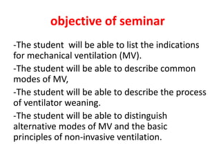
- 1. objective of seminar -The student will be able to list the indications for mechanical ventilation (MV). -The student will be able to describe common modes of MV, -The student will be able to describe the process of ventilator weaning. -The student will be able to distinguish alternative modes of MV and the basic principles of non-invasive ventilation.
- 3. Mechanical ventilation is a positive- or negative-pressure breathing device that can maintain ventilation and oxygen delivery for a prolonged period. Caring for a patient on mechanical ventilation has become an very important part of nursing care in critical care or general medical-surgical units
- 4. Purposes: • To maintain or improve ventilation, & tissue oxygenation. • To decrease the work of breathing & improve patient’s comfort.
- 5. Indications: -decrease in oxygenation (PaO2) -increase in arterial carbon dioxide levels (PaCO2) -persistent acidosis (decreased pH) -Conditions such as thoracic or abdominal Surgery -drug overdose, neuromuscular disorders, inhalation injury,
- 6. Indications: -COPD. -Multiple trauma -Shock -multisystem failure -Infectious diseases of the lung such as pneumonia, tuberculosis. -Acute respiratory failure
- 7. Classification of Ventilators Mechanical ventilators are classified according to the method by which they support ventilation. The two general categories are 1-negative-pressure 2- positive-pressure ventilators.
- 8. Negative-Pressure Ventilators Negative-pressure ventilators exert a negative pressure on external chest. Decreasing the intrathoracic pressure during inspiration Physiologically, this type of assisted ventilation is similar to spontaneous ventilation. It is used mainly in chronic respiratory failure associated with neuromuscular conditions, such as poliomyelitis, muscular dystrophy, lateral sclerosis, and myasthenia gravis.
- 9. types of negative-pressure ventilators Iron Lung The iron lung is a negative-pressure chamber used for ventilation. It was used extensively during polio in the past and currently is used and patients with other neuromuscular disorders (e.g., lateral sclerosis, muscular dystrophy).
- 11. types of negative-pressure ventilators Body Wrap The body wrap and chest cuirass are portable devices that require a rigid cage or shell to create a negative-pressure chamber around the thorax and abdomen.
- 12. Contraindication of NIPPV 1-experienced respiratory arrest, 2 serious dysrhythmias, 3-cognitive impairment, 4-head or facial trauma.
- 13. Positive-Pressure Ventilators Today, the most common ventilators use positive pressure. Positive-pressure ventilators inflate the lungs by exerting positive pressure on the airway, pushing air in, similar to a bellows mechanism, forcing the alveoli to expand during inspiration and Expiration occurs passively
- 14. Positive-Pressure Ventilators Pressure-Cycled Ventilators flow of air (inspiration) until it reaches a preset pressure, and then cycles off, and Expiration occurs passively. Time-Cycled Ventilators control inspiration after a preset time. The volume of air the patient receives is regulated by the length of inspiration and the flow rate of the air.
- 15. Positive-Pressure Ventilators Volume-Cycled Ventilators most commonly used positive-pressure ventilators today mandatory ventilation, pressure support ventilation, and airway pressure release ventilation
- 17. noninvasive positive-pressure ventilation is a method of positive-pressure ventilation that can be given via face masks that cover the nose and mouth, nasal masks, or other oral or nasal devices It eliminates the need for endotracheal intubation or tracheostomy and decreases the risk of nosocomial infections such as pneumonia
- 18. noninvasive positive-pressure ventilation if they have acute or chronic respiratory failure, 1-acute pulmonary edema, 2-COPD,3- chronic heart failure, or a 4- sleep-related breathing disorder. The technique also may be used at home to improve tissue oxygenation and to rest the respiratory muscles while patients sleep at night.
- 19. Continuous positive airway pressure ( (CPAP -(CPAP) provides positive pressure to the airways throughout the respiratory cycle. Although it can be used as an assistant to mechanical ventilation with a cuffed endotracheal tube or tracheostomy tube to open the alveoli -CPAP is the most effective treatment for obstructive sleep apnea,
- 20. Ventilator Modes Ventilator mode refers to how breaths are The most commonly used modes are 1-assist–control. 2-intermittent mandatory ventilation. 3-synchronized intermittent mandatory ventilation. 4- pressure support ventilation. 5-airway pressure release ventilation.
- 21. Assist–control (A/C) ventilation provides full ventilatory support by delivering a preset tidal volume and respiratory rate. If the patient initiates a breath between the machine’s breaths, the ventilator delivers at the preset volume Client can breathe at a higher rate than the preset number of breaths/minute -The total respiratory rate is determined by the number of spontaneous inspiration initiated by the patient plus the number of breaths set.
- 22. Synchronized intermittent mandatory ventilation (SIMV) delivers a preset tidal volume and number of breaths per minute. Between ventilator- delivered breaths, the patient can breathe spontaneously with no assistance from the Ventilator Used to when wean the patient from the mechanical ventilator.
- 23. Intermittent mandatory ventilation (IMV) provides a combination of mechanically assisted breaths and spontaneous breaths. Mechanical breaths are delivered at preset intervals and a preselected tidal volume, Although the patient can increase the respiratory rate by initiating inspiration between ventilator-delivered breaths, efforts. regardless of the patient’s
- 24. Pressure support ventilation (PSV) patient-triggered inspiration to decrease resistance within the tracheal tube and ventilator tubing. The inspiratory pressure level, respiratory rate, and inspiratory–expiratory (I:E) ratio must be selected. Pressure support is reduced gradually as the patient’s strength used when the risk of barotrauma is high. The nurse must closely observe the patient’s respiratory rate and tidal volumes on initiation of PSV.
- 25. Airway pressure release ventilation (APRV time triggered, pressure-limited, time-cycled mode of mechanical ventilation that allows unrestricted, spontaneous breathing throughout the ventilatory cycle. The inflation period is long, and breaths may be initiated spontaneously as well as by the ventilator.
- 26. Ventilator Settings 1-tidal volume required(10 to 15 mL/kg). 2-lower FiO2 to maintain normal PaO2 (80 to 100 mm Hg). A/C). - APRV - 3-Set mode(SIMV-PCV-IMV 4-And rate as pt. condition 5-Adjust sensitivity so that the patient can trigger the ventilator with a minimal effort (usually 2 mm Hg negative inspiratory force).
- 27. Ventilator Settings 6-set the PEEP 7-Record minute volume and obtain ABGs to measure carbon dioxide (PaCO2), pH, and PaO2 after 20 minutes of continuous mechanical ventilation. 8-Adjust setting (FiO2 and rate) according to results of arterial blood gas analysis
- 28. PEEP advantage • Prevent atelectasis or collapse of alveoli • Improve gas exchange & oxygenation • Treat pulmonary edema ( pressure help fluids out from alveoli )
- 29. ● Fraction of inspired oxygen (FIO2) • The percent of oxygen concentration that the patient is receiving from the ventilator. (Between 21% & 100%) (room air has 21% oxygen content). • Initially a patient is placed on a high level of FIO2 . • Subsequent changes in FIO2 are based on ABGs • For infants, and especially in premature infants, high levels of FiO2 (>60%) should be avoided.
- 30. ● Tidal Volume (VT) • The volume of air delivered to a patient during a ventilator breath. • The amount of air inspired and expired with each breath. • Usual volume selected is between 5 to 15 ml/ kg body weight)
- 31. ● I:E Ratio (Inspiration to Expiration Ratio):- • The ratio of inspiratory time to expiratory time during a breath (Usually = 1:2)
- 32. Signs and symptoms of oxygen toxicity :- 1- Flushed face 2- Dry cough 3- Dyspnea 4- Chest pain 5- Tightness of chest 6- Sore throat
- 33. ● Respiratory Rate/ Breath Rate / Frequency ( F) • The number of breaths the ventilator will deliver/minute (10-16 b/m). • Total respiratory rate equals patient rate plus ventilator rate. • The nurse double-checks the functioning of the ventilator by observing the patient’s respiratory rate.
- 34. ● Sensitivity(trigger Sensitivity) • The sensitivity function controls the amount of patient effort needed to initiate an inspiration • Increasing the sensitivity (requiring less negative force) decreases the amount of work the patient must do to initiate a ventilator breath. • Decreasing the sensitivity increases the amount of negative pressure that the patient needs to initiate inspiration and increases the work of breathing.
- 35. Complications of Mechanical Ventilation:- I- Airway Complications, II- Mechanical complications, III- Physiological Complications, IV- Artificial Airway Complications.
- 36. I- Airway Complications 1- Aspiration 2- Decreased clearance of secretions 3- Nosocomial or ventilator-acquired pneumonia
- 37. II- Mechanical complications 1- Hypoventilation with atelectasis with respiratory acidosis or hypoxemia. 2- Hyperventilation with hypocapnia and respiratory alkalosis 3- Barotrauma a- Closed pneumothorax, b- Tension pneumothorax, c- Subcutaneous emphysema. 4- Inadequate nebulization or humidification 5- Overheated inspired air, resulting in hyperthermia
- 38. III- Physiological Complications 1- Fluid overload with humidified air and sodium chloride (NaCl) retention 2- Depressed cardiac function and hypotension 3- Stress ulcers 4- Paralytic ileus 5- Gastric distension 6- Starvation
- 39. Causes of Ventilator Alarms High pressure alarm • Increased secretions • Kinked ventilator tubing or endotracheal tube (ETT) • Patient biting the ETT • Water in the ventilator tubing. • ETT advanced into right mainstem bronchus.
- 40. Causes of Ventilator Alarms High pressure alarm • Increased secretions • Kinked ventilator tubing or endotracheal tube (ETT) • Patient biting the ETT • Water in the ventilator tubing. • ETT advanced into right mainstem bronchus.
- 41. High respiratory rate alarm • Episodes of tachypnea, • Anxiety, • Pain, • Hypoxia, • Fever. -
- 42. Nursing process of patients on mechanical ventilation Assessment: 1- Assess the patient 2- Assess the artificial airway (tracheostomy or endotracheal tube) 3- Assess the ventilator
- 43. Nursing Diagnoses 1-Impaired gas exchange related to underlying illness 2-Ineffective airway clearance related to 3_increased mucus Production 4-Risk for trauma and infection 5-Impaired physical mobility 6-Impaired verbal communication
- 44. Planning and Goals The major goals for the patient may include 1-achievement of optimal gas exchange, maintenance of a patent airway, 2- absence of trauma or infection, optimal mobility 3-adjustment to nonverbal methods of communication 4-absence of complications.
- 45. Nursing Interventions 1-Maintain airway patency & oxygenation 2- Promote comfort 3- Maintain fluid & electrolytes balance 4- Maintain nutritional state 5- Maintain urinary & bowel elimination 6- Maintain eye , mouth and cleanliness and integrity 7- Maintain mobility/ musculoskeletal function
- 46. Nursing Interventions 8- Maintain safety:- 9- Provide psychological support 10- Facilitate communication 11- Provide psychological support & information to family 12- Responding to ventilator alarms /Troublshooting ventilator alarms 13- Prevent nosocomial infection 14- Documentation
- 47. Respiratory weaning, the process of withdrawing the patient from dependence on the ventilator, takes place in three stages: the patient is gradually removed from the ventilator, then from the tube, and finally from oxygen.
- 48. Criteria for Weaning Careful assessment determine whether the patient is ready to be removed from mechanical ventilation Stable vital signs and arterial blood gases are also important predictors of successful weaning. Once readiness has been determined, the nurse records baseline measurements of weaning indices to monitor progress.
- 49. Criteria for weaning • Awake and alert • Hemodynamically stable, • Arterial blood gases (ABGs) normalized or at patient’s baseline - PaCO2 acceptable - PH of 7.35 – 7.45 - PaO2 > 60 mm Hg , - SaO2 >92% - FIO2 ≤40%
- 50. • Chest x-ray reviewed for correctable factors; treated as indicated, • Major electrolytes within normal range, • Hematocrit >25%, • Core temperature >36°C and <39°C, • Adequate management of pain/anxiety/agitation, • Adequate analgesia/ sedation (record scores on flow sheet), • No residual neuromuscular blockade.
- 51. Methods of Weaning 1- T-piece trial, 2- Continuous Positive Airway Pressure (CPAP) weaning, 3- Synchronized Intermittent Mandatory Ventilation (SIMV) weaning, 4- Pressure Support Ventilation (PSV) weaning.
- 52. 1- T-Piece trial • It consists of removing the patient from the ventilator and having him / her breathe spontaneously on a T-tube connected to oxygen source. • During T-piece weaning, periods of ventilator support are alternated with spontaneous breathing. • The goal is to progressively increase the time spent off the ventilator.
- 53. 2-Synchronized Intermittent Mandatory Ventilation ( SIMV) Weaning • SIMV is the most common method of weaning. • It consists of gradually decreasing the number of breaths delivered by the ventilator to allow the patient to increase number of spontaneous breaths
- 54. 3-Continuous Positive Airway Pressure ( CPAP) Weaning • When placed on CPAP, the patient does all the work of breathing without the aid of a back up rate or tidal volume. • No mandatory all ventilation is spontaneously initiated by the patient. • Weaning by gradual decrease in pressure value
- 55. 4- Pressure Support Ventilation (PSV) Weaning • The patient must initiate all pressure support breaths. • During weaning using the PSV mode the level of pressure support is gradually decreased based on the patient maintaining an adequate tidal volume (8 to 12 mL/kg) and a respiratory rate of less than 25 breaths/minute. • PSV weaning is indicated for :- - Difficult to wean patients - Small spontaneous tidal volume.