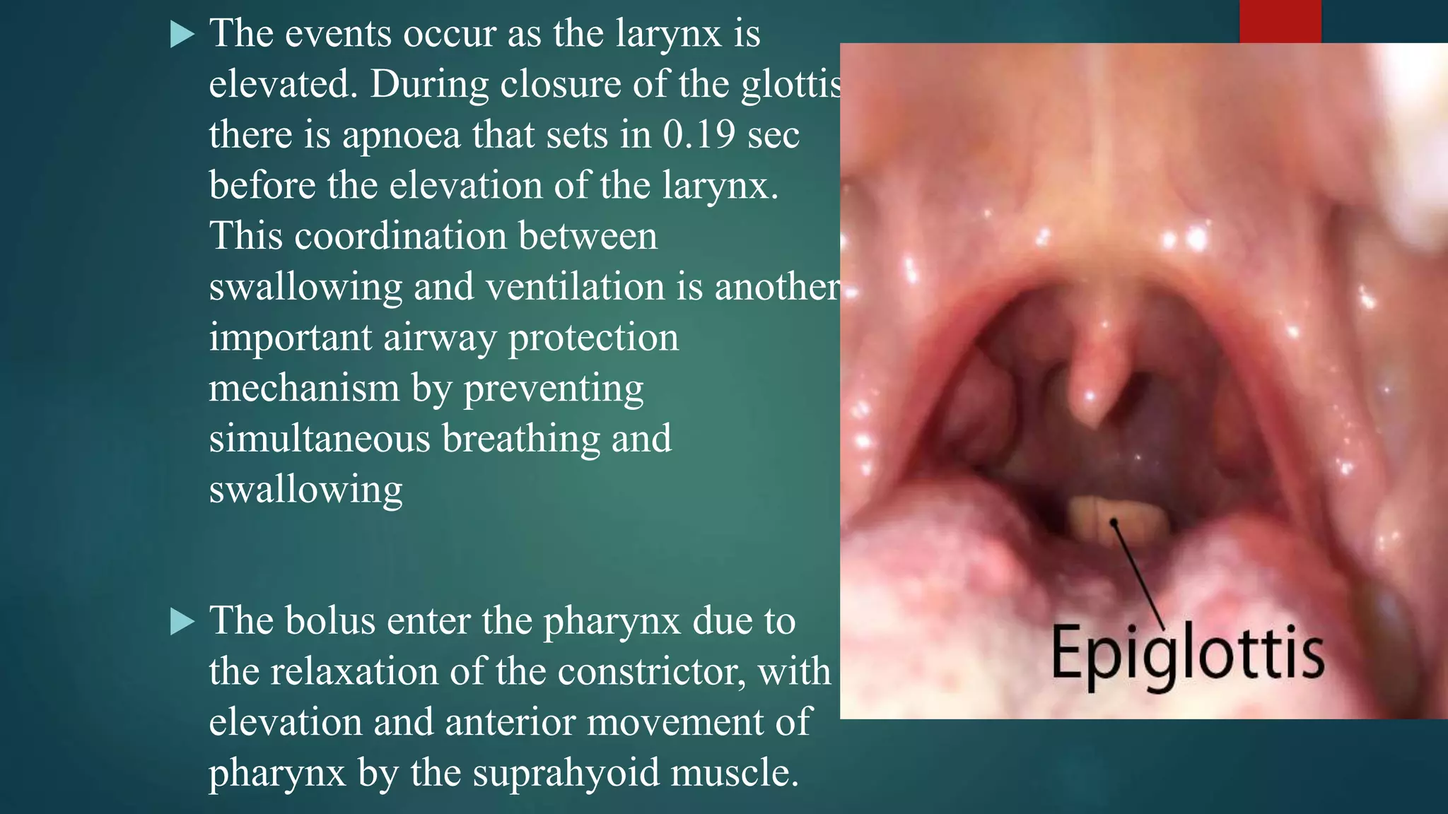
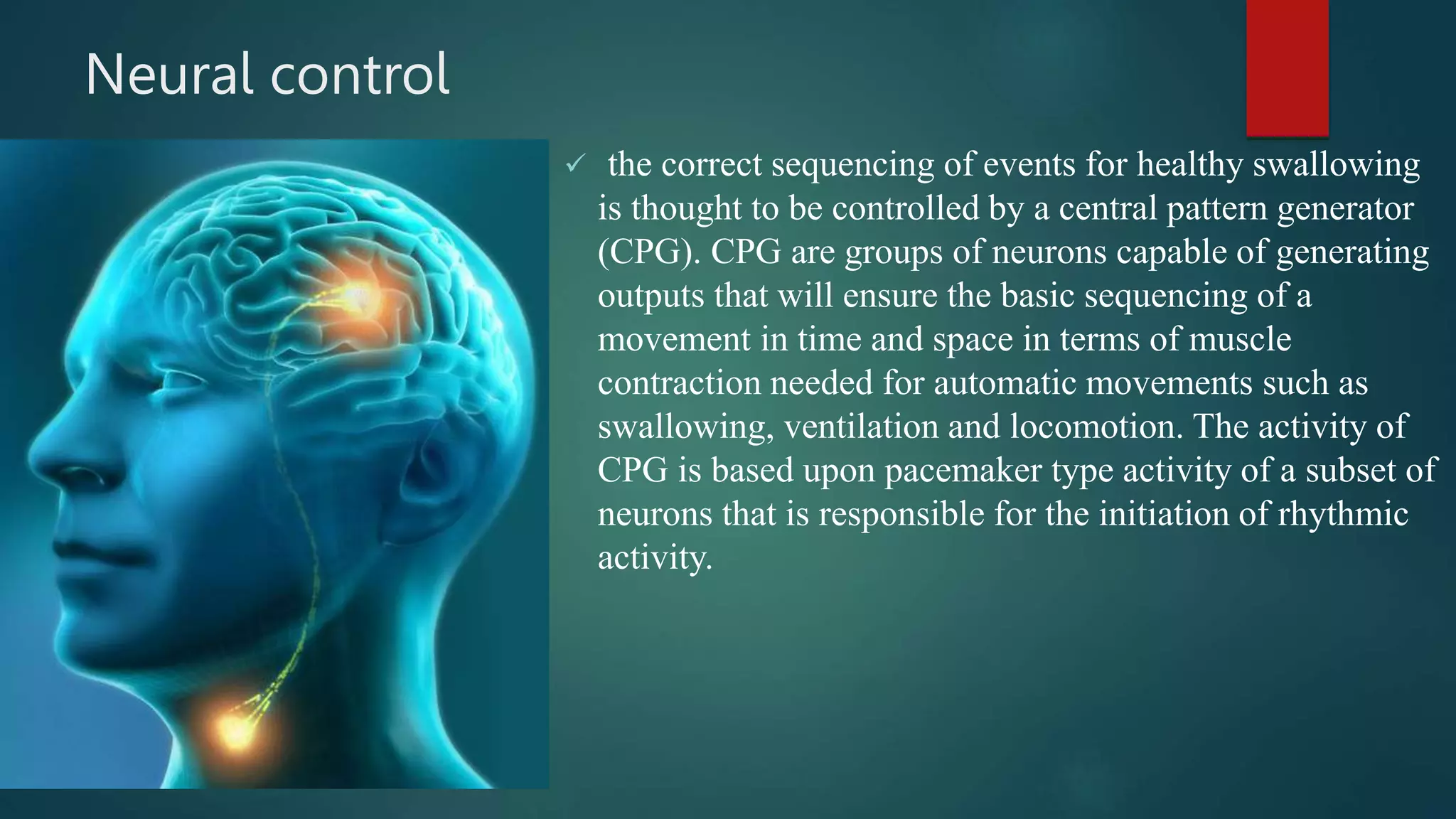
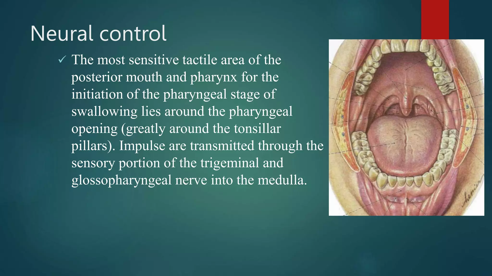
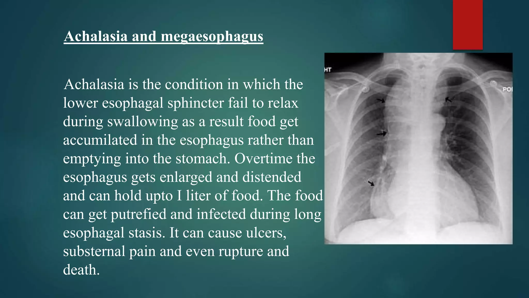
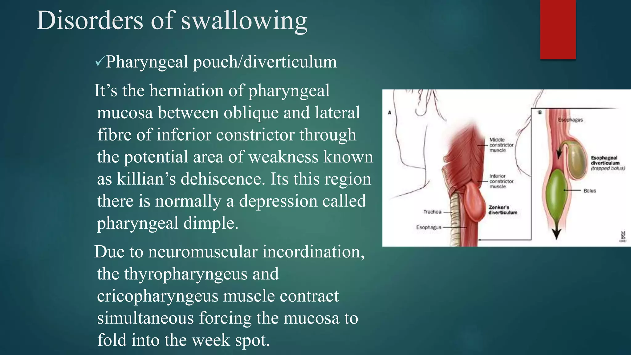
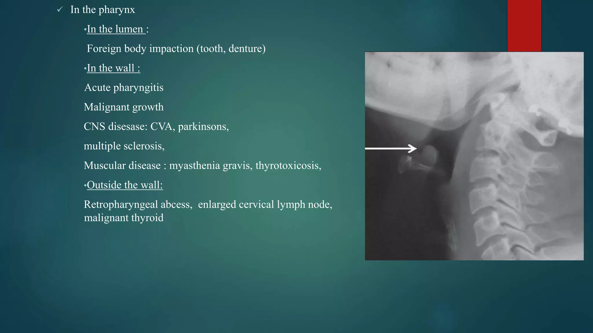
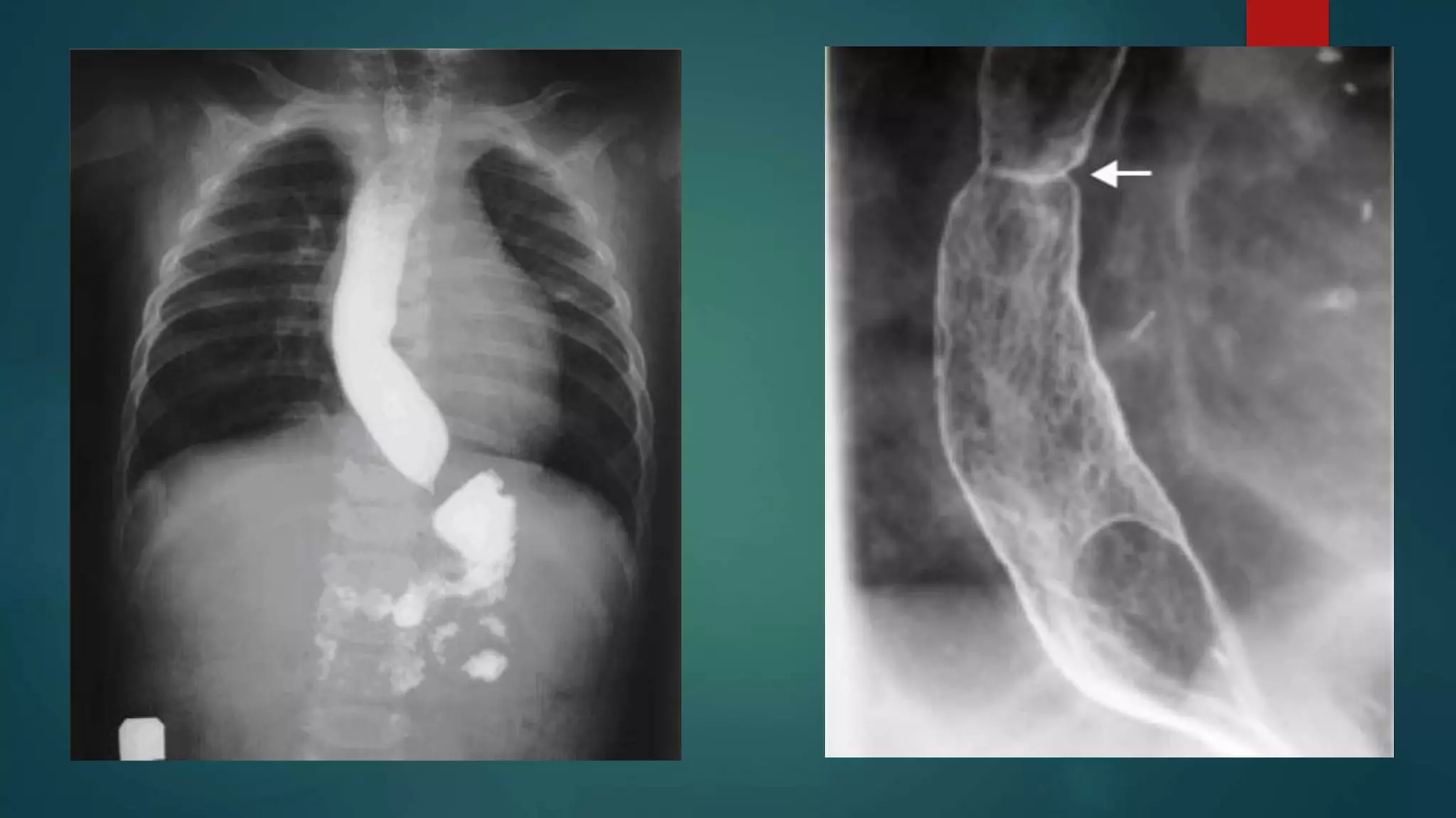
Deglutition
Contents
 Introduction
 Structures Involved
 Sequence Of Normal Deglutition
 Neural Control
 Respiration And Swallowing
 Disorders
 Conclusion
 Bibliography
Introduction
 The amount of food a person ingests is determined by the intrinsic
desire for food is called hunger while the type of food the person
preferentially seeks is determined by appetite.
 In case of solids, deglutition is usually preceded by mastication and
bolus formation. Its in turn is determined by the movement of the
tongue, buccinators, orbicularis oris etc.
 Swallowing require the coordinated activity of the muscles in 3 regions
: oral cavity, pharynx & larynx and oesophagus. The complex sequence
is part reflex and part under voluntary control.
Structures involved in deglutition
 Deglutition involves the passage
of bolus of food/liquid from oral
cavity to the stomach via
pharynx and oesophagus,
passing over the entrance to the
laryngeal vestibule, coordinated
by tongue, pharynx and larynx
Tongue :
The elevator and depresser muscle of the jaw
helps in bolus formation by grinding and reducing
food between the teeth. The intrinsic and extrinsic
muscle of the tongue form and guide the bolus.
The shape of the bolus is formed by the alteration
in the shape of the tongue. The intrinsic muscle
change the shape of the tongue while the extrinsic
muscle change the position of the tongue.
The alteration in the shape and position of the tongue guide the bolus into the
pharynx. Its aided by the lips, buccinator of cheek and soft palate.
The lips seal the oral cavity, buccinators move the food from the vestibule to
the teeth and soft palate prevent nasal regurgitation and premature movement
 Pharynx
On leaving the oral cavity food enters the pharynx. Pharynx is 15 cm long
and continuous with the oesophagus below and nasal cavity above and
larynx that open into its anterior wall. The anterior wall of pharynx is formed
by posterior border of the tongue and the larynx. Thus pharynx is divided
into nasopharynx, oropharynx and laryngopharynx (according to the
structure present anteriorly).
The wall of the pharynx has 4 layers
1- areolar,
2- muscular,
3- submucous,
4-mucous.
The muscular layer consists of circular
and longitudinal muscles. The circular
muscles are the 3 constrictors. The
inferior constrictor has
thyropharyngeus and cricopharyngeus
part. Except for the cricopharyngeus
all the constrictor muscle of pharynx
are paired and attached to the posterior
midline raphe. The two longitudinal
muscle are the palatopharyngeus and
stylopharyngeus.
At the point where the laryngopharynx
meet the oesophagus, the
cricopharyngeus forms a distinct
sphincter.
 Larynx
Larynx is a series of cartilage in the
wall of the upper part of trachea. The
main being thyroid, cricoid and
arytenoid. Larynx is suspended from
the hyoid by the thyrohyoid
membrane and thyrohyoid muscle.
The movement of the hyoid bone
due to supra and infrahyoid muscle
alter the size of the larynx. The
epiglottis is attached to the posterior
aspect of the thyroid cartilage and
project over the hyoid.
The quadrangular membrane extend
from the epiglottis in the anterior
and the arytenoid cartilage in the
posterior. The superior border of
which forms the boundary of the
laryngeal inlet. The aryepiglottic
muscle and the thyroepiglotic
muscle prevent aspiration by
depressing the epiglottis. The
second line of defence is provided
by adduction of the vocal cords.
Sequence of events in normal swallowing
 A variety of mechanism has evolved to ensure that during normal
swallowing no liquid or fluid can be aspirated into the lung through
larynx.
 Swallowing has 2 main components: passage of bolus from the oral
cavity to the stomach and airway protection. The same mechanism
also serve to inhibit the ingestion of air into the stomach.
Oral phase (voluntary)
 The oral preparatory phase is where
food is readied for deglutition by
reducing and mixing it with saliva. The
jaws are closed by the temporalis,
massater and medial pterygoid. The lips
are closed to make a tight seal by action
of orbicularis oris and buccinators help
to move the food from the vestibule
into occlusion during mastication.
 The soft palate is kept lowered by the
action of palatoglossus and
palatopharyngeus muscle to the arches
of the same name. the airway remains
open.
 The tongue is moved by the action of intrinsic muscle and
genioglossus. The tongue tip and blade is elevated toward the hard
palate. Keeping the mandible in a fixed almost closed position the
hyoid bone can be elevated for swallowing. The floor of the mouth
is elevated by stylohyoid muscle. The tongue is flattened and raised
pushing the bolus toward the oropharynx. The bolus reaches the
back of the tongue. The soft palate is elevated and the nasopharynx
is protected from the entry of food with the help of levator and
tenser vili palatini.
Pharyngeal phase (involuntary)
 As the bolus is moved back by the tongue
to enter the pharynx, a sequence of events
is initiated that ensure the airways are
protected during bolus transport.
1.Diaphramatic contraction is inhibited
making simultaneous breathing and
swallowing impossible
2.Soft palate is elevated to ensure
spincteric closure of the nasopharynx.
Larynx is elevated.
3.Vocal cord start to close to protect the
airway
As the bolus enter the oropharynx, food comes
in contact with the facial arches and the
mucosa over the posterior pharynx. This
activates the glossopharyngeal nerve for
initiation of the reflex. The constrictors relax
to dilate the pharynx. The larynx and pharynx
are raised by the longitudinal muscle. The
constrictors contract in sequence to propel the
bolus over the epiglottis.
 As the food bolus enter the oropharynx there are preparatory
movement in the vocal cord and the laryngeal inlet prior to full
closure to larynx. The full closure of the larynx require full
adduction of the vocal folds and medial movement of the
arytenoid cartilage. Above the glottis there is approximation of
the ventricular folds and lowering of the epiglottis.
 The events occur as the larynx is
elevated. During closure of the glottis
there is apnoea that sets in 0.19 sec
before the elevation of the larynx.
This coordination between
swallowing and ventilation is another
important airway protection
mechanism by preventing
simultaneous breathing and
swallowing
 The bolus enter the pharynx due to
the relaxation of the constrictor, with
elevation and anterior movement of
pharynx by the suprahyoid muscle.
 The laryngeal inlet is never actually fully closed but should food enter
the laryngeal vestibule the true and false fold guard the entrance of the
airway and true vocal fold are adducted to prevent ingress of foreign
bodies. The protective cough reflex can then be used to remove the
object.
 The pharynx constrict behind
the bolus as the superior
constrictor contracts. The bolus
is transported downward due to
the simultaneous coordinated
movement of the constrictor
muscle. After passing over the
laryngeal aditus and arytenoid
cartilage it enter the lower part
of laryngo pharynx. The inferior
constrictor then move the bolus
toward the esophagus.
Oesphageal phase
 The cricopharyngeus relaxes and anterior
superior movement of the larynx-thyroid
complex open the upper oesophageal
sphincter. The bolus pass the sphincter by
peristalisis and through the oesophagus.
The levator and tensor veli palatini relax
and soft palate is lowered. The laryngeal
vestibule open and hyoid bone drops and
vocal cords open.
 The oesophagus functions primarily to
conduct food rapidly from the pharynx to
the stomach. It exhibits two types of
peristaltic movements : primary and
secondary peristalitic movements.
Primary peristalsis
Its simply a continuation of peristaltic
wave that begin in the pharynx and spread
into the oesophagus during the
pharyngeal stage of swallowing. This
wave passes all the way from pharynx to
the stomach in about 8-10 sec. food
swallowed by the person who is in the
upright position is usually transmitted to
the lower end of the oesophagus even
more rapidly than peristaltic wave itself
due to the additional effect of gravity. If
primary peristalsis fails to empty all the
food into the oesophagus to the stomach
the secondary peristalisis comes to play.
Neural control
The initiation of deglutition can either be as a
voluntary act or as a reflex as the result of
stimulation of the oral mucosa. The control is
divided between 2 major regions : the cerebral
cortex and the brainstem. Due to the close
association of deglutition, ventilation and
mastication there is extensive overlap in the
brainstem area controlling these functions.
 The voluntary initiation of swallowing involve
bilateral area of pre-frontal, frontal and parietal
cortices. In most people swallowing control is
assymetrical with projection from one
hemisphere being larger than the other. This
explains the prevelance of swallowing problem
following stroke and recovery that occur in
most patients over a period of weeks.
Recovery occur by the intact projection from
the undamaged hemisphere being reorganized.
Neural control
 There are important areas within the brainstem
necessary for the control of swallowing that are
located primarily in the medulla. The swallowing
is initiated by touch and pressure sensation from
the posterior oral cavity, epiglottis or
oropharynx, jaw, muscle of mastication, lips and
tongue.
The efferent pathway from the medulla and
pons to the muscle of swallowing involve
several cranial motor nuclei. The most
important are
1. Nucleus Ambigus: for muscle of
palate, pharynx and larynx
2. Hypoglossal Nuclei : for muscle of
tongue
3. motor Nuclei : of trigeminal and
facial nerve for muscle of jaws and lips.
- Via- 5th , 9th, 10th and 12th nerve.
Neural control
 the correct sequencing of events for healthy swallowing
is thought to be controlled by a central pattern generator
(CPG). CPG are groups of neurons capable of generating
outputs that will ensure the basic sequencing of a
movement in time and space in terms of muscle
contraction needed for automatic movements such as
swallowing, ventilation and locomotion. The activity of
CPG is based upon pacemaker type activity of a subset of
neurons that is responsible for the initiation of rhythmic
activity.
Neural control
 The most sensitive tactile area of the
posterior mouth and pharynx for the
initiation of the pharyngeal stage of
swallowing lies around the pharyngeal
opening (greatly around the tonsillar
pillars). Impulse are transmitted through the
sensory portion of the trigeminal and
glossopharyngeal nerve into the medulla.
 The reticular substance of the
medulla and lower portion of the
pons automatically control the
successive stages of swallowing
and is known as the deglutition or
the swallowing centre.
 In summary, deglutition is a reflex
act, initiated by voluntary
movement of food in to the back
of the mouth which inturn excites
involuntary pharyngeal sensory
receptor to elicit the swallowing
reflex.
Respiration and swallowing
 Efficient transport of food and drink to the
esophagus has to occur with maintenance of a safe
airway and prevention of material entering the
lower respiratory tract. There appears to be an
individual swallowing respiration pattern that
matures in the teenage years and is remarkably
consistent there after.
 The existence of such an individual pattern may
present a risk for aspiration if it is disturbed.
Disease or injury may upset this delicate balance
whether due to neurological insult or common
otolaryngological conditions such as posterior
laryngitis.
Swallow Apnoea
 Ventilation has to be suspended during pharyngeal
transport of bolus. This is known as the period of
swallow apnoea. The duration is dependent on the
bolus volume and bolus consistency. Its been reported
that the increase in the bolus volume over 15ml
corresponds to with increased swallow apnoea.
 Solids also increase the duration of swallow apnoea.
Phase of respiration and swallowing
 Swallowing tend to occur in expiration phase of
respiration. Expiration occur after 80-100 percent of
healthy swallows. This is likely to be a protection
mechanism. Material left in the laryngeal vestibule
post swallow will be moved to the pharynx rather
than sucked into the lung. Post swallow inspiration is
more common in population with impaired
swallowing.
Disorders of swallowing
 Paralysis of swallowing
mechanism:
Damage to the 5th, 9th and 10th
nerve can cause paralysis of
significant portion of the
swallowing mechanism. Normal
swallowing can be hindered due to
factors affecting the local area or
that affecting the higher centre like
muscle dystrophy, myasthenia
gravis, polio myelitis and
encephalitis.
Swallowing abnormalities include:
Complete abolision of swallowing reflex
Failure of glottis to close so that food passes into the lungs rather than
the esophagus
Failure of soft palate and uvula to close the posterior nares so that
food reflexes into the nose during swallowing.
Disorders of swallowing
During general anesthesia there is complete abolision of the
swallowing procedure, while on the table if the patient vomits there
are chances that the vomitus going into the trachea rather than the
esophagus due to abolition of swallowing reflex.
Achalasia and megaesophagus
Achalasia is the condition in which the
lower esophagal sphincter fail to relax
during swallowing as a result food get
accumilated in the esophagus rather than
emptying into the stomach. Overtime the
esophagus gets enlarged and distended
and can hold upto I liter of food. The food
can get putrefied and infected during long
esophagal stasis. It can cause ulcers,
substernal pain and even rupture and
death.
 It can be treated by using an
esophagal balloon at the end
that help to stretch the lower
end of the esophagus.
Antispasmotic drug that
cause smooth muscle
relaxation can also be
used(chlordiazepoxide-
librium, dicyclomine).
Disorders of swallowing
Pharyngeal pouch/diverticulum
It’s the herniation of pharyngeal
mucosa between oblique and lateral
fibre of inferior constrictor through
the potential area of weakness known
as killian’s dehiscence. Its this region
there is normally a depression called
pharyngeal dimple.
Due to neuromuscular incordination,
the thyropharyngeus and
cricopharyngeus muscle contract
simultaneous forcing the mucosa to
fold into the week spot.
Diagnosis :
History: foreign body sensation in the
throat. Gurgling sound in the neck
during swallowing
Regurgitation of undigested old food
and foul eructation due to food getting
rotten in pouch.
Untreated case can cause dysphagia
and starvation
Barium swallow in semilateral
position delinate the pouch.
Oesophageal atresia
 oesophageal atresia is a congenital
medical condition that affects
the alimentary tract. It causes
the esophagus to end in a blind-ended
pouch rather than connecting normally to
the stomach.
Any attempt at feeding could
cause aspiration pneumonia as the milk
collects in the blind pouch and overflows
into the trachea and lungs. Furthermore, a
fistula between the lower esophagus and
trachea may allow stomach acid to flow
into the lungs and cause damage. Because
of these dangers, the condition must be
treated as soon as possible after birth.
Dysphagia
Dysphagia: It refers to the difficulty in
swallowing while odynophagia is painful
swallowing.
It may be caused due to
Causes of dysphagia
 In the mouth :
• Tonsilitis
• Quincy
• Ca tongue
• Paralysis of soft palate (diphtheria and bulbar palsy)
 In the pharynx
•In the lumen :
Foreign body impaction (tooth, denture)
•In the wall :
Acute pharyngitis
Malignant growth
CNS disesase: CVA, parkinsons,
multiple sclerosis,
Muscular disease : myasthenia gravis, thyrotoxicosis,
•Outside the wall:
Retropharyngeal abcess, enlarged cervical lymph node,
malignant thyroid
Dysphagia
 In the oesophagus
 In the lumen: foreign body impaction
 In the wall :
benign stricture (swallowed
corrosive, tuberculosis,
scleroderma, radiotherapy),
Spasm : peterson-kelly syndrome,
achlasia
Diverticulum and cyst
Neoplasm
Nervous disesase : bulbar paralysis
Miscellaneous : crohns disease
 In the oesophagus
 Outside the wall:
Malignant or large thyroid
swelling
Retrosternal goitre
Pharyngeal diverticulum
Aneursym of aorta
Conclusion
 Deglutition is a complex mechanism
that is still under study. Deglutition
had been studied using radiography,
ultrasound, electromyography,
videofluroscopy and endoscopy. There
is surprising amount of variation in the
swallowing pattern in each individual.
BIBLIOGRAPHY
 Textbook of Medical Physiology :Guyton and Hall
 Ganong ‘s review of Medical Physiology
 A Textbook of Surgery : S Das
 Scott-Brown’s Otorhinolaryngology, Head and Neck Surgery

Deglutition.pptx

  • 1.
  • 2.
    Contents  Introduction  StructuresInvolved  Sequence Of Normal Deglutition  Neural Control  Respiration And Swallowing  Disorders  Conclusion  Bibliography
  • 3.
    Introduction  The amountof food a person ingests is determined by the intrinsic desire for food is called hunger while the type of food the person preferentially seeks is determined by appetite.  In case of solids, deglutition is usually preceded by mastication and bolus formation. Its in turn is determined by the movement of the tongue, buccinators, orbicularis oris etc.  Swallowing require the coordinated activity of the muscles in 3 regions : oral cavity, pharynx & larynx and oesophagus. The complex sequence is part reflex and part under voluntary control.
  • 4.
    Structures involved indeglutition  Deglutition involves the passage of bolus of food/liquid from oral cavity to the stomach via pharynx and oesophagus, passing over the entrance to the laryngeal vestibule, coordinated by tongue, pharynx and larynx
  • 5.
    Tongue : The elevatorand depresser muscle of the jaw helps in bolus formation by grinding and reducing food between the teeth. The intrinsic and extrinsic muscle of the tongue form and guide the bolus. The shape of the bolus is formed by the alteration in the shape of the tongue. The intrinsic muscle change the shape of the tongue while the extrinsic muscle change the position of the tongue. The alteration in the shape and position of the tongue guide the bolus into the pharynx. Its aided by the lips, buccinator of cheek and soft palate. The lips seal the oral cavity, buccinators move the food from the vestibule to the teeth and soft palate prevent nasal regurgitation and premature movement
  • 6.
     Pharynx On leavingthe oral cavity food enters the pharynx. Pharynx is 15 cm long and continuous with the oesophagus below and nasal cavity above and larynx that open into its anterior wall. The anterior wall of pharynx is formed by posterior border of the tongue and the larynx. Thus pharynx is divided into nasopharynx, oropharynx and laryngopharynx (according to the structure present anteriorly). The wall of the pharynx has 4 layers 1- areolar, 2- muscular, 3- submucous, 4-mucous.
  • 7.
    The muscular layerconsists of circular and longitudinal muscles. The circular muscles are the 3 constrictors. The inferior constrictor has thyropharyngeus and cricopharyngeus part. Except for the cricopharyngeus all the constrictor muscle of pharynx are paired and attached to the posterior midline raphe. The two longitudinal muscle are the palatopharyngeus and stylopharyngeus. At the point where the laryngopharynx meet the oesophagus, the cricopharyngeus forms a distinct sphincter.
  • 9.
     Larynx Larynx isa series of cartilage in the wall of the upper part of trachea. The main being thyroid, cricoid and arytenoid. Larynx is suspended from the hyoid by the thyrohyoid membrane and thyrohyoid muscle. The movement of the hyoid bone due to supra and infrahyoid muscle alter the size of the larynx. The epiglottis is attached to the posterior aspect of the thyroid cartilage and project over the hyoid.
  • 10.
    The quadrangular membraneextend from the epiglottis in the anterior and the arytenoid cartilage in the posterior. The superior border of which forms the boundary of the laryngeal inlet. The aryepiglottic muscle and the thyroepiglotic muscle prevent aspiration by depressing the epiglottis. The second line of defence is provided by adduction of the vocal cords.
  • 11.
    Sequence of eventsin normal swallowing  A variety of mechanism has evolved to ensure that during normal swallowing no liquid or fluid can be aspirated into the lung through larynx.  Swallowing has 2 main components: passage of bolus from the oral cavity to the stomach and airway protection. The same mechanism also serve to inhibit the ingestion of air into the stomach.
  • 12.
    Oral phase (voluntary) The oral preparatory phase is where food is readied for deglutition by reducing and mixing it with saliva. The jaws are closed by the temporalis, massater and medial pterygoid. The lips are closed to make a tight seal by action of orbicularis oris and buccinators help to move the food from the vestibule into occlusion during mastication.  The soft palate is kept lowered by the action of palatoglossus and palatopharyngeus muscle to the arches of the same name. the airway remains open.
  • 13.
     The tongueis moved by the action of intrinsic muscle and genioglossus. The tongue tip and blade is elevated toward the hard palate. Keeping the mandible in a fixed almost closed position the hyoid bone can be elevated for swallowing. The floor of the mouth is elevated by stylohyoid muscle. The tongue is flattened and raised pushing the bolus toward the oropharynx. The bolus reaches the back of the tongue. The soft palate is elevated and the nasopharynx is protected from the entry of food with the help of levator and tenser vili palatini.
  • 14.
    Pharyngeal phase (involuntary) As the bolus is moved back by the tongue to enter the pharynx, a sequence of events is initiated that ensure the airways are protected during bolus transport. 1.Diaphramatic contraction is inhibited making simultaneous breathing and swallowing impossible 2.Soft palate is elevated to ensure spincteric closure of the nasopharynx. Larynx is elevated. 3.Vocal cord start to close to protect the airway
  • 15.
    As the bolusenter the oropharynx, food comes in contact with the facial arches and the mucosa over the posterior pharynx. This activates the glossopharyngeal nerve for initiation of the reflex. The constrictors relax to dilate the pharynx. The larynx and pharynx are raised by the longitudinal muscle. The constrictors contract in sequence to propel the bolus over the epiglottis.
  • 16.
     As thefood bolus enter the oropharynx there are preparatory movement in the vocal cord and the laryngeal inlet prior to full closure to larynx. The full closure of the larynx require full adduction of the vocal folds and medial movement of the arytenoid cartilage. Above the glottis there is approximation of the ventricular folds and lowering of the epiglottis.
  • 17.
     The eventsoccur as the larynx is elevated. During closure of the glottis there is apnoea that sets in 0.19 sec before the elevation of the larynx. This coordination between swallowing and ventilation is another important airway protection mechanism by preventing simultaneous breathing and swallowing  The bolus enter the pharynx due to the relaxation of the constrictor, with elevation and anterior movement of pharynx by the suprahyoid muscle.
  • 18.
     The laryngealinlet is never actually fully closed but should food enter the laryngeal vestibule the true and false fold guard the entrance of the airway and true vocal fold are adducted to prevent ingress of foreign bodies. The protective cough reflex can then be used to remove the object.
  • 19.
     The pharynxconstrict behind the bolus as the superior constrictor contracts. The bolus is transported downward due to the simultaneous coordinated movement of the constrictor muscle. After passing over the laryngeal aditus and arytenoid cartilage it enter the lower part of laryngo pharynx. The inferior constrictor then move the bolus toward the esophagus.
  • 20.
    Oesphageal phase  Thecricopharyngeus relaxes and anterior superior movement of the larynx-thyroid complex open the upper oesophageal sphincter. The bolus pass the sphincter by peristalisis and through the oesophagus. The levator and tensor veli palatini relax and soft palate is lowered. The laryngeal vestibule open and hyoid bone drops and vocal cords open.  The oesophagus functions primarily to conduct food rapidly from the pharynx to the stomach. It exhibits two types of peristaltic movements : primary and secondary peristalitic movements.
  • 21.
    Primary peristalsis Its simplya continuation of peristaltic wave that begin in the pharynx and spread into the oesophagus during the pharyngeal stage of swallowing. This wave passes all the way from pharynx to the stomach in about 8-10 sec. food swallowed by the person who is in the upright position is usually transmitted to the lower end of the oesophagus even more rapidly than peristaltic wave itself due to the additional effect of gravity. If primary peristalsis fails to empty all the food into the oesophagus to the stomach the secondary peristalisis comes to play.
  • 22.
    Neural control The initiationof deglutition can either be as a voluntary act or as a reflex as the result of stimulation of the oral mucosa. The control is divided between 2 major regions : the cerebral cortex and the brainstem. Due to the close association of deglutition, ventilation and mastication there is extensive overlap in the brainstem area controlling these functions.
  • 23.
     The voluntaryinitiation of swallowing involve bilateral area of pre-frontal, frontal and parietal cortices. In most people swallowing control is assymetrical with projection from one hemisphere being larger than the other. This explains the prevelance of swallowing problem following stroke and recovery that occur in most patients over a period of weeks. Recovery occur by the intact projection from the undamaged hemisphere being reorganized.
  • 24.
    Neural control  Thereare important areas within the brainstem necessary for the control of swallowing that are located primarily in the medulla. The swallowing is initiated by touch and pressure sensation from the posterior oral cavity, epiglottis or oropharynx, jaw, muscle of mastication, lips and tongue.
  • 25.
    The efferent pathwayfrom the medulla and pons to the muscle of swallowing involve several cranial motor nuclei. The most important are 1. Nucleus Ambigus: for muscle of palate, pharynx and larynx 2. Hypoglossal Nuclei : for muscle of tongue 3. motor Nuclei : of trigeminal and facial nerve for muscle of jaws and lips. - Via- 5th , 9th, 10th and 12th nerve.
  • 26.
    Neural control  thecorrect sequencing of events for healthy swallowing is thought to be controlled by a central pattern generator (CPG). CPG are groups of neurons capable of generating outputs that will ensure the basic sequencing of a movement in time and space in terms of muscle contraction needed for automatic movements such as swallowing, ventilation and locomotion. The activity of CPG is based upon pacemaker type activity of a subset of neurons that is responsible for the initiation of rhythmic activity.
  • 27.
    Neural control  Themost sensitive tactile area of the posterior mouth and pharynx for the initiation of the pharyngeal stage of swallowing lies around the pharyngeal opening (greatly around the tonsillar pillars). Impulse are transmitted through the sensory portion of the trigeminal and glossopharyngeal nerve into the medulla.
  • 28.
     The reticularsubstance of the medulla and lower portion of the pons automatically control the successive stages of swallowing and is known as the deglutition or the swallowing centre.  In summary, deglutition is a reflex act, initiated by voluntary movement of food in to the back of the mouth which inturn excites involuntary pharyngeal sensory receptor to elicit the swallowing reflex.
  • 29.
    Respiration and swallowing Efficient transport of food and drink to the esophagus has to occur with maintenance of a safe airway and prevention of material entering the lower respiratory tract. There appears to be an individual swallowing respiration pattern that matures in the teenage years and is remarkably consistent there after.  The existence of such an individual pattern may present a risk for aspiration if it is disturbed. Disease or injury may upset this delicate balance whether due to neurological insult or common otolaryngological conditions such as posterior laryngitis.
  • 30.
    Swallow Apnoea  Ventilationhas to be suspended during pharyngeal transport of bolus. This is known as the period of swallow apnoea. The duration is dependent on the bolus volume and bolus consistency. Its been reported that the increase in the bolus volume over 15ml corresponds to with increased swallow apnoea.  Solids also increase the duration of swallow apnoea.
  • 31.
    Phase of respirationand swallowing  Swallowing tend to occur in expiration phase of respiration. Expiration occur after 80-100 percent of healthy swallows. This is likely to be a protection mechanism. Material left in the laryngeal vestibule post swallow will be moved to the pharynx rather than sucked into the lung. Post swallow inspiration is more common in population with impaired swallowing.
  • 32.
    Disorders of swallowing Paralysis of swallowing mechanism: Damage to the 5th, 9th and 10th nerve can cause paralysis of significant portion of the swallowing mechanism. Normal swallowing can be hindered due to factors affecting the local area or that affecting the higher centre like muscle dystrophy, myasthenia gravis, polio myelitis and encephalitis.
  • 33.
    Swallowing abnormalities include: Completeabolision of swallowing reflex Failure of glottis to close so that food passes into the lungs rather than the esophagus Failure of soft palate and uvula to close the posterior nares so that food reflexes into the nose during swallowing.
  • 34.
    Disorders of swallowing Duringgeneral anesthesia there is complete abolision of the swallowing procedure, while on the table if the patient vomits there are chances that the vomitus going into the trachea rather than the esophagus due to abolition of swallowing reflex.
  • 35.
    Achalasia and megaesophagus Achalasiais the condition in which the lower esophagal sphincter fail to relax during swallowing as a result food get accumilated in the esophagus rather than emptying into the stomach. Overtime the esophagus gets enlarged and distended and can hold upto I liter of food. The food can get putrefied and infected during long esophagal stasis. It can cause ulcers, substernal pain and even rupture and death.
  • 36.
     It canbe treated by using an esophagal balloon at the end that help to stretch the lower end of the esophagus. Antispasmotic drug that cause smooth muscle relaxation can also be used(chlordiazepoxide- librium, dicyclomine).
  • 37.
    Disorders of swallowing Pharyngealpouch/diverticulum It’s the herniation of pharyngeal mucosa between oblique and lateral fibre of inferior constrictor through the potential area of weakness known as killian’s dehiscence. Its this region there is normally a depression called pharyngeal dimple. Due to neuromuscular incordination, the thyropharyngeus and cricopharyngeus muscle contract simultaneous forcing the mucosa to fold into the week spot.
  • 38.
    Diagnosis : History: foreignbody sensation in the throat. Gurgling sound in the neck during swallowing Regurgitation of undigested old food and foul eructation due to food getting rotten in pouch. Untreated case can cause dysphagia and starvation Barium swallow in semilateral position delinate the pouch.
  • 39.
    Oesophageal atresia  oesophagealatresia is a congenital medical condition that affects the alimentary tract. It causes the esophagus to end in a blind-ended pouch rather than connecting normally to the stomach. Any attempt at feeding could cause aspiration pneumonia as the milk collects in the blind pouch and overflows into the trachea and lungs. Furthermore, a fistula between the lower esophagus and trachea may allow stomach acid to flow into the lungs and cause damage. Because of these dangers, the condition must be treated as soon as possible after birth.
  • 40.
    Dysphagia Dysphagia: It refersto the difficulty in swallowing while odynophagia is painful swallowing. It may be caused due to Causes of dysphagia  In the mouth : • Tonsilitis • Quincy • Ca tongue • Paralysis of soft palate (diphtheria and bulbar palsy)
  • 41.
     In thepharynx •In the lumen : Foreign body impaction (tooth, denture) •In the wall : Acute pharyngitis Malignant growth CNS disesase: CVA, parkinsons, multiple sclerosis, Muscular disease : myasthenia gravis, thyrotoxicosis, •Outside the wall: Retropharyngeal abcess, enlarged cervical lymph node, malignant thyroid
  • 42.
    Dysphagia  In theoesophagus  In the lumen: foreign body impaction  In the wall : benign stricture (swallowed corrosive, tuberculosis, scleroderma, radiotherapy), Spasm : peterson-kelly syndrome, achlasia Diverticulum and cyst Neoplasm Nervous disesase : bulbar paralysis Miscellaneous : crohns disease
  • 44.
     In theoesophagus  Outside the wall: Malignant or large thyroid swelling Retrosternal goitre Pharyngeal diverticulum Aneursym of aorta
  • 45.
    Conclusion  Deglutition isa complex mechanism that is still under study. Deglutition had been studied using radiography, ultrasound, electromyography, videofluroscopy and endoscopy. There is surprising amount of variation in the swallowing pattern in each individual.
  • 46.
    BIBLIOGRAPHY  Textbook ofMedical Physiology :Guyton and Hall  Ganong ‘s review of Medical Physiology  A Textbook of Surgery : S Das  Scott-Brown’s Otorhinolaryngology, Head and Neck Surgery