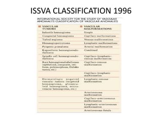
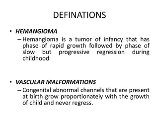
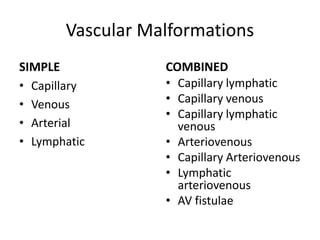
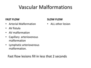
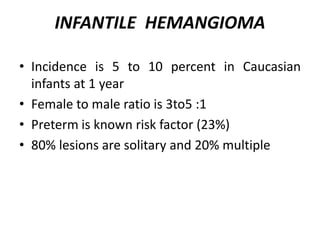
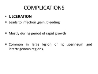
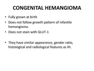
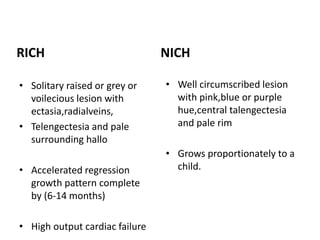
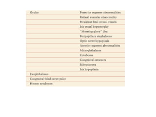
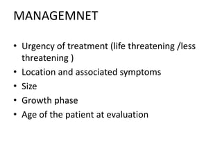
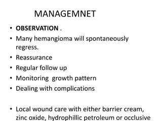
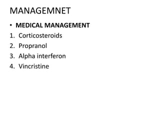
VASCULAR ANOMALIES
CLASSIFICATION
 Mullekin and Glowacki classified vascular anamolies
into hemangiomas and vascular malformation based
on
 Clinical course
 Biologic behavior
 Histological features
confusing terminology and maltreatment based on misdiagnosis has been
great impediment in the filed of vascular anomalies in past
ISSVA CLASSIFICATION 1996
• Vascular tumors have growth potential like tumors by
endothelial proliferation.

 hemangioma is most common type of vascular
tumors
 vascular malformations develop by abnormal
development of vascular channels during embryonic
life
DEFINATIONS
• HEMANGIOMA
– Hemangioma is a tumor of infancy that has
phase of rapid growth followed by phase of
slow but progressive regression during
childhood
• VASCULAR MALFORMATIONS
– Congenital abnormal channels that are present
at birth grow proportionately with the growth
of child and never regress.
Vascular Malformations
SIMPLE
• Capillary
• Venous
• Arterial
• Lymphatic
COMBINED
• Capillary lymphatic
• Capillary venous
• Capillary lymphatic
venous
• Arteriovenous
• Capillary Arteriovenous
• Lymphatic
arteriovenous
• AV fistulae
Vascular Malformations
FAST FLOW
• Arterial Malformation
• AV fistula
• AV malformation
• Capillary arteriovenous
malformation
• Lymphatic arteriovenous
malformation.
SLOW FLOW
• ALL other lesion
Fast flow lesions fill in less that 2 seconds
INFANTILE HEMANGIOMA
• Incidence is 5 to 10 percent in Caucasian
infants at 1 year
• Female to male ratio is 3to5 :1
• Preterm is known risk factor (23%)
• 80% lesions are solitary and 20% multiple
PATHOGENESIS
• Clonal expansion of hemangioma initiating multipotent cells
• These cells form blood vessels which express GLUT-1 and
merosin
• Low expression of VEGFR-1 and missense mutation in VEGFR_2
TEM-8.
• Hemagioma is basically abnormal endothelial cells that are
derived from mutated stem cells
• Characteristic of infantile hemagioma is GLUT_1 which stain
with immunostains
CLINICAL COURSE
• Characteristic 3 phase growth pattern .
• Proliferative phase
• Involuting phase
• involuted phase
CLINICAL COURSE
• PROLIFERATIVE PHASE :
• Proliferation occurs in rapid growth phase in
first 8-10 years with cessation by one year of
life
• Superficial component: bright red well
demarcated non compressible plaque
• deep components: ill defined subcutaneous
mass that has bluish hue.
• INVOLUTING PHASE
• Color changes to purplish with increased pallor
and decreased turgor
• This phase can last somewhere between 2 to 10
years
• INVOLUTED PHASE
• Roughly 50% regression at 5 years
• 70% regression at 7 years with continued
COMPLICATIONS
• ULCERATION
 Leads to infection ,pain ,bleeding
 Mostly during period of rapid growth
 Common in large lesion of lip ,perineum and
intertrigenous regions.
• Eyelid hemangioma can
cause visual disturbance
strabismus and even
amblypia
• Hemangioma in neck and
mandible can be assocaited
CONGENITAL HEMANGIOMA
• Fully grown at birth
• Does not follow growth pattern of infantile
hemangioma.
• Does not stain with GLUT-1
• They have similar appearance, gender ratio,
histological and radiological features as IH.
RICH
• Solitary raised or grey or
voilecious lesion with
ectasia,radialveins,
• Telengectesia and pale
surrounding hallo
• Accelerated regression
growth pattern complete
by (6-14 months)
• High output cardiac failure
NICH
• Well circumscribed lesion
with pink,blue or purple
hue,central talengectesia
and pale rim
• Grows proportionately to a
child.
DIAGNOSIS
• Detailed clinical history
• Examination
• U/s (operator depends)
• MRI (gold standard)
ANOMALIES WITH HEMANGIOMA
MANAGEMNET
• Urgency of treatment (life threatening /less
threatening )
• Location and associated symptoms
• Size
• Growth phase
• Age of the patient at evaluation
MANAGEMNET
• OBSERVATION .
• Many hemangioma will spontaneously
regress.
• Reassurance
• Regular follow up
• Monitoring growth pattern
• Dealing with complications
• Local wound care with either barrier cream,
zinc oxide, hydrophillic petroleum or occlusive
MANAGEMNET
• MEDICAL MANAGEMENT
1. Corticosteroids
2. Propranol
3. Alpha interferon
4. Vincristine
Corticosteroids
• Overall response rate of 85%
• Inhibition of vasculogenic potential of
hemangioma derived stem cells
• Down regulation of VEGF-A,urokinase
plasminogenactivator, monocyte
chemoattractant protein 1 ,interlukin-6,MMP-1
• Route of administration can be intralesional
Corticosteroids
• Recommended dose is 2-3mg/kg of oral
prednisolone
• Intralesional dose is repeated every 6-8 weeks
• Oral dose is given as single morning dose for 6-8
weeks and then tapered gradually .
• Common side effects are cushingoid faces.
• Irritabbility,hypertension.immunosuppresion,hir
sutism,myopathy,cardiomyopathy and
premature thelarche .
PROPRANOLOL
• Remarkable literature on propranolol
treatment was published in 2008
• First line of treatment in many centers .
• Mechanism of action is not exactly known but
a possible mechanism is inhibition of hypoxia
inducible factor 1-alpha-VEGF signaling
pathway
PROPRANOLOL
• Adverse effects are hypotenstion, hypoglycemia,
bradycardia
• gradual dose increment and tapering and patient
education regarding side effects is important part
of propranolol treatment
INTERFERON ALPHA
• Interferron alpha 2a and 2b is second line agent
for life threatening and vital function
compromising hemangiomas.
• Indications
• Failure to respond to corticosteroids
• Side effects of corticosteroids
• parental refusal to corticosteriods use
• Dose is 2-3mU/m2 and increased with child
growth
• Interferron is effective in kassabach-merritt
KASSABACH-MERRITT
PHENONMENON
• Consumption coagulopathy as platelets are trapped in
vascular channels and destroyed with thromboctopenia,loss
of clotting factors ,DIC and even death
• Classically its assocaited with IH but also with kaposifrom
hemangioendothelioma and tufted angioma
• No platelete transfusion untill bleeding or surgical procedure
indicated
• Do not use heparin it aggravates situation .
• Fever is common side effect (acetaminophen)
LASERS IN HEMANGIOMA Rx
• PDL is used in relatively superficial flat lesions
• Most useful for residual talengectasia after
regression
• Nd-YAG and KPT lasers are also used in
hemangiomas but have higher rate of scarring
.
SURGICAL MANAGMENT
• Indications for surgery are
• For residual scarring
• Localized lesions for cosmetic reasons
• Persistent ulceration and bleeding
• School going children for normal appearance
VASULAR MALFORMATIONS
• 0.3 to 0.5%population with no gender predilection
• Each of the 4 basic types have characteristic
histopathological appearance
• Multidisciplinary team approach is best whenever
warranted .
INVESTIGATIONS
• MRI is gold standard with superb details of
soft tissue.
• MRA and MRV helps further in slow and fast
flow lesions
• Plain radiograph can help in skeletal growth
abnormalities and venous phlebitis
• US and Doppler ultrasound is help but
operator dependent
• Role of CT is limited except for intraosseous
anomalies
CAPILLARY MALFORMATIONS
• Most common anomalies with 3 in 1000 live
births and equal gender distribution
• Present at birth like red or pink ntradermal
discoloration
• True CMs thicken ,darken and becmoes
nodular with age
• A variant called macular stain and named
SYNDROMES ASSOCIATED WITH
CMs
• Klippel-trenauny syndrome (slow flow CLVM
with limb hemi hypertrophy and axial elongation)
• Parkes-webber syndrome
• (AVM, cutaneous CM and skeletal and soft tissue
hypertrophy of limb)
• Cobbs syndrome (AVM of spinal cord with CM of
back )
TREATMENT
• Flashlamp pumped PDL is treatment of choice
for CMs
• Those not responding to PDL , Nd-YAG,
alexendrite and IPL are other options
• Surgical option can be considered in selected
patients
VENOUS MALFORMATIONS
• Soft ,compressible blue subcutaneous masses
• Enlarge with physical activity in dependent
portions
• Lesions are typically painful in morning due to
stasis and microthrombi
• Head and neck is most common site
Management
• MRI is extremely useful for diagnosis and
combined with venography helps in surgical
planning
• Coagulation profile should be checked due to
coagulopathy and rsik of DIC is there folowing
trauma and intervention
• Percutaneous sclerotherapy is first line treatment
• Compression stockings and aspirin for phlebitis
are adjuncts
MANAGEMENT
• SURGERY is useful for
• cosmetic reasons specially in head and neck .
• symptomatic patients with bleeding.
• Painful lesions .
• Well localized lesions.
• Debulking is considered in hand and feet
lesion and intramuscular lesions in thigh and
leg.
AV MALFORMATIONS
• Fast flow connection without capillary bed.
• 40-60% patient present at birth ,equal gender
predilection
• Epicenter of AVM is called nidus
• Consists of feeding vessels ,micro and macro
fistulas and ectatic veins
SCHOBINGER STAGING SYSTEM
• Stage 1 (quiescent phase) from birth to adolescence,
asymptomatic
• Stage 2 (progressive phase)
AVM enlarged ,darkens with thrill and palpable pulse and
murmur on auscultation
Trauma, puberty and pregnancy leads to this stage
• Stage 3 (destructive phase)
• Ulceration, pain, bleeding and bone erosions
• Stage 4 (decompensation phase )
• Cardiac decomposition with heart failure
Treatment
• Localized lesions can be excised and
reconstructed
• Large and diffuse lesions can be embolised or
super selected embolized and resected 24-48
hours later
• Counseling for recurrence and monitoring for
years for recurrence
LYMPHATIC MALFORMATION
• Anomalous channels, vesicles and pouches filled
with lymph fluid.
• LMs never regress but expand and contract
depending on ebb n flow of lymphatic fluid
• Classified
• Microcystic
• Macrocystic
• Combined micro-macrocystic
• Macrocytic lesions are mostly located at head,
neck and axilla reffered to as cystic hygroma
• Head and neck LMs are also characterized by
skeletal hypertrophy
• Microcytic LMs are usually located on proximal
extremities.chest and axilla termed as
lymphangioma circumscriptum
Treatment
SURGICAL RESECTION
 Only way to potentially cure LMs
 Complete excision may not be possible in may
areas
PERCUTANEOUS SCLEROTHERAPY
 Recently gained popularity.
 Mainly effective in macrocystic variety.
VASCULAR ANOMALIES.pptx

VASCULAR ANOMALIES.pptx

  • 1.
  • 2.
    CLASSIFICATION  Mullekin andGlowacki classified vascular anamolies into hemangiomas and vascular malformation based on  Clinical course  Biologic behavior  Histological features confusing terminology and maltreatment based on misdiagnosis has been great impediment in the filed of vascular anomalies in past
  • 3.
  • 4.
    • Vascular tumorshave growth potential like tumors by endothelial proliferation.   hemangioma is most common type of vascular tumors  vascular malformations develop by abnormal development of vascular channels during embryonic life
  • 5.
    DEFINATIONS • HEMANGIOMA – Hemangiomais a tumor of infancy that has phase of rapid growth followed by phase of slow but progressive regression during childhood • VASCULAR MALFORMATIONS – Congenital abnormal channels that are present at birth grow proportionately with the growth of child and never regress.
  • 6.
    Vascular Malformations SIMPLE • Capillary •Venous • Arterial • Lymphatic COMBINED • Capillary lymphatic • Capillary venous • Capillary lymphatic venous • Arteriovenous • Capillary Arteriovenous • Lymphatic arteriovenous • AV fistulae
  • 7.
    Vascular Malformations FAST FLOW •Arterial Malformation • AV fistula • AV malformation • Capillary arteriovenous malformation • Lymphatic arteriovenous malformation. SLOW FLOW • ALL other lesion Fast flow lesions fill in less that 2 seconds
  • 8.
    INFANTILE HEMANGIOMA • Incidenceis 5 to 10 percent in Caucasian infants at 1 year • Female to male ratio is 3to5 :1 • Preterm is known risk factor (23%) • 80% lesions are solitary and 20% multiple
  • 9.
    PATHOGENESIS • Clonal expansionof hemangioma initiating multipotent cells • These cells form blood vessels which express GLUT-1 and merosin • Low expression of VEGFR-1 and missense mutation in VEGFR_2 TEM-8. • Hemagioma is basically abnormal endothelial cells that are derived from mutated stem cells • Characteristic of infantile hemagioma is GLUT_1 which stain with immunostains
  • 10.
    CLINICAL COURSE • Characteristic3 phase growth pattern . • Proliferative phase • Involuting phase • involuted phase
  • 11.
    CLINICAL COURSE • PROLIFERATIVEPHASE : • Proliferation occurs in rapid growth phase in first 8-10 years with cessation by one year of life • Superficial component: bright red well demarcated non compressible plaque • deep components: ill defined subcutaneous mass that has bluish hue.
  • 12.
    • INVOLUTING PHASE •Color changes to purplish with increased pallor and decreased turgor • This phase can last somewhere between 2 to 10 years • INVOLUTED PHASE • Roughly 50% regression at 5 years • 70% regression at 7 years with continued
  • 13.
    COMPLICATIONS • ULCERATION  Leadsto infection ,pain ,bleeding  Mostly during period of rapid growth  Common in large lesion of lip ,perineum and intertrigenous regions.
  • 14.
    • Eyelid hemangiomacan cause visual disturbance strabismus and even amblypia • Hemangioma in neck and mandible can be assocaited
  • 15.
    CONGENITAL HEMANGIOMA • Fullygrown at birth • Does not follow growth pattern of infantile hemangioma. • Does not stain with GLUT-1 • They have similar appearance, gender ratio, histological and radiological features as IH.
  • 16.
    RICH • Solitary raisedor grey or voilecious lesion with ectasia,radialveins, • Telengectesia and pale surrounding hallo • Accelerated regression growth pattern complete by (6-14 months) • High output cardiac failure NICH • Well circumscribed lesion with pink,blue or purple hue,central talengectesia and pale rim • Grows proportionately to a child.
  • 17.
    DIAGNOSIS • Detailed clinicalhistory • Examination • U/s (operator depends) • MRI (gold standard)
  • 18.
  • 22.
    MANAGEMNET • Urgency oftreatment (life threatening /less threatening ) • Location and associated symptoms • Size • Growth phase • Age of the patient at evaluation
  • 23.
    MANAGEMNET • OBSERVATION . •Many hemangioma will spontaneously regress. • Reassurance • Regular follow up • Monitoring growth pattern • Dealing with complications • Local wound care with either barrier cream, zinc oxide, hydrophillic petroleum or occlusive
  • 24.
    MANAGEMNET • MEDICAL MANAGEMENT 1.Corticosteroids 2. Propranol 3. Alpha interferon 4. Vincristine
  • 25.
    Corticosteroids • Overall responserate of 85% • Inhibition of vasculogenic potential of hemangioma derived stem cells • Down regulation of VEGF-A,urokinase plasminogenactivator, monocyte chemoattractant protein 1 ,interlukin-6,MMP-1 • Route of administration can be intralesional
  • 26.
    Corticosteroids • Recommended doseis 2-3mg/kg of oral prednisolone • Intralesional dose is repeated every 6-8 weeks • Oral dose is given as single morning dose for 6-8 weeks and then tapered gradually . • Common side effects are cushingoid faces. • Irritabbility,hypertension.immunosuppresion,hir sutism,myopathy,cardiomyopathy and premature thelarche .
  • 27.
    PROPRANOLOL • Remarkable literatureon propranolol treatment was published in 2008 • First line of treatment in many centers . • Mechanism of action is not exactly known but a possible mechanism is inhibition of hypoxia inducible factor 1-alpha-VEGF signaling pathway
  • 28.
    PROPRANOLOL • Adverse effectsare hypotenstion, hypoglycemia, bradycardia • gradual dose increment and tapering and patient education regarding side effects is important part of propranolol treatment
  • 29.
    INTERFERON ALPHA • Interferronalpha 2a and 2b is second line agent for life threatening and vital function compromising hemangiomas. • Indications • Failure to respond to corticosteroids • Side effects of corticosteroids • parental refusal to corticosteriods use • Dose is 2-3mU/m2 and increased with child growth • Interferron is effective in kassabach-merritt
  • 30.
    KASSABACH-MERRITT PHENONMENON • Consumption coagulopathyas platelets are trapped in vascular channels and destroyed with thromboctopenia,loss of clotting factors ,DIC and even death • Classically its assocaited with IH but also with kaposifrom hemangioendothelioma and tufted angioma • No platelete transfusion untill bleeding or surgical procedure indicated • Do not use heparin it aggravates situation . • Fever is common side effect (acetaminophen)
  • 31.
    LASERS IN HEMANGIOMARx • PDL is used in relatively superficial flat lesions • Most useful for residual talengectasia after regression • Nd-YAG and KPT lasers are also used in hemangiomas but have higher rate of scarring .
  • 32.
    SURGICAL MANAGMENT • Indicationsfor surgery are • For residual scarring • Localized lesions for cosmetic reasons • Persistent ulceration and bleeding • School going children for normal appearance
  • 33.
    VASULAR MALFORMATIONS • 0.3to 0.5%population with no gender predilection • Each of the 4 basic types have characteristic histopathological appearance • Multidisciplinary team approach is best whenever warranted .
  • 34.
    INVESTIGATIONS • MRI isgold standard with superb details of soft tissue. • MRA and MRV helps further in slow and fast flow lesions • Plain radiograph can help in skeletal growth abnormalities and venous phlebitis • US and Doppler ultrasound is help but operator dependent • Role of CT is limited except for intraosseous anomalies
  • 35.
    CAPILLARY MALFORMATIONS • Mostcommon anomalies with 3 in 1000 live births and equal gender distribution • Present at birth like red or pink ntradermal discoloration • True CMs thicken ,darken and becmoes nodular with age • A variant called macular stain and named
  • 36.
    SYNDROMES ASSOCIATED WITH CMs •Klippel-trenauny syndrome (slow flow CLVM with limb hemi hypertrophy and axial elongation) • Parkes-webber syndrome • (AVM, cutaneous CM and skeletal and soft tissue hypertrophy of limb) • Cobbs syndrome (AVM of spinal cord with CM of back )
  • 37.
    TREATMENT • Flashlamp pumpedPDL is treatment of choice for CMs • Those not responding to PDL , Nd-YAG, alexendrite and IPL are other options • Surgical option can be considered in selected patients
  • 38.
    VENOUS MALFORMATIONS • Soft,compressible blue subcutaneous masses • Enlarge with physical activity in dependent portions • Lesions are typically painful in morning due to stasis and microthrombi • Head and neck is most common site
  • 39.
    Management • MRI isextremely useful for diagnosis and combined with venography helps in surgical planning • Coagulation profile should be checked due to coagulopathy and rsik of DIC is there folowing trauma and intervention • Percutaneous sclerotherapy is first line treatment • Compression stockings and aspirin for phlebitis are adjuncts
  • 40.
    MANAGEMENT • SURGERY isuseful for • cosmetic reasons specially in head and neck . • symptomatic patients with bleeding. • Painful lesions . • Well localized lesions. • Debulking is considered in hand and feet lesion and intramuscular lesions in thigh and leg.
  • 41.
    AV MALFORMATIONS • Fastflow connection without capillary bed. • 40-60% patient present at birth ,equal gender predilection • Epicenter of AVM is called nidus • Consists of feeding vessels ,micro and macro fistulas and ectatic veins
  • 42.
    SCHOBINGER STAGING SYSTEM •Stage 1 (quiescent phase) from birth to adolescence, asymptomatic • Stage 2 (progressive phase) AVM enlarged ,darkens with thrill and palpable pulse and murmur on auscultation Trauma, puberty and pregnancy leads to this stage • Stage 3 (destructive phase) • Ulceration, pain, bleeding and bone erosions • Stage 4 (decompensation phase ) • Cardiac decomposition with heart failure
  • 43.
    Treatment • Localized lesionscan be excised and reconstructed • Large and diffuse lesions can be embolised or super selected embolized and resected 24-48 hours later • Counseling for recurrence and monitoring for years for recurrence
  • 44.
    LYMPHATIC MALFORMATION • Anomalouschannels, vesicles and pouches filled with lymph fluid. • LMs never regress but expand and contract depending on ebb n flow of lymphatic fluid • Classified • Microcystic • Macrocystic • Combined micro-macrocystic
  • 45.
    • Macrocytic lesionsare mostly located at head, neck and axilla reffered to as cystic hygroma • Head and neck LMs are also characterized by skeletal hypertrophy • Microcytic LMs are usually located on proximal extremities.chest and axilla termed as lymphangioma circumscriptum
  • 46.
    Treatment SURGICAL RESECTION  Onlyway to potentially cure LMs  Complete excision may not be possible in may areas PERCUTANEOUS SCLEROTHERAPY  Recently gained popularity.  Mainly effective in macrocystic variety.