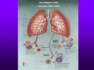
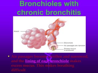
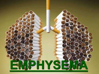
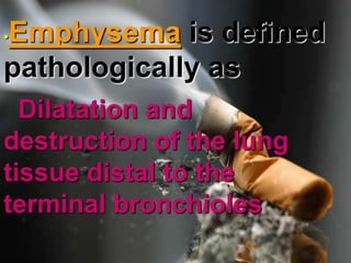
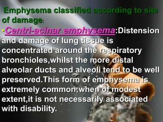
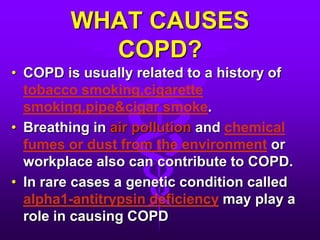
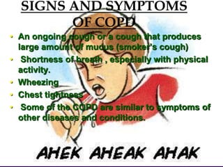
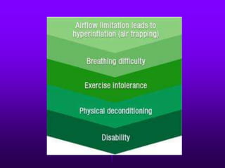
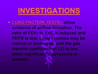
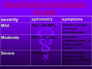
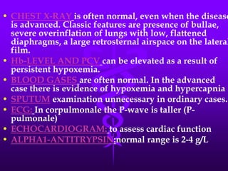
COPDCHRONIC OBSTRUCTIVE PULMONARY DISEASE
According to American Thoracic Society,  COPD is defined as  A disorder characterized by abnormal test of expiratory flow (structural or functional) that do not change markedly over periods of several months of obstruction.It is a progressive disease that makes it hard to breath. Progressive means this gets worse over time
COPD is a very frequent respiratory disorder affecting millions of people in India. It forms the most important cause of chronic cor pulmonale.
SYNONYMSChronic obstructive lung disease (COLD)Chronic obstructive airways disease (COAD)Chronic airflow obstruction (CAO)
COPD is a common term applied toCHRONIC BRONCHITISEMPHYSEMASmall airways diseaseASTHMA NOT included in COPD
CHRONIC BRONCHITIS
Chronic bronchitis is defined on the basis of the history as  Cough productive of sputum on most days for at least three months of the year for more than one year.
Bronchioles with chronic bronchitisAir passages become swollen and narrowed, and thelining of each bronchiolemakes excess mucus. This makes breathing difficult
EMPHYSEMA
.•Emphysemais defined pathologically asDilatation and destruction of the lung tissue distal to the terminal bronchioles
Emphysema classified according to site of damage:•Centri-acinar emphysema:Distension and damage of lung tissue is concentrated around the respiratory bronchioles,whilst the more distal alveolar ducts and alveoli tend to be well preserved.This form of emphysema is extremely common;when of modest extent,it is not necessarily associated with disability.   
•Pan-acinar emphysema:. This is less common.Here,distension and destruction appear to involve the whole of the acinus,and in the extreme form the lung becomes a mass of bullae. Severe airflow limitation and VA/Q mismatch occur. •Occurs in alpha1-antitrypsin deficiency
Irregular emphysema:There is                                                                                                                scarring and damage affecting the lung parenchyma patchily without particular regard for acinar structure. 
Clinical observations led to suggestions that there were two distinct type of patientsTYPE-Afighter ispink and puffing.Although the person is breathless,arterial tensions of oxygen and carbon dioxide are normal and there isno cor pulmonale.These individuals were thought to be suffering predominantly from emphysema withlittleemphysema.
TYPE-B non-fighter,on the otherhand,isblue and bloated  .The person does not appear to be breathless but has marked arterial hypoxemia,carbon dioxide retention,secondary polycythemia and cor pulmonale.these patients were thought to be suffering predominantly from chronic bronchitis.
WHAT CAUSES COPD?COPD is usually related to a history of tobacco smoking,cigarette smoking,pipe&cigar smoke.Breathing in air pollution and chemical fumes or dust from the environment or workplace also can contribute to COPD.In rare cases a genetic condition called alpha1-antitrypsin deficiency may play a role in causing COPD
SIGNS AND SYMPTOMS OF COPDAn ongoing cough or a cough that produces large amount of mucus (smoker’s cough) Shortness of breath , especially with physical activity.WheezingChest tightness Some of the COPD are similar to symptoms of other diseases and conditions.
INVESTIGATIONSLUNG FNCTION TESTS : show evidence of airflow limitation. The ratio of FEV1 to FVC is reduced and PEFR is low. Lung volumes may be normal or increased, and the gas transfer coefficient of CO is low when significant emphysema is present.
Classification and diagnosis of copd
CHEST X-RAYis often normal, even when the disease is advanced. Classic features are presence of bullae, severe overinflation of lungs with low, flattened diaphragms, a large retrosternal airspace on the lateral film.Hb-LEVEL AND PCVcan be elevated as a result of persistent hypoxemia.BLOOD GASESare often normal. In the advanced case there is evidence of hypoxemia and hypercapnia . SPUTUM examination unnecessary in ordinary cases.ECG:In corpulmonale the P-wave is taller (P-pulmonale)ECHOCARDIOGRAM:to assess cardiac functionALPHA1-ANTITRYPSIN:normal range is 2-4 g/L
TREATMENTThe single most important aspect in management of COPD is to persuade the patient to stop smoking.
DRUG THERAPY  This is used both for the short-term management of exacerbations and for long term relief of symptoms.
PREVENTION
IF NOT PREVENTED
PRESENTED BY:RENU SHARDA2008 BATCH
Copd

Copd

  • 1.
  • 2.
    According to AmericanThoracic Society, COPD is defined as A disorder characterized by abnormal test of expiratory flow (structural or functional) that do not change markedly over periods of several months of obstruction.It is a progressive disease that makes it hard to breath. Progressive means this gets worse over time
  • 3.
    COPD is avery frequent respiratory disorder affecting millions of people in India. It forms the most important cause of chronic cor pulmonale.
  • 5.
    SYNONYMSChronic obstructive lungdisease (COLD)Chronic obstructive airways disease (COAD)Chronic airflow obstruction (CAO)
  • 6.
    COPD is acommon term applied toCHRONIC BRONCHITISEMPHYSEMASmall airways diseaseASTHMA NOT included in COPD
  • 7.
  • 8.
    Chronic bronchitis isdefined on the basis of the history as Cough productive of sputum on most days for at least three months of the year for more than one year.
  • 9.
    Bronchioles with chronicbronchitisAir passages become swollen and narrowed, and thelining of each bronchiolemakes excess mucus. This makes breathing difficult
  • 10.
  • 11.
    .•Emphysemais defined pathologicallyasDilatation and destruction of the lung tissue distal to the terminal bronchioles
  • 12.
    Emphysema classified accordingto site of damage:•Centri-acinar emphysema:Distension and damage of lung tissue is concentrated around the respiratory bronchioles,whilst the more distal alveolar ducts and alveoli tend to be well preserved.This form of emphysema is extremely common;when of modest extent,it is not necessarily associated with disability.  
  • 13.
    •Pan-acinar emphysema:. Thisis less common.Here,distension and destruction appear to involve the whole of the acinus,and in the extreme form the lung becomes a mass of bullae. Severe airflow limitation and VA/Q mismatch occur. •Occurs in alpha1-antitrypsin deficiency
  • 14.
    Irregular emphysema:There is scarring and damage affecting the lung parenchyma patchily without particular regard for acinar structure. 
  • 16.
    Clinical observations ledto suggestions that there were two distinct type of patientsTYPE-Afighter ispink and puffing.Although the person is breathless,arterial tensions of oxygen and carbon dioxide are normal and there isno cor pulmonale.These individuals were thought to be suffering predominantly from emphysema withlittleemphysema.
  • 17.
    TYPE-B non-fighter,on theotherhand,isblue and bloated .The person does not appear to be breathless but has marked arterial hypoxemia,carbon dioxide retention,secondary polycythemia and cor pulmonale.these patients were thought to be suffering predominantly from chronic bronchitis.
  • 18.
    WHAT CAUSES COPD?COPDis usually related to a history of tobacco smoking,cigarette smoking,pipe&cigar smoke.Breathing in air pollution and chemical fumes or dust from the environment or workplace also can contribute to COPD.In rare cases a genetic condition called alpha1-antitrypsin deficiency may play a role in causing COPD
  • 20.
    SIGNS AND SYMPTOMSOF COPDAn ongoing cough or a cough that produces large amount of mucus (smoker’s cough) Shortness of breath , especially with physical activity.WheezingChest tightness Some of the COPD are similar to symptoms of other diseases and conditions.
  • 22.
    INVESTIGATIONSLUNG FNCTION TESTS: show evidence of airflow limitation. The ratio of FEV1 to FVC is reduced and PEFR is low. Lung volumes may be normal or increased, and the gas transfer coefficient of CO is low when significant emphysema is present.
  • 23.
  • 24.
    CHEST X-RAYis oftennormal, even when the disease is advanced. Classic features are presence of bullae, severe overinflation of lungs with low, flattened diaphragms, a large retrosternal airspace on the lateral film.Hb-LEVEL AND PCVcan be elevated as a result of persistent hypoxemia.BLOOD GASESare often normal. In the advanced case there is evidence of hypoxemia and hypercapnia . SPUTUM examination unnecessary in ordinary cases.ECG:In corpulmonale the P-wave is taller (P-pulmonale)ECHOCARDIOGRAM:to assess cardiac functionALPHA1-ANTITRYPSIN:normal range is 2-4 g/L
  • 25.
    TREATMENTThe single mostimportant aspect in management of COPD is to persuade the patient to stop smoking.
  • 26.
    DRUG THERAPY This is used both for the short-term management of exacerbations and for long term relief of symptoms.
  • 27.
  • 30.
  • 32.