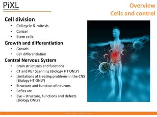
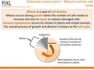
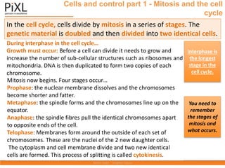
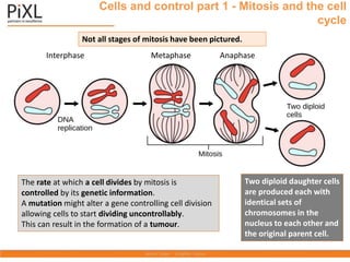
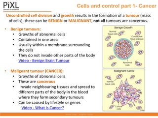
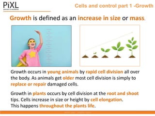
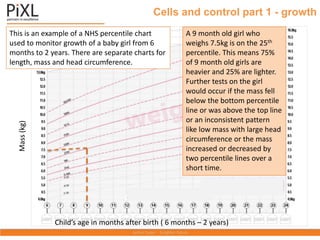
The PiXL Club The PiXL Club The PiXL Club The PiXL Club The PiXL Club The PiXL Club The PiXL Club The PiXL Club The PiXL Club The PiXL Club The PiXL Club The PiXL Club The PiXL Club The PiXL Club
The PiXL Club The PiXL Club The PiXL Club XL Club The PiXL Club The PiXL Club The PiXL Club The PiXL Club The PiXL Club The PiXL Club The PiXL Club The PiXL Club The PiXL Club The PiXL Club The
PiXL Club The PiXL Club The PiXL Club The PiXL Club The PiXL Club The PiXL Club The PiXL Club The PiXL Club The PiXL Club The PiXL Club The PiXL Club The PiXL Club The PiXL Club The PiXL Club The
This resource is strictly for the use of member schools for as long as they remain members of The PiXL Club. It may not be copied, sold nor transferred to
a third party or used by the school after membership ceases. Until such time it may be freely used within the member school.
All opinions and contributions are those of the authors. The contents of this resource are not connected with nor endorsed by any other company,
organisation or institution.
www.pixl.org.uk The PiXL Club Ltd, Company number 07321607
© Copyright The PiXL Club Ltd, 2017
PiXL KnowIT!
GCSE Biology
Edexcel Topic – Cells and Control
Overview
Cells and control
Cell division
• Cell cycle & mitosis
• Cancer
• Stem cells
Growth and differentiation
• Growth
• Cell differentiation
Central Nervous System
• Brain structures and functions
• CT and PET Scanning (Biology HT ONLY)
• Limitations of treating problems in the CNS
(Biology HT ONLY)
• Structure and function of neurons
• Reflex arc
• Eye – structure, functions and defects
(Biology ONLY)
LearnIT!
KnowIT!
Cell division
(2.1-2.9)
• Mitosis and the cell cycle
• Cancer
• Stem cells
• Growth and cell differentiation
Cells and control part 1 - Mitosis and the cell
cycle
Mitosis is a type of cell division.
Mitosis occurs during growth when the number of cells needs to
increase and also to repair or replace damaged cells.
Asexual reproduction occurs by mitosis in plants and simple animals.
The overall process of growth and division is known as the cell cycle.
Mitosis Growth of the cell and
increase in number of
sub-cellular structures
DNA replication occurs. Each
chromosome is copied.
Interphase
Cytokinesis
Cells and control part 1 - Mitosis and the cell
cycle
During interphase in the cell cycle…
Growth must occur: Before a cell can divide it needs to grow and
increase the number of sub-cellular structures such as ribosomes and
mitochondria. DNA is then duplicated to form two copies of each
chromosome.
Mitosis now begins. Four stages occur…
Prophase: the nuclear membrane dissolves and the chromosomes
become shorter and fatter.
Metaphase: the spindle forms and the chromosomes line up on the
equator.
Anaphase: the spindle fibres pull the identical chromosomes apart
to opposite ends of the cell.
Telophase: Membranes form around the outside of each set of
chromosomes. These are the nuclei of the 2 new daughter cells.
The cytoplasm and cell membrane divide and two new identical
cells are formed. This process of splitting is called cytokinesis.
In the cell cycle, cells divide by mitosis in a series of stages. The
genetic material is doubled and then divided into two identical cells.
Interphase is
the longest
stage in the
cell cycle.
You need to
remember
the stages of
mitosis and
what occurs.
Cells and control part 1 - Mitosis and the cell
cycle
Metaphase Anaphase
Two diploid daughter cells
are produced each with
identical sets of
chromosomes in the
nucleus to each other and
the original parent cell.
Interphase
Not all stages of mitosis have been pictured.
The rate at which a cell divides by mitosis is
controlled by its genetic information.
A mutation might alter a gene controlling cell division
allowing cells to start dividing uncontrollably.
This can result in the formation of a tumour.
Uncontrolled cell division and growth results in the formation of a tumour (mass
of cells), these can be BENIGN or MALIGNANT, not all tumours are cancerous.
• Benign tumours:
• Growths of abnormal cells
• Contained in one area
• Usually within a membrane surrounding
the cells
• They do not invade other parts of the body
Video - Benign Brain Tumour
• Malignant tumour (CANCER):
• Growths of abnormal cells
• These are cancerous
• Invade neighbouring tissues and spread to
different parts of the body in the blood
where they form secondary tumours
• Can be caused by lifestyle or genes
Video - What is Cancer?
Cells and control part 1- Cancer
Cells and control part 1 -Growth
Growth is defined as an increase in size or mass.
Growth occurs in young animals by rapid cell division all over
the body. As animals get older most cell division is simply to
replace or repair damaged cells.
Growth in plants occurs by cell division at the root and shoot
tips. Cells increase in size or height by cell elongation.
This happens throughout the plants life.
Cells and control part 1 - growth
Child’s age in months after birth ( 6 months – 2 years)
Mass
(kg)
This is an example of a NHS percentile chart
used to monitor growth of a baby girl from 6
months to 2 years. There are separate charts for
length, mass and head circumference.
A 9 month old girl who
weighs 7.5kg is on the 25th
percentile. This means 75%
of 9 month old girls are
heavier and 25% are lighter.
Further tests on the girl
would occur if the mass fell
below the bottom percentile
line or was above the top line
or an inconsistent pattern
like low mass with large head
circumference or the mass
increased or decreased by
two percentile lines over a
short time.
Cells and control part 1 - Cell differentiation
Most types of ANIMAL cells
differentiate in the early stage
of development.
Cell differentiation occurs as organisms develop and the cell
changes to become specialised. As the cell differentiates, it
forms different sub-cellular structures, e.g. the tail on a sperm
cell or the hairs on a root hair cell.
Most types of PLANT cells
can differentiate
throughout their life cycle.
Human embryo stem cells can be cloned and made to
differentiate into most different types of human cells.
Human adult stem cells can form many (but not all)
types of cells including blood cells.
Human stem cells can be used to help treat diseases
like diabetes and paralysis.
Stem cells are undifferentiated cells within an organism. They can produce
other stem cells that can then differentiate into many different types of cells.
Embryos produced by therapeutic cloning have the same genes as the patient.
This means stem cells from the embryo are not rejected by the patient’s body.
This is why they can be used for medical treatments.
The risks of using stem cells risks such as the transfer of viral infections.
Some people have objections to stem cell use for ethical and religious reasons.
During fertility treatment doctors usually fertilise many more eggs than are going to
be used. The embryos formed are used to obtain stem cells.
In the UK, scientists can use these embryos for research but only under very strict
guidelines.
Cells and control part 1 –Stem cells
Cells and control part 1- Stem Cells (plants)
Most types of PLANT cells can differentiate
throughout their life cycle.
Undifferentiated stem cells in plants are grouped
together in structures called meristems.
The undifferentiated cells can then specialise e.g.
root hair cell, xylem or phloem cells.
Stem cells from meristems in plants can be used to
produce clones of plants quickly and economically.
• Rare species can be cloned to protect from
extinction.
• Crop plants with special features, such as disease
resistance, can be cloned to produce large numbers
of identical plants for farmers
e.g. potatoes, strawberries and dates.
QuestionIT!
Cell division
• Mitosis and the cell cycle
• Cancer
• Stem cells
• Growth and differentiation
1. What is mitosis?
2. What are the three main stages of the cell cycle?
3. List the 4 stages involved in mitosis, starting with prophase.
4. Why is mitosis important to the organism?
5. How many daughter cells are formed as a result of mitosis?
6. How many chromosomes are present in a human diploid cell?
7. What is cancer?
8. What is the definition of growth?
9. Does cell elongation occur in animal or plant growth?
10. What is cell differentiation?
11. If a baby was described as being on the 50th percentile for head
circumference– what does that mean?
12. What is a stem cell?
13. How is a human embryonic stem cell different to an adult stem cell?
14. Where is a meristem found?
15. What is a meristem?
16. Name a benefit and a risk to using stem cells in medicine.
Cells and control part 1 - Question IT
17. In what stage of an animal’s life cycle do most cells differentiate?
18. In mature animals when do cells still need to differentiate?
19. In what stage of their life cycle do plant cells differentiate?
Cells and control part 1 - Question IT
AnswerIT!
Cell division
• Mitosis and the cell cycle
• Cancer
• Stem cells
• Growth and differentiation
1. What is mitosis? A type of cell division
2. What are the three stages of the cell cycle? Interphase, Mitosis,
Cytokinesis
3. List the stages involved in mitosis, starting with prophase.
Prophase, metaphase, anaphase, telophase
4. Why is mitosis important to the organism?
For growth
Repair or replacement of damaged cells
Asexual reproduction in some animals
5. How many daughter cells are formed as a result of mitosis? 2
6. How many chromosomes are present in a human diploid cell? 46
7. What is cancer? Uncontrolled cell division as a result of changes
in cells.
8. What is the definition of growth? Increase in size or number of
cells.
Cells and control part 1 – AnswerIT
9. Does cell elongation occur in animal or plant growth? Plant
10. What is cell differentiation? The process by which a cell
changes to become specialised for a specific role.
11 If a baby was described as being on the 50th percentile for
head circumference– what does that mean? 50% of children have
a bigger head and 50% have a smaller head.
12. What is a stem cell? An undifferentiated cell.
13. How is a human embryonic stem cell different to an adult
stem cell? Embryonic stem cells have the potential to divide and
produce any type of body cell.
Adult stem cells can only differentiate to form a few types of
cells e.g. red blood cells.
14. Where is a meristem found? In a plant shoot or root tip
15. What is a meristem? This is where the plant produces
undifferentiated cells .
Cells and control part 1 – AnswerIT
16. Name a benefit and a risk to using stem cells in medicine.
Benefits: Already in use to cure some diseases by bone marrow
transplant.
Can be used to create specialised cells to replace damaged ones.
Potential for being able to find new cures to life changing
conditions such as spinal injuries or CNS diseases.
Risks: Diseases can be passed on as viruses are found inside cells
and may go undetected in the stem cells used.
Tumour development- stem cells divide very quickly and if this is
not controlled then a tumour may develop.
Rejection – stem cells which are not from the patient are likely to
trigger an immune response.
A patient undergoing stem cell treatment will have to take
immunosuppressive drugs which makes them more susceptible to
disease.
17. In what stage of an animal’s life cycle do most cells differentiate?
In the early stages.
18. In mature animals when do cells still need to differentiate?
For repair and replacement of cells.
19. In what stage of their life cycle do plant cells differentiate?
They differentiate throughout their lifecycle.
Cells and control part 1 – AnswerIT
LearnIT!
KnowIT!
The Central
Nervous system
Part 2 (2.10B -2.12B)
• Brain structures and
functions
• CT and PET Scanning
(Biology HT ONLY)
• Limitations of treating
problems in the CNS
(Biology HT ONLY)
The central nervous system Part 2 - The brain (biology only)
The brain controls complex behaviour. It is made of billions of
interconnected neurones and has different regions that carry out
different functions.
cerebral hemispheres
cerebellum
Three of the main structures in the brain are:
• Cerebral hemispheres – the outer
‘wrinkled’ layers of the brain. It is
responsible for intelligence, language,
memory and consciousness.
• Cerebellum – is located at the lower
part of the back of the brain. It is
responsible for voluntary coordination of
the muscles and balance.
• Medulla oblongata – is located in the
lower part of the brain stem. It is
responsible for involuntary (unconscious)
coordination such as breathing,
swallowing and heart rate.
medulla
oblongata
front
back
Video - The Brain
The central nervous system Part 2 - The brain (HT biology only)
A neuroscientist is a scientist that studies the structure and function
of the brain. The complexity and delicacy of the brain makes
investigating the brain and treating brain disorders very difficult.
Accessing the brain tissue
inside the hard, protective
skull can be difficult.
Neuroscientists use CT or
PET scanning to investigate
brain function.
Studying brain damage
Damage to different brain areas produces different behavioural
and psychological effects. For example, damage in the front end
of the brain disturbs the ability to make decisions whereas
damage to the back of the brain disturbs vision.
A CT (computerised tomography)
scan uses X-rays to produce
detailed 2-d images of the brain.
A CT scan results in the body being
subjected to a dose of ionising
radiation that is much greater than
for a simple X-ray as it takes
hundreds of images.
CT scans are commonly performed
and generally safe. Any increased
risk of cancer usually outweighs
the benefits of having the scan.
A CT scan shows the main structures of the brain. The function of
the brain cannot be tested using this equipment. A CT scan will
show diseased or damaged areas and this can then give a good idea
of why the brain is not functioning effectively.
The central nervous system Part 2 - The brain
(HT biology only)
A PET (positron emission
tomography) scan uses
radioactive tracer
chemicals to show which
parts of the brain are
functioning whilst in the
scanner.
It measures oxygen use
and blood flow. More
active parts of the brain
show in red or orange
colours. Green shows low
activity.
A PET scan shows both the main structures of the brain and the
function of the brain in real time. PET scans are useful for studying
disorders like Alzheimer’s disease where activity in parts of the
brain are reduced compared to a healthy brain.
The central nervous system Part 2 - The brain
(HT biology only)
The central nervous system Part 2 - The brain (biology only)
Treatment of brain or spinal cord injuries, tumours or CNS diseases
can be difficult.
Drugs do not always pass through the protective membrane which
surrounds the brain and spinal cord.
The skull and bony vertebrae means it is difficult to access the brain
and spinal cord tissue without risky surgery which can itself lead to
permanent damage being caused. Not all areas of the brain are fully
understood yet.
Neurones do not automatically repair themselves unlike some other
cells in the body. Stem cells are currently being pioneered as one
method of treating spinal cord injuries.
QuestionIT!
The Central Nervous system
Part 2
• Brain structures and functions
• CT and PET Scanning (Biology HT ONLY)
• Limitations of treating problems in the
CNS (Biology HT ONLY)
The central nervous system Part 2 - Question IT
1. Name the three main parts of the brain and state the role of
each.
2. What is a CT scan?
3. What is a PET scan?
4. What does a PET scan do?
5. What are the limitations in treating damage and disease in the
brain and nervous system?
AnswerIT!
The Central Nervous
system
Part 2
• Brain structures and functions
• CT and PET Scanning (Biology HT
ONLY)
• Limitations of treating problems in
the CNS (Biology HT ONLY)
The central nervous system Part 2 - AnswerIT
1. Name the three main parts of the brain and state the role of each.
cerebral hemispheres– the outer ‘wrinkled’ layer of the brain. It is
responsible for intelligence, language, memory and consciousness;
cerebellum – is located at the lower part of the back of the brain and is
responsible for voluntary coordination of the muscles; medulla oblongata – is
located in the lower part of the brain stem. It is responsible for involuntary
coordination such as breathing, swallowing and heart rate.
2. What is a CT scan? A scanner which uses X-rays to produce images of the brain
3. What is a PET scan? A scanner which uses radioactive chemicals to produce
images of the brain
4. What does a PET scan do? The scanner can show both structure and function
of the brain which can show if parts of the brain are abnormally active or
inactive compared to a healthy brain.
5. What are the limitations in treating damage and disease in the brain and
nervous system?
Drugs do not always pass through the protective membrane which surrounds
the brain and spinal cord. The skull and bony vertebrae means it is difficult to
access the brain and spinal cord tissue without risky surgery which can itself
lead to permanent damage being caused.
LearnIT!
KnowIT!
The Central
Nervous system
Part 3
(2.13-2.14)
• Structure and function
of neurons
• Reflex arc
The central nervous system Part 3 - Structure and function
The nervous system enables humans to react to their surroundings and
to coordinate their behaviour.
A stimulus is any change in the
soundings. These are detected by
receptors (cells that detect a change)
and information passes along cells
(neurones) as electrical impulses to the
central nervous system (CNS).
The CNS is the brain and spinal cord.
The CNS coordinates the response of
effectors which may be muscles
contracting or glands secreting
hormones.
stimulus  receptor  coordinator  effector  response
CNS
Peripheral
Nervous
System
The central nervous system Part 3 - Structure and function
There are three main types of neurones:
A. Sensory neurones – these carry impulses from the receptors to the central
nervous system (CNS).
B. Relay neurones – these connect the sensory neurones to the motor neurones in
the CNS.
C. Motor neurones – these carry impulses from the CNS to an effector.
B. Relay neurone
A . Sensory neurone
C. Motor neurone
receptor endings
dendrites
cell body
axon
axon terminals
axon
dendrites
axon
dendron
Myelin
sheath
axon
The central nervous system Part 3 - Structure and function
Neurones are not joined together. They have small gap between them.
• When an impulse reaches the end of a
neurone a chemical (neurotransmitter)
is released across the gap.
• The chemical then diffuses across the
synapse.
• When the chemical reaches the next
neurone in sufficient quantities this
starts another impulse.
The gap is called
a synapse
The central nervous system Part 3 - Structure and function
Structure Function
Receptor Specialised cells which detect stimuli and turn them into electrical
impulses
Axon Carries electrical impulses away from the cell body
Dendron Carries electrical impulses from receptor towards cell body
Dendrite Branched ends which receive electrical impulses
Myelin sheath Layer of fatty insulating material around motor and sensory neurones
which speeds up transmission of the impulse
Synapse Gap between neurones which ensures impulses travel in one direction
Neurotransmitter Chemical involved in passing impulses from one nerve cell to another
across a synapse
Effector Organ, tissue or cell which produces a response
The central nervous system Part 3 - Structure and function
Reflex action
Source of heat
Receptor cells in
the finger
Effector – muscles in
the arm
Motor neurone
Sensory neurone
Spinal cord
Reflex actions
are important as
they protect the
body from injury.
Reflex actions are automatic and rapid; they do not involve the
conscious part of the brain. The path that a reflex action takes is
called a REFLEX ARC.
stimulus  receptor  sensory neurone  relay neurone  motor neurone  effector
CNS
Relay neurone
QuestionIT!
The Central Nervous system
Part 3
• Structure and function
of neurons
• Reflex arc
1. What is a stimulus?
2. What is a receptor?
3. Name the two parts of the central nervous system.
4. What is an effector?
5. What does the CNS coordinate?
6. Put these in the correct order: receptor, stimulus, response,
coordinator, effector.
7. What is the role of the sensory neurone?
8. What is the role of the relay neurone?
9. What is the role of the motor neurone?
10. What is a synapse?
11. Describe what happens at the synapse.
12. Why are reflex actions important?
13. Recall the pathway of the reflex arc.
The central nervous system Part 3 - Question IT
The central nervous system Part 3 - Structure and function
Structure Function
Receptor
Carries electrical impulses away from the cell body
Carries electrical impulses from receptor towards cell body
Dendrite Branched ends which receive electrical impulses
Myelin sheath
Gap between neurones which ensures impulses travel in one direction.
Neurotransmitter
Organ, tissue or cell which produces a response
14. Complete the missing parts of the table below:
AnswerIT!
The Central Nervous
system
Part 3
• Structure and function
of neurons
• Reflex arc
The central nervous system Part 3 - AnswerIT
1. What is a stimulus? Any change in the surroundings
2. What is a receptor? Cells that detect a change
3. Name the two parts of the central nervous system. Brain and spinal
cord
4. What is an effector? A muscle or gland
5. What does the CNS coordinate? The response of effectors
6. Put these in the correct order: receptor, stimulus, response,
coordinator, effector.
stimulus  receptor  coordinator  effector  response
7. What is the role of the sensory neurone? Carry impulses from the
receptors to the Central nervous system (CNS)
The central nervous system Part 3 - AnswerIT
8. What is the role of the relay neurone? Connect the sensory neurones
to the motor neurones in the CNS.
9.What is the role of the motor neurone? These carry impulses from
the CNS to an effector.
10. What is a synapse? A gap between two neurones
11. Describe what happens at the synapse An impulse reaches the end
of a neurone; neurotransmitter is released across the gap. It then
diffuses across the synapse and when it reaches the next neurone this
starts another impulse.
12. Why are reflex actions important? They protect the body from
injury
13. Recall the pathway of the reflex arc. receptor  sensory neurone
 relay neurone (CNS)  motor neurone  effector
The central nervous system Part 3 - Structure and function
Structure Function
Receptor Specialised cells which detect stimuli and turn them into electrical
impulses
Axon Carries electrical impulses away from the cell body
Dendron Carries electrical impulses from receptor towards cell body
Dendrite Branched ends which receive electrical impulses
Myelin sheath Layer of fatty insulating material around motor and sensory neurones
which speeds up transmission of the impulse
Synapse Gap between neurones which ensures impulses travel in one direction.
Neurotransmitter Chemical involved in passing impulses from one nerve cell to another
across a synapse
Effector Organ, tissue or cell which produces a response
14. Complete the missing parts of the table below:
LearnIT!
KnowIT!
The Central
Nervous system
Part 4
(2.15B - 2.17B)
• Eye – structure,
functions and defects
(Biology ONLY)
The eye is a sense organ containing receptors
sensitive to light intensity and colour.
sclera
cornea
iris
ciliary muscles
suspensory ligaments
retina
optic nerve
pupil
lens
Video - The eye
The central nervous system Part 4 - The eye (biology only)
Part of the eye Description and function
cornea Transparent layer at the front of the eye. It refracts light
into the eye.
iris The coloured part of the eye that contains muscles that
control the amount of light entering the eye.
pupil The hole in the middle of the iris that lets light in. Its
diameter is controlled by the iris.
lens A transparent, biconvex structure in the eye that refracts
light onto the retina.
retina Thin layer of tissue at the back of the eye that contains
receptor cells called rods and cones for light and colour.
sclera Tough white supporting wall of the eye.
ciliary muscles Muscles that are connected to the lens by the suspensory
ligaments; they change the shape of the lens.
suspensory ligaments These connect the lens to the ciliary muscles.
optic nerve Carries impulses from the retina to the brain.
The central nervous system Part 4 - The eye (biology only)
Accommodation is the process of changing the shape of the lens to
focus on near or distant objects.
cornea
retina
lens
To focus on a near object:
• the ciliary muscles contract
• the suspensory ligaments loosen
• the lens is then thicker and
refracts light rays strongly.
To focus on a distant object:
• the ciliary muscles relax
• the suspensory ligaments are
pulled tight
• the lens is then pulled thin and
only slightly refracts light rays. Video - accommodation
The central nervous system Part 4 - The eye (biology only)
Two common defects of the eyes are myopia (short sightedness) and
hyperopia (long sightedness) in which rays of light do not focus on the retina.
Myopia (short sightedness)
• People who are short
sighted cannot focus on
objects that are far away.
• It usually occurs when the
eyeball is too long.
• The light rays from distant
objects focus in the eyeball
in front of the retina.
• Myopia can be corrected
with concave lenses so that
the light rays focus on the
retina.
Concave lens
The central nervous system Part 4 - The eye (biology only)
Convex lens
Hyperopia (long sightedness)
• People who are long sighted
cannot focus on objects
that are near.
• It usually occurs when the
eyeball is too short.
• The light rays from distant
objects focus in the eyeball
behind the retina.
• Hyperopia can be corrected
with convex lenses so that
the light rays focus on the
retina.
Using lenses to correct vision
Short Long
Colour blindness is a condition that a person is born with and
is inherited from their parents. More men are colour blind
because the gene is carried on the X chromosome.
A colour blind person is unable to tell the difference between
certain colours. The most common condition is red green
colour blindness. The red and green cones in the retina are
not functioning normally. There is no cure for this condition.
Rod cells enable us to see
objects in dim light. They do not
detect colour.
Cone cells enable us to see
objects clearly in bright light
and in colour. There are three
types of cone – red, green and
blue. They do not function well
in dim light.
The central nervous system Part 4 - The eye (biology only)
Due to a cloudy patch
on the lens, light is
unable to enter the eye
normally.
People who have
cataracts often have
blurred vision and find
it difficult to see in
bright light or in
accurate colour.
Cataracts can be cured
by replacing the lens
with an artificial one.
The central nervous system Part 4 - The eye (biology only)
There are new technologies available to correct vision including:
Contact lenses – these can be hard or soft and are
inserted directly into the eye and sit on the cornea.
This then refracts the light rays. Advantages -
lightweight, almost invisible, good for people playing
sports. Disadvantages – not a permanent solution
and they can cause irritation and infections.
Hard
contact lens
Soft
contact lens
Inserting a soft
contact lens
Laser surgery – the other layer of the cornea is peeled back
and lasers are used to change the shape of the cornea so
that the light rays will refract and form an image on the
retina. Advantages – permanent solution to correct vision.
Disadvantages – a surgical procedure, can cause infections.
Lens replacement – surgery refers to a medical procedure
where an artificial acrylic or silicone lens is implanted into
the eye to correct problems with vision. Advantages –
permanent solution to correct vision. Disadvantages – a
surgical procedure, can cause infections.
QuestionIT!
The Central Nervous system
Part 4
• Eye – structure,
functions and defects
(Biology ONLY)
1. Label parts A – I on the diagram below:
The central nervous system Part 4 - Question IT
F.
G.
A.
I.
H.
D.
B.
E.
C.
The central nervous system Part 4 - Question IT
2. Write the name of each part of the eye in the table below:
Part of the eye Description and function
Transparent layer at the front of the eye. It refracts light into the eye.
The coloured part of the eye; contains muscles that control the amount of
light entering the eye.
The hole in the middle of the iris that lets light in. Its diameter is
controlled by the iris.
A transparent, biconvex structure in the eye that refracts light onto the
retina.
Thin layer of tissue at the back of the eye that contains receptor cells for
light and colour.
Tough white supporting wall of the eye.
Muscles that are connected to the lens by the suspensory ligaments; they
change the shape of the lens.
These connect the lens to the ciliary muscles.
Carries impulses fro the retina to the brain.
The central nervous system Part 4 - Question IT
3. What is accommodation?
4. How does the eye focus on distant objects?
5. How does the eye focus on near objects?
6. What is myopia? How can it be corrected?
7. What is hyperopia? How can it be corrected?
8. What is a cataract? How can it be treated?
9. What is the most common form of colour blindness?
10. What causes colour blindness?
AnswerIT!
The Central Nervous
system
Part 4
• Eye – structure,
functions and defects
(Biology ONLY)
The central nervous system Part 4 - AnswerIT
1. Label parts A – I on the diagram below:
G. ciliary muscles
A. suspensoryligaments
I. retina
H. optic nerve
B. lens
C. cornea
D. pupil
E. iris
F. sclera
The central nervous system Part 4 - AnswerIT
2. Write the name of each part of the eye in the table below:
Part of the eye Description and function
cornea Transparent layer at the front of the eye. It refracts light into the
eye.
iris The coloured part of the eye; contains muscles that control the
amount of light entering the eye.
pupil The hole in the middle of the iris that lets light in. Its diameter is
controlled by the iris.
lens A transparent, biconvex structure in the eye that refracts light
onto the retina.
retina Thin layer of tissue at the back of the eye that contains receptor
cells for light and colour.
sclera Tough white supporting wall of the eye.
ciliary muscles Muscles that are connected to the lens by the suspensory
ligaments; they change the shape of the lens.
suspensory ligaments These connect the lens to the ciliary muscles.
optic nerve Carries impulses fro the retina to the brain.
The central nervous system Part 4 - AnswerIT
3. What is accommodation? The process of changing the shape of the lens to focus
on near or distant objects
4. How does the eye focus on distant objects? The ciliary muscles relax, the
suspensory ligaments are pulled tight, the lens is then pulled thin and only slightly
refracts light rays.
5. How does the eye focus on near objects? The ciliary muscles contract, the
suspensory ligaments loosen, the lens is then thicker and refracts light rays strongly
6. What is myopia? How can it be corrected? Short-sightedness, it can be corrected
with concave lenses.
7. What is hyperopia? How can it be corrected? Long-sightedness, it can be
corrected with convex lenses.
8. What is a cataract? How can it be treated? A cloudy patch on the lens. Treated by
replacement with an artificial lens.
9. What is the most common form of colour blindness? Red green
10. What causes colour blindness? Cones not functioning normally- it is an inherited
condition which cannot be cured.

Control-Know

  • 1.
    The PiXL ClubThe PiXL Club The PiXL Club The PiXL Club The PiXL Club The PiXL Club The PiXL Club The PiXL Club The PiXL Club The PiXL Club The PiXL Club The PiXL Club The PiXL Club The PiXL Club The PiXL Club The PiXL Club The PiXL Club The PiXL Club The PiXL Club The PiXL Club The PiXL Club The PiXL Club The PiXL Club The PiXL Club The PiXL Club The PiXL Club The PiXL Club The PiXL Club The PiXL Club The PiXL Club The PiXL Club The PiXL Club The PiXL Club The PiXL Club The PiXL Club The PiXL Club The PiXL Club The PiXL Club The PiXL Club The PiXL Club The PiXL Club The PiXL Club The PiXL Club The PiXL Club The PiXL Club XL Club The PiXL Club The PiXL Club The PiXL Club The PiXL Club The PiXL Club The PiXL Club The PiXL Club The PiXL Club The PiXL Club The PiXL Club The PiXL Club The PiXL Club The PiXL Club The PiXL Club The PiXL Club The PiXL Club The PiXL Club The PiXL Club The PiXL Club The PiXL Club The PiXL Club The PiXL Club The PiXL Club The PiXL Club The PiXL Club The PiXL Club The PiXL Club The PiXL Club The PiXL Club The PiXL Club The PiXL Club The PiXL Club The PiXL Club The PiXL Club The PiXL Club The PiXL Club The PiXL Club The PiXL Club The PiXL Club The PiXL Club The PiXL Club The PiXL Club The PiXL Club The PiXL Club The PiXL Club The PiXL Club The PiXL Club The PiXL Club The PiXL Club The PiXL Club The PiXL Club The PiXL Club The PiXL Club The PiXL Club The PiXL Club The PiXL Club The PiXL Club The PiXL Club The PiXL Club The PiXL Club The PiXL Club The PiXL Club The PiXL Club The PiXL Club The PiXL Club The PiXL Club The PiXL Club The PiXL Club The PiXL Club The PiXL Club The PiXL Club The PiXL Club The PiXL Club The PiXL Club The PiXL Club The PiXL Club The PiXL Club The PiXL Club The PiXL Club The PiXL Club The PiXL Club The PiXL Club The PiXL Club The PiXL Club The PiXL Club The PiXL Club The PiXL Club The PiXL Club The PiXL Club The PiXL Club The PiXL Club The PiXL Club The PiXL Club The PiXL Club The PiXL Club The PiXL Club The PiXL Club The PiXL Club The PiXL Club The PiXL Club The PiXL Club The PiXL Club The PiXL Club The PiXL Club The PiXL Club The PiXL Club The PiXL Club The PiXL Club The PiXL Club The PiXL Club The PiXL Club The PiXL Club The PiXL Club The PiXL Club The PiXL Club The PiXL Club The PiXL Club The PiXL Club The PiXL Club The PiXL Club The PiXL Club The PiXL Club The PiXL Club The PiXL Club The PiXL Club The PiXL Club The PiXL Club The PiXL Club The PiXL Club The PiXL Club The PiXL Club The PiXL Club The PiXL Club The PiXL Club The PiXL Club The PiXL Club The PiXL Club The PiXL Club The PiXL Club The PiXL Club The PiXL Club The PiXL Club The PiXL Club The PiXL Club The PiXL Club The PiXL Club The PiXL Club The PiXL Club The PiXL Club The PiXL Club The PiXL Club The PiXL Club The PiXL Club The PiXL Club The PiXL Club The PiXL Club The PiXL Club The PiXL Club The PiXL Club The PiXL Club The PiXL Club The PiXL Club The PiXL Club The PiXL Club The PiXL Club The PiXL Club The PiXL Club The PiXL Club The PiXL Club The PiXL Club The PiXL Club The PiXL Club The PiXL Club The PiXL Club The PiXL Club The PiXL Club The PiXL Club The PiXL Club The PiXL Club The PiXL Club The PiXL Club The PiXL Club The PiXL Club The PiXL Club The PiXL Club The PiXL Club The PiXL Club The PiXL Club The PiXL Club The PiXL Club The PiXL Club The PiXL Club The PiXL Club The PiXL Club The PiXL Club The PiXL Club The PiXL Club The PiXL Club The PiXL Club The PiXL Club The PiXL Club The PiXL Club The PiXL Club The PiXL Club The PiXL Club The PiXL Club The PiXL Club The PiXL Club The PiXL Club The PiXL Club The PiXL Club The PiXL Club The PiXL Club The PiXL Club The PiXL Club The PiXL Club The PiXL Club The PiXL Club The PiXL Club The PiXL Club The PiXL Club The PiXL Club The PiXL Club The PiXL Club The PiXL Club The PiXL Club The PiXL Club The PiXL Club The PiXL Club The PiXL Club The PiXL Club The PiXL Club The PiXL Club The PiXL Club The PiXL Club The PiXL Club The PiXL Club The PiXL Club The PiXL Club The PiXL Club The PiXL Club The PiXL Club The PiXL Club The PiXL Club The PiXL Club The PiXL Club The PiXL Club The PiXL Club The PiXL Club The PiXL Club The PiXL Club The PiXL Club The PiXL Club The PiXL Club The PiXL Club The PiXL Club The PiXL Club The PiXL Club The PiXL Club The PiXL Club The PiXL Club The PiXL Club The PiXL Club The PiXL Club The PiXL Club The PiXL Club The PiXL Club The PiXL Club The PiXL Club The PiXL Club The PiXL Club The PiXL Club The PiXL Club The PiXL Club The PiXL Club The PiXL Club The PiXL Club The PiXL Club The PiXL Club The PiXL Club The PiXL Club The PiXL Club The PiXL Club The PiXL Club The PiXL Club The PiXL Club The PiXL Club The PiXL Club The PiXL Club The PiXL Club The PiXL Club The PiXL Club The PiXL Club The PiXL Club The PiXL Club The PiXL Club The PiXL Club The PiXL Club The PiXL Club The PiXL Club The PiXL Club The PiXL Club The PiXL Club The PiXL Club The PiXL Club The PiXL Club The PiXL Club The PiXL Club The PiXL Club The PiXL Club The PiXL Club The PiXL Club The PiXL Club The PiXL Club The PiXL Club The PiXL Club The PiXL Club The PiXL Club The PiXL Club The PiXL Club The PiXL Club The PiXL Club The PiXL Club The PiXL Club The PiXL Club The PiXL Club The PiXL Club The PiXL Club The PiXL Club The PiXL Club The PiXL Club The PiXL Club The PiXL Club The PiXL Club The PiXL Club The PiXL Club The PiXL Club The PiXL Club The PiXL Club The PiXL Club The PiXL Club The PiXL Club The PiXL Club The PiXL Club The PiXL Club The PiXL Club The PiXL Club The PiXL Club The PiXL Club The PiXL Club The PiXL Club The PiXL Club The PiXL Club The PiXL Club The PiXL Club The PiXL Club The PiXL Club The PiXL Club The PiXL Club The PiXL Club The PiXL Club The PiXL Club The PiXL Club The PiXL Club The PiXL Club The PiXL Club The PiXL Club The PiXL Club The PiXL Club The PiXL Club The PiXL Club The PiXL Club The PiXL Club The PiXL Club The PiXL Club The PiXL Club The PiXL Club The PiXL Club The PiXL Club The PiXL Club The PiXL Club The PiXL Club The PiXL Club The PiXL Club The PiXL Club The PiXL Club The PiXL Club The PiXL Club The PiXL Club The PiXL Club The PiXL Club The PiXL Club The PiXL Club The PiXL Club The PiXL Club The PiXL Club The PiXL Club The PiXL Club The PiXL Club The PiXL Club The PiXL Club The PiXL Club The PiXL Club The PiXL Club The PiXL Club The PiXL Club The PiXL Club The PiXL Club The PiXL Club The PiXL Club The PiXL Club The PiXL Club The PiXL Club The PiXL Club The PiXL Club The PiXL Club The PiXL Club The PiXL Club The PiXL Club The PiXL Club The PiXL Club The PiXL Club The PiXL Club The PiXL Club The PiXL Club The PiXL Club The PiXL Club The PiXL Club The PiXL Club The PiXL Club The PiXL Club The PiXL Club The PiXL Club The PiXL Club The PiXL Club The PiXL Club The PiXL Club The PiXL Club The PiXL Club The PiXL Club The PiXL Club The PiXL Club The PiXL Club The PiXL Club The PiXL Club The PiXL Club The PiXL Club The PiXL Club The PiXL Club The PiXL Club The PiXL Club The PiXL Club The PiXL Club The PiXL Club The PiXL Club The PiXL Club The PiXL Club The PiXL Club The PiXL Club The PiXL Club The PiXL Club The PiXL Club The PiXL Club The PiXL Club The PiXL Club The PiXL Club The PiXL Club The PiXL Club The PiXL Club The PiXL Club The PiXL Club The PiXL Club The PiXL Club The PiXL Club The PiXL Club The PiXL Club The PiXL Club The PiXL Club The PiXL Club The PiXL Club The PiXL Club The PiXL Club The PiXL Club The PiXL Club The PiXL Club The PiXL Club The PiXL Club The PiXL Club The PiXL Club The PiXL Club The PiXL Club The PiXL Club The PiXL Club The PiXL Club The PiXL Club The PiXL Club The PiXL Club The PiXL Club The PiXL Club The PiXL Club The PiXL Club The PiXL Club The PiXL Club The PiXL Club The PiXL Club The PiXL Club The PiXL Club The PiXL Club The PiXL Club The PiXL Club The PiXL Club The PiXL Club The PiXL Club The PiXL Club The PiXL Club The PiXL Club The PiXL Club The PiXL Club The PiXL Club The PiXL Club The PiXL Club The PiXL Club The PiXL Club The PiXL Club The PiXL Club The PiXL Club The PiXL Club The PiXL Club The PiXL Club The PiXL Club The PiXL Club The PiXL Club The PiXL Club The PiXL Club The PiXL Club The PiXL Club The PiXL Club The PiXL Club The PiXL Club The PiXL Club The PiXL Club The PiXL Club The PiXL Club The PiXL Club The PiXL Club The PiXL Club The PiXL Club The PiXL Club The PiXL Club The PiXL Club The PiXL Club The PiXL Club The PiXL Club The PiXL Club The PiXL Club The PiXL Club The PiXL Club The PiXL Club The PiXL Club The PiXL Club The PiXL Club The PiXL Club The PiXL Club The PiXL Club The PiXL Club The PiXL Club The PiXL Club The PiXL Club The PiXL Club The PiXL Club The PiXL Club The PiXL Club The PiXL Club The PiXL Club The PiXL Club The PiXL Club The PiXL Club The PiXL Club The PiXL Club The PiXL Club The PiXL Club The PiXL Club The PiXL Club The PiXL Club The PiXL Club The PiXL Club The PiXL Club The PiXL Club The PiXL Club The PiXL Club The PiXL Club The PiXL Club The PiXL Club The PiXL Club The PiXL Club The PiXL Club The PiXL Club The PiXL Club The PiXL Club The PiXL Club The PiXL Club The PiXL Club The PiXL Club The PiXL Club The PiXL Club The PiXL Club The PiXL Club The PiXL Club The PiXL Club The PiXL Club The PiXL Club The PiXL Club The PiXL Club The PiXL Club The PiXL Club The PiXL Club The PiXL Club The PiXL Club The PiXL Club The PiXL Club The PiXL Club The PiXL Club The PiXL Club The PiXL Club The PiXL Club The PiXL Club The PiXL Club The PiXL Club The PiXL Club The PiXL Club The PiXL Club The PiXL Club The PiXL Club The PiXL Club The PiXL Club The PiXL Club The PiXL Club The PiXL Club The PiXL Club The PiXL Club The This resource is strictly for the use of member schools for as long as they remain members of The PiXL Club. It may not be copied, sold nor transferred to a third party or used by the school after membership ceases. Until such time it may be freely used within the member school. All opinions and contributions are those of the authors. The contents of this resource are not connected with nor endorsed by any other company, organisation or institution. www.pixl.org.uk The PiXL Club Ltd, Company number 07321607 © Copyright The PiXL Club Ltd, 2017 PiXL KnowIT! GCSE Biology Edexcel Topic – Cells and Control
  • 2.
    Overview Cells and control Celldivision • Cell cycle & mitosis • Cancer • Stem cells Growth and differentiation • Growth • Cell differentiation Central Nervous System • Brain structures and functions • CT and PET Scanning (Biology HT ONLY) • Limitations of treating problems in the CNS (Biology HT ONLY) • Structure and function of neurons • Reflex arc • Eye – structure, functions and defects (Biology ONLY)
  • 3.
    LearnIT! KnowIT! Cell division (2.1-2.9) • Mitosisand the cell cycle • Cancer • Stem cells • Growth and cell differentiation
  • 4.
    Cells and controlpart 1 - Mitosis and the cell cycle Mitosis is a type of cell division. Mitosis occurs during growth when the number of cells needs to increase and also to repair or replace damaged cells. Asexual reproduction occurs by mitosis in plants and simple animals. The overall process of growth and division is known as the cell cycle. Mitosis Growth of the cell and increase in number of sub-cellular structures DNA replication occurs. Each chromosome is copied. Interphase Cytokinesis
  • 5.
    Cells and controlpart 1 - Mitosis and the cell cycle During interphase in the cell cycle… Growth must occur: Before a cell can divide it needs to grow and increase the number of sub-cellular structures such as ribosomes and mitochondria. DNA is then duplicated to form two copies of each chromosome. Mitosis now begins. Four stages occur… Prophase: the nuclear membrane dissolves and the chromosomes become shorter and fatter. Metaphase: the spindle forms and the chromosomes line up on the equator. Anaphase: the spindle fibres pull the identical chromosomes apart to opposite ends of the cell. Telophase: Membranes form around the outside of each set of chromosomes. These are the nuclei of the 2 new daughter cells. The cytoplasm and cell membrane divide and two new identical cells are formed. This process of splitting is called cytokinesis. In the cell cycle, cells divide by mitosis in a series of stages. The genetic material is doubled and then divided into two identical cells. Interphase is the longest stage in the cell cycle. You need to remember the stages of mitosis and what occurs.
  • 6.
    Cells and controlpart 1 - Mitosis and the cell cycle Metaphase Anaphase Two diploid daughter cells are produced each with identical sets of chromosomes in the nucleus to each other and the original parent cell. Interphase Not all stages of mitosis have been pictured. The rate at which a cell divides by mitosis is controlled by its genetic information. A mutation might alter a gene controlling cell division allowing cells to start dividing uncontrollably. This can result in the formation of a tumour.
  • 7.
    Uncontrolled cell divisionand growth results in the formation of a tumour (mass of cells), these can be BENIGN or MALIGNANT, not all tumours are cancerous. • Benign tumours: • Growths of abnormal cells • Contained in one area • Usually within a membrane surrounding the cells • They do not invade other parts of the body Video - Benign Brain Tumour • Malignant tumour (CANCER): • Growths of abnormal cells • These are cancerous • Invade neighbouring tissues and spread to different parts of the body in the blood where they form secondary tumours • Can be caused by lifestyle or genes Video - What is Cancer? Cells and control part 1- Cancer
  • 8.
    Cells and controlpart 1 -Growth Growth is defined as an increase in size or mass. Growth occurs in young animals by rapid cell division all over the body. As animals get older most cell division is simply to replace or repair damaged cells. Growth in plants occurs by cell division at the root and shoot tips. Cells increase in size or height by cell elongation. This happens throughout the plants life.
  • 9.
    Cells and controlpart 1 - growth Child’s age in months after birth ( 6 months – 2 years) Mass (kg) This is an example of a NHS percentile chart used to monitor growth of a baby girl from 6 months to 2 years. There are separate charts for length, mass and head circumference. A 9 month old girl who weighs 7.5kg is on the 25th percentile. This means 75% of 9 month old girls are heavier and 25% are lighter. Further tests on the girl would occur if the mass fell below the bottom percentile line or was above the top line or an inconsistent pattern like low mass with large head circumference or the mass increased or decreased by two percentile lines over a short time.
  • 10.
    Cells and controlpart 1 - Cell differentiation Most types of ANIMAL cells differentiate in the early stage of development. Cell differentiation occurs as organisms develop and the cell changes to become specialised. As the cell differentiates, it forms different sub-cellular structures, e.g. the tail on a sperm cell or the hairs on a root hair cell. Most types of PLANT cells can differentiate throughout their life cycle.
  • 11.
    Human embryo stemcells can be cloned and made to differentiate into most different types of human cells. Human adult stem cells can form many (but not all) types of cells including blood cells. Human stem cells can be used to help treat diseases like diabetes and paralysis. Stem cells are undifferentiated cells within an organism. They can produce other stem cells that can then differentiate into many different types of cells. Embryos produced by therapeutic cloning have the same genes as the patient. This means stem cells from the embryo are not rejected by the patient’s body. This is why they can be used for medical treatments. The risks of using stem cells risks such as the transfer of viral infections. Some people have objections to stem cell use for ethical and religious reasons. During fertility treatment doctors usually fertilise many more eggs than are going to be used. The embryos formed are used to obtain stem cells. In the UK, scientists can use these embryos for research but only under very strict guidelines. Cells and control part 1 –Stem cells
  • 12.
    Cells and controlpart 1- Stem Cells (plants) Most types of PLANT cells can differentiate throughout their life cycle. Undifferentiated stem cells in plants are grouped together in structures called meristems. The undifferentiated cells can then specialise e.g. root hair cell, xylem or phloem cells. Stem cells from meristems in plants can be used to produce clones of plants quickly and economically. • Rare species can be cloned to protect from extinction. • Crop plants with special features, such as disease resistance, can be cloned to produce large numbers of identical plants for farmers e.g. potatoes, strawberries and dates.
  • 13.
    QuestionIT! Cell division • Mitosisand the cell cycle • Cancer • Stem cells • Growth and differentiation
  • 14.
    1. What ismitosis? 2. What are the three main stages of the cell cycle? 3. List the 4 stages involved in mitosis, starting with prophase. 4. Why is mitosis important to the organism? 5. How many daughter cells are formed as a result of mitosis? 6. How many chromosomes are present in a human diploid cell? 7. What is cancer? 8. What is the definition of growth? 9. Does cell elongation occur in animal or plant growth? 10. What is cell differentiation? 11. If a baby was described as being on the 50th percentile for head circumference– what does that mean? 12. What is a stem cell? 13. How is a human embryonic stem cell different to an adult stem cell? 14. Where is a meristem found? 15. What is a meristem? 16. Name a benefit and a risk to using stem cells in medicine. Cells and control part 1 - Question IT
  • 15.
    17. In whatstage of an animal’s life cycle do most cells differentiate? 18. In mature animals when do cells still need to differentiate? 19. In what stage of their life cycle do plant cells differentiate? Cells and control part 1 - Question IT
  • 16.
    AnswerIT! Cell division • Mitosisand the cell cycle • Cancer • Stem cells • Growth and differentiation
  • 17.
    1. What ismitosis? A type of cell division 2. What are the three stages of the cell cycle? Interphase, Mitosis, Cytokinesis 3. List the stages involved in mitosis, starting with prophase. Prophase, metaphase, anaphase, telophase 4. Why is mitosis important to the organism? For growth Repair or replacement of damaged cells Asexual reproduction in some animals 5. How many daughter cells are formed as a result of mitosis? 2 6. How many chromosomes are present in a human diploid cell? 46 7. What is cancer? Uncontrolled cell division as a result of changes in cells. 8. What is the definition of growth? Increase in size or number of cells. Cells and control part 1 – AnswerIT
  • 18.
    Cells and controlpart 1 – AnswerIT 9. Does cell elongation occur in animal or plant growth? Plant 10. What is cell differentiation? The process by which a cell changes to become specialised for a specific role. 11 If a baby was described as being on the 50th percentile for head circumference– what does that mean? 50% of children have a bigger head and 50% have a smaller head. 12. What is a stem cell? An undifferentiated cell. 13. How is a human embryonic stem cell different to an adult stem cell? Embryonic stem cells have the potential to divide and produce any type of body cell. Adult stem cells can only differentiate to form a few types of cells e.g. red blood cells. 14. Where is a meristem found? In a plant shoot or root tip 15. What is a meristem? This is where the plant produces undifferentiated cells .
  • 19.
    Cells and controlpart 1 – AnswerIT 16. Name a benefit and a risk to using stem cells in medicine. Benefits: Already in use to cure some diseases by bone marrow transplant. Can be used to create specialised cells to replace damaged ones. Potential for being able to find new cures to life changing conditions such as spinal injuries or CNS diseases. Risks: Diseases can be passed on as viruses are found inside cells and may go undetected in the stem cells used. Tumour development- stem cells divide very quickly and if this is not controlled then a tumour may develop. Rejection – stem cells which are not from the patient are likely to trigger an immune response. A patient undergoing stem cell treatment will have to take immunosuppressive drugs which makes them more susceptible to disease.
  • 20.
    17. In whatstage of an animal’s life cycle do most cells differentiate? In the early stages. 18. In mature animals when do cells still need to differentiate? For repair and replacement of cells. 19. In what stage of their life cycle do plant cells differentiate? They differentiate throughout their lifecycle. Cells and control part 1 – AnswerIT
  • 21.
    LearnIT! KnowIT! The Central Nervous system Part2 (2.10B -2.12B) • Brain structures and functions • CT and PET Scanning (Biology HT ONLY) • Limitations of treating problems in the CNS (Biology HT ONLY)
  • 22.
    The central nervoussystem Part 2 - The brain (biology only) The brain controls complex behaviour. It is made of billions of interconnected neurones and has different regions that carry out different functions. cerebral hemispheres cerebellum Three of the main structures in the brain are: • Cerebral hemispheres – the outer ‘wrinkled’ layers of the brain. It is responsible for intelligence, language, memory and consciousness. • Cerebellum – is located at the lower part of the back of the brain. It is responsible for voluntary coordination of the muscles and balance. • Medulla oblongata – is located in the lower part of the brain stem. It is responsible for involuntary (unconscious) coordination such as breathing, swallowing and heart rate. medulla oblongata front back Video - The Brain
  • 23.
    The central nervoussystem Part 2 - The brain (HT biology only) A neuroscientist is a scientist that studies the structure and function of the brain. The complexity and delicacy of the brain makes investigating the brain and treating brain disorders very difficult. Accessing the brain tissue inside the hard, protective skull can be difficult. Neuroscientists use CT or PET scanning to investigate brain function. Studying brain damage Damage to different brain areas produces different behavioural and psychological effects. For example, damage in the front end of the brain disturbs the ability to make decisions whereas damage to the back of the brain disturbs vision.
  • 24.
    A CT (computerisedtomography) scan uses X-rays to produce detailed 2-d images of the brain. A CT scan results in the body being subjected to a dose of ionising radiation that is much greater than for a simple X-ray as it takes hundreds of images. CT scans are commonly performed and generally safe. Any increased risk of cancer usually outweighs the benefits of having the scan. A CT scan shows the main structures of the brain. The function of the brain cannot be tested using this equipment. A CT scan will show diseased or damaged areas and this can then give a good idea of why the brain is not functioning effectively. The central nervous system Part 2 - The brain (HT biology only)
  • 25.
    A PET (positronemission tomography) scan uses radioactive tracer chemicals to show which parts of the brain are functioning whilst in the scanner. It measures oxygen use and blood flow. More active parts of the brain show in red or orange colours. Green shows low activity. A PET scan shows both the main structures of the brain and the function of the brain in real time. PET scans are useful for studying disorders like Alzheimer’s disease where activity in parts of the brain are reduced compared to a healthy brain. The central nervous system Part 2 - The brain (HT biology only)
  • 26.
    The central nervoussystem Part 2 - The brain (biology only) Treatment of brain or spinal cord injuries, tumours or CNS diseases can be difficult. Drugs do not always pass through the protective membrane which surrounds the brain and spinal cord. The skull and bony vertebrae means it is difficult to access the brain and spinal cord tissue without risky surgery which can itself lead to permanent damage being caused. Not all areas of the brain are fully understood yet. Neurones do not automatically repair themselves unlike some other cells in the body. Stem cells are currently being pioneered as one method of treating spinal cord injuries.
  • 27.
    QuestionIT! The Central Nervoussystem Part 2 • Brain structures and functions • CT and PET Scanning (Biology HT ONLY) • Limitations of treating problems in the CNS (Biology HT ONLY)
  • 28.
    The central nervoussystem Part 2 - Question IT 1. Name the three main parts of the brain and state the role of each. 2. What is a CT scan? 3. What is a PET scan? 4. What does a PET scan do? 5. What are the limitations in treating damage and disease in the brain and nervous system?
  • 29.
    AnswerIT! The Central Nervous system Part2 • Brain structures and functions • CT and PET Scanning (Biology HT ONLY) • Limitations of treating problems in the CNS (Biology HT ONLY)
  • 30.
    The central nervoussystem Part 2 - AnswerIT 1. Name the three main parts of the brain and state the role of each. cerebral hemispheres– the outer ‘wrinkled’ layer of the brain. It is responsible for intelligence, language, memory and consciousness; cerebellum – is located at the lower part of the back of the brain and is responsible for voluntary coordination of the muscles; medulla oblongata – is located in the lower part of the brain stem. It is responsible for involuntary coordination such as breathing, swallowing and heart rate. 2. What is a CT scan? A scanner which uses X-rays to produce images of the brain 3. What is a PET scan? A scanner which uses radioactive chemicals to produce images of the brain 4. What does a PET scan do? The scanner can show both structure and function of the brain which can show if parts of the brain are abnormally active or inactive compared to a healthy brain. 5. What are the limitations in treating damage and disease in the brain and nervous system? Drugs do not always pass through the protective membrane which surrounds the brain and spinal cord. The skull and bony vertebrae means it is difficult to access the brain and spinal cord tissue without risky surgery which can itself lead to permanent damage being caused.
  • 31.
    LearnIT! KnowIT! The Central Nervous system Part3 (2.13-2.14) • Structure and function of neurons • Reflex arc
  • 32.
    The central nervoussystem Part 3 - Structure and function The nervous system enables humans to react to their surroundings and to coordinate their behaviour. A stimulus is any change in the soundings. These are detected by receptors (cells that detect a change) and information passes along cells (neurones) as electrical impulses to the central nervous system (CNS). The CNS is the brain and spinal cord. The CNS coordinates the response of effectors which may be muscles contracting or glands secreting hormones. stimulus  receptor  coordinator  effector  response CNS Peripheral Nervous System
  • 33.
    The central nervoussystem Part 3 - Structure and function There are three main types of neurones: A. Sensory neurones – these carry impulses from the receptors to the central nervous system (CNS). B. Relay neurones – these connect the sensory neurones to the motor neurones in the CNS. C. Motor neurones – these carry impulses from the CNS to an effector. B. Relay neurone A . Sensory neurone C. Motor neurone receptor endings dendrites cell body axon axon terminals axon dendrites axon dendron Myelin sheath axon
  • 34.
    The central nervoussystem Part 3 - Structure and function Neurones are not joined together. They have small gap between them. • When an impulse reaches the end of a neurone a chemical (neurotransmitter) is released across the gap. • The chemical then diffuses across the synapse. • When the chemical reaches the next neurone in sufficient quantities this starts another impulse. The gap is called a synapse
  • 35.
    The central nervoussystem Part 3 - Structure and function Structure Function Receptor Specialised cells which detect stimuli and turn them into electrical impulses Axon Carries electrical impulses away from the cell body Dendron Carries electrical impulses from receptor towards cell body Dendrite Branched ends which receive electrical impulses Myelin sheath Layer of fatty insulating material around motor and sensory neurones which speeds up transmission of the impulse Synapse Gap between neurones which ensures impulses travel in one direction Neurotransmitter Chemical involved in passing impulses from one nerve cell to another across a synapse Effector Organ, tissue or cell which produces a response
  • 36.
    The central nervoussystem Part 3 - Structure and function Reflex action Source of heat Receptor cells in the finger Effector – muscles in the arm Motor neurone Sensory neurone Spinal cord Reflex actions are important as they protect the body from injury. Reflex actions are automatic and rapid; they do not involve the conscious part of the brain. The path that a reflex action takes is called a REFLEX ARC. stimulus  receptor  sensory neurone  relay neurone  motor neurone  effector CNS Relay neurone
  • 37.
    QuestionIT! The Central Nervoussystem Part 3 • Structure and function of neurons • Reflex arc
  • 38.
    1. What isa stimulus? 2. What is a receptor? 3. Name the two parts of the central nervous system. 4. What is an effector? 5. What does the CNS coordinate? 6. Put these in the correct order: receptor, stimulus, response, coordinator, effector. 7. What is the role of the sensory neurone? 8. What is the role of the relay neurone? 9. What is the role of the motor neurone? 10. What is a synapse? 11. Describe what happens at the synapse. 12. Why are reflex actions important? 13. Recall the pathway of the reflex arc. The central nervous system Part 3 - Question IT
  • 39.
    The central nervoussystem Part 3 - Structure and function Structure Function Receptor Carries electrical impulses away from the cell body Carries electrical impulses from receptor towards cell body Dendrite Branched ends which receive electrical impulses Myelin sheath Gap between neurones which ensures impulses travel in one direction. Neurotransmitter Organ, tissue or cell which produces a response 14. Complete the missing parts of the table below:
  • 40.
    AnswerIT! The Central Nervous system Part3 • Structure and function of neurons • Reflex arc
  • 41.
    The central nervoussystem Part 3 - AnswerIT 1. What is a stimulus? Any change in the surroundings 2. What is a receptor? Cells that detect a change 3. Name the two parts of the central nervous system. Brain and spinal cord 4. What is an effector? A muscle or gland 5. What does the CNS coordinate? The response of effectors 6. Put these in the correct order: receptor, stimulus, response, coordinator, effector. stimulus  receptor  coordinator  effector  response 7. What is the role of the sensory neurone? Carry impulses from the receptors to the Central nervous system (CNS)
  • 42.
    The central nervoussystem Part 3 - AnswerIT 8. What is the role of the relay neurone? Connect the sensory neurones to the motor neurones in the CNS. 9.What is the role of the motor neurone? These carry impulses from the CNS to an effector. 10. What is a synapse? A gap between two neurones 11. Describe what happens at the synapse An impulse reaches the end of a neurone; neurotransmitter is released across the gap. It then diffuses across the synapse and when it reaches the next neurone this starts another impulse. 12. Why are reflex actions important? They protect the body from injury 13. Recall the pathway of the reflex arc. receptor  sensory neurone  relay neurone (CNS)  motor neurone  effector
  • 43.
    The central nervoussystem Part 3 - Structure and function Structure Function Receptor Specialised cells which detect stimuli and turn them into electrical impulses Axon Carries electrical impulses away from the cell body Dendron Carries electrical impulses from receptor towards cell body Dendrite Branched ends which receive electrical impulses Myelin sheath Layer of fatty insulating material around motor and sensory neurones which speeds up transmission of the impulse Synapse Gap between neurones which ensures impulses travel in one direction. Neurotransmitter Chemical involved in passing impulses from one nerve cell to another across a synapse Effector Organ, tissue or cell which produces a response 14. Complete the missing parts of the table below:
  • 44.
    LearnIT! KnowIT! The Central Nervous system Part4 (2.15B - 2.17B) • Eye – structure, functions and defects (Biology ONLY)
  • 45.
    The eye isa sense organ containing receptors sensitive to light intensity and colour. sclera cornea iris ciliary muscles suspensory ligaments retina optic nerve pupil lens Video - The eye The central nervous system Part 4 - The eye (biology only)
  • 46.
    The central nervoussystem Part 4 - The eye (biology only) Part of the eye Description and function cornea Transparent layer at the front of the eye. It refracts light into the eye. iris The coloured part of the eye that contains muscles that control the amount of light entering the eye. pupil The hole in the middle of the iris that lets light in. Its diameter is controlled by the iris. lens A transparent, biconvex structure in the eye that refracts light onto the retina. retina Thin layer of tissue at the back of the eye that contains receptor cells called rods and cones for light and colour. sclera Tough white supporting wall of the eye. ciliary muscles Muscles that are connected to the lens by the suspensory ligaments; they change the shape of the lens. suspensory ligaments These connect the lens to the ciliary muscles. optic nerve Carries impulses from the retina to the brain.
  • 47.
    The central nervoussystem Part 4 - The eye (biology only) Accommodation is the process of changing the shape of the lens to focus on near or distant objects. cornea retina lens To focus on a near object: • the ciliary muscles contract • the suspensory ligaments loosen • the lens is then thicker and refracts light rays strongly. To focus on a distant object: • the ciliary muscles relax • the suspensory ligaments are pulled tight • the lens is then pulled thin and only slightly refracts light rays. Video - accommodation
  • 48.
    The central nervoussystem Part 4 - The eye (biology only) Two common defects of the eyes are myopia (short sightedness) and hyperopia (long sightedness) in which rays of light do not focus on the retina. Myopia (short sightedness) • People who are short sighted cannot focus on objects that are far away. • It usually occurs when the eyeball is too long. • The light rays from distant objects focus in the eyeball in front of the retina. • Myopia can be corrected with concave lenses so that the light rays focus on the retina. Concave lens
  • 49.
    The central nervoussystem Part 4 - The eye (biology only) Convex lens Hyperopia (long sightedness) • People who are long sighted cannot focus on objects that are near. • It usually occurs when the eyeball is too short. • The light rays from distant objects focus in the eyeball behind the retina. • Hyperopia can be corrected with convex lenses so that the light rays focus on the retina.
  • 50.
    Using lenses tocorrect vision Short Long
  • 51.
    Colour blindness isa condition that a person is born with and is inherited from their parents. More men are colour blind because the gene is carried on the X chromosome. A colour blind person is unable to tell the difference between certain colours. The most common condition is red green colour blindness. The red and green cones in the retina are not functioning normally. There is no cure for this condition. Rod cells enable us to see objects in dim light. They do not detect colour. Cone cells enable us to see objects clearly in bright light and in colour. There are three types of cone – red, green and blue. They do not function well in dim light. The central nervous system Part 4 - The eye (biology only)
  • 52.
    The central nervoussystem Part 4 - The eye (biology only) Due to a cloudy patch on the lens, light is unable to enter the eye normally. People who have cataracts often have blurred vision and find it difficult to see in bright light or in accurate colour. Cataracts can be cured by replacing the lens with an artificial one.
  • 53.
    The central nervoussystem Part 4 - The eye (biology only) There are new technologies available to correct vision including: Contact lenses – these can be hard or soft and are inserted directly into the eye and sit on the cornea. This then refracts the light rays. Advantages - lightweight, almost invisible, good for people playing sports. Disadvantages – not a permanent solution and they can cause irritation and infections. Hard contact lens Soft contact lens Inserting a soft contact lens Laser surgery – the other layer of the cornea is peeled back and lasers are used to change the shape of the cornea so that the light rays will refract and form an image on the retina. Advantages – permanent solution to correct vision. Disadvantages – a surgical procedure, can cause infections. Lens replacement – surgery refers to a medical procedure where an artificial acrylic or silicone lens is implanted into the eye to correct problems with vision. Advantages – permanent solution to correct vision. Disadvantages – a surgical procedure, can cause infections.
  • 54.
    QuestionIT! The Central Nervoussystem Part 4 • Eye – structure, functions and defects (Biology ONLY)
  • 55.
    1. Label partsA – I on the diagram below: The central nervous system Part 4 - Question IT F. G. A. I. H. D. B. E. C.
  • 56.
    The central nervoussystem Part 4 - Question IT 2. Write the name of each part of the eye in the table below: Part of the eye Description and function Transparent layer at the front of the eye. It refracts light into the eye. The coloured part of the eye; contains muscles that control the amount of light entering the eye. The hole in the middle of the iris that lets light in. Its diameter is controlled by the iris. A transparent, biconvex structure in the eye that refracts light onto the retina. Thin layer of tissue at the back of the eye that contains receptor cells for light and colour. Tough white supporting wall of the eye. Muscles that are connected to the lens by the suspensory ligaments; they change the shape of the lens. These connect the lens to the ciliary muscles. Carries impulses fro the retina to the brain.
  • 57.
    The central nervoussystem Part 4 - Question IT 3. What is accommodation? 4. How does the eye focus on distant objects? 5. How does the eye focus on near objects? 6. What is myopia? How can it be corrected? 7. What is hyperopia? How can it be corrected? 8. What is a cataract? How can it be treated? 9. What is the most common form of colour blindness? 10. What causes colour blindness?
  • 58.
    AnswerIT! The Central Nervous system Part4 • Eye – structure, functions and defects (Biology ONLY)
  • 59.
    The central nervoussystem Part 4 - AnswerIT 1. Label parts A – I on the diagram below: G. ciliary muscles A. suspensoryligaments I. retina H. optic nerve B. lens C. cornea D. pupil E. iris F. sclera
  • 60.
    The central nervoussystem Part 4 - AnswerIT 2. Write the name of each part of the eye in the table below: Part of the eye Description and function cornea Transparent layer at the front of the eye. It refracts light into the eye. iris The coloured part of the eye; contains muscles that control the amount of light entering the eye. pupil The hole in the middle of the iris that lets light in. Its diameter is controlled by the iris. lens A transparent, biconvex structure in the eye that refracts light onto the retina. retina Thin layer of tissue at the back of the eye that contains receptor cells for light and colour. sclera Tough white supporting wall of the eye. ciliary muscles Muscles that are connected to the lens by the suspensory ligaments; they change the shape of the lens. suspensory ligaments These connect the lens to the ciliary muscles. optic nerve Carries impulses fro the retina to the brain.
  • 61.
    The central nervoussystem Part 4 - AnswerIT 3. What is accommodation? The process of changing the shape of the lens to focus on near or distant objects 4. How does the eye focus on distant objects? The ciliary muscles relax, the suspensory ligaments are pulled tight, the lens is then pulled thin and only slightly refracts light rays. 5. How does the eye focus on near objects? The ciliary muscles contract, the suspensory ligaments loosen, the lens is then thicker and refracts light rays strongly 6. What is myopia? How can it be corrected? Short-sightedness, it can be corrected with concave lenses. 7. What is hyperopia? How can it be corrected? Long-sightedness, it can be corrected with convex lenses. 8. What is a cataract? How can it be treated? A cloudy patch on the lens. Treated by replacement with an artificial lens. 9. What is the most common form of colour blindness? Red green 10. What causes colour blindness? Cones not functioning normally- it is an inherited condition which cannot be cured.

Editor's Notes

  • #5  https://www.youtube.com/watch?v=lpAa4TWjHQ4
  • #8 https://www.youtube.com/watch?v=UopUxkeC4Ls
  • #10  Growth chart examples https://www.rcpch.ac.uk/system/files/protected/page/GIRLS%20NICM%20(4th%20Jan%202013).pdf
  • #23 https://www.youtube.com/watch?v=5_vT_mnKomY
  • #46 http://www.bbc.co.uk/programmes/p011n9qk
  • #48 https://www.youtube.com/watch?v=43hu11ah28Q