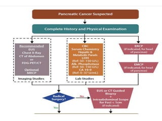
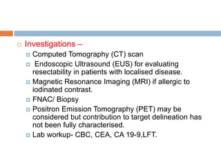
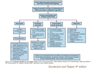
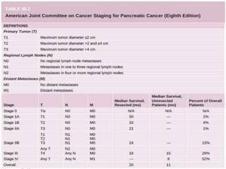
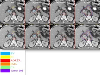
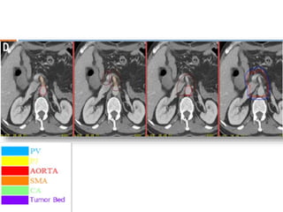
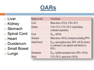
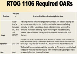
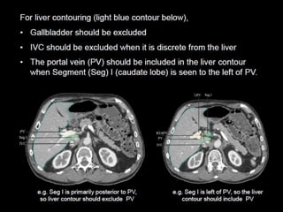
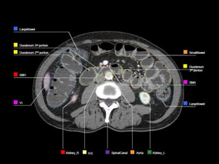
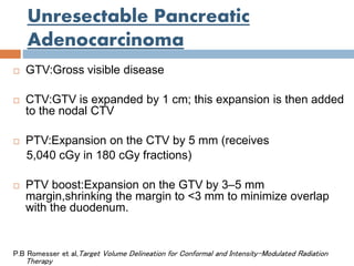
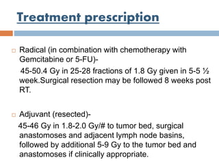
CONTOURING GUIDELINES-
PANCREATIC MALIGNANCIES
DR. P. MANASA/ DR. ASTHA
SRIVASTAVA
MODERATOR – PROF. S. PATHY
Introduction
 The number of new cases diagnosed per year
is 10860 (0.94%) and mortality rate is about
10528 (1.3%) in 2018.
 Seventh leading cause of cancer mortality ,
although only the twelfth most common cancer
worldwide.
Globocan 2018
Anatomy
 Risk factors –
 Chronic pancreatitis, smoking, alcohol consumption,
Helicobacter pylori infection.
 Factors associated with metabolic syndrome such as
obesity and glucose intolerance.
 Familial - BRCA1/2 and PALB2 mutations, Peutz-
Jeghers syndrome, FAP, HNPCC.
Clinical presentation &
Evaluation
 Pain, weight loss, jaundice.
 Recent onset/ exacerbation of diabetes mellitus,
migratory thrombophlebitis(Trousseau’s sign).
 Palpable abdominal mass, ascites,
supraclavicular nodes.
 Investigations –
 Computed Tomography (CT) scan
 Endoscopic Ultrasound (EUS) for evaluating
resectability in patients with localised disease.
 Magnetic Resonance Imaging (MRI) if allergic to
iodinated contrast.
 FNAC/ Biopsy
 Positron Emission Tomography (PET) may be
considered but contribution to target delineation has
not been fully characterised.
 Lab workup- CBC, CEA, CA 19-9,LFT.
Gunderson and Tepper 4th edition
CT Simulation
 CT simulation with oral and IV contrast (unless
contraindicated)
 Patients are generally asked not to eat any
large meals 2–3 h prior to simulation and
treatment to avoid significant interfraction
variation in stomach distention.
 Arms should be above the head, orifit /vaclock
for immobilisation.
 Planning scan should be done(minimum of 3
mm cuts) with a scan from carina to iliac crest.
 For patients treated with respiratory gating or
without a specific motion-management
technique, a four dimensional (4D) CT is
necessary to evaluate the degree of tumor
motion.
Target volumes: GTV
 By definition there is no gross tumor volume (GTV) at
time of radiotherapy because the tumor has been
resected.
Treatment Volumes: CTV
 Typical anatomical landmarks that can help in CTV
delineation include:
1. Pancreas - L1-L2
2. Celiac axis - T12
3. SMA - L1
 CTV is the area with the highest likelihood for residual
subclinical tumor.
 Imperative to review pre-op imaging , operative notes
and histopathology report at the time of treatment
planning.
1) Post op tumor bed
2 ) Anastomoses
 Pancreaticojejunostomy(PJ)
 Choledochal or hepaticojejunostomy
3) Abdominal nodal regions
 Peripancreatic
 Celiac
 Superior mesenteric
 Porta hepatis
 Para-aortic
ROI Delineation:Post-op Bed
 Location of tumor should be reviewed and contoured
based on pre-op imaging.
 Surgical clips placed should only be included if there is
documentation from the surgeon intraoperatively such
as close margins or specific tumor related .
ROI Delineation:PJ and HJ
 PJ is identified by following pancreatic remnant medially
and until the junction with the jejunal loop is noted.
 HJ is identified by following air in biliary tree to CHD or
CBD remnant to jejunal loop , or by following PV out of
liver to jejunal loop region.
ROI Delineation:Ao and PV
 Aorta(Ao) extending from top of the upper most PV,CA or
SMA slice to the bottom of L2 or L3.
 Portal vein(PV) is contoured from the bifurcation of the PV .
The PV bifurcation can be extrahepatic or almost
intrahepatic.
(portion of portal vein running anterior and medial to IVC
and stop prior to confluence of SMV or splenic vein).
ROI Delineation:CA and SMA
 Celiac artery (CA): The most proximal 1.0-1.5 cm of the
CA and should include up to the first branching.
 Superior mesenteric artery (SMA): The most proximal
2.5-3.0 cm of SMA.
ROI Expansions
 Pancreaticojejunostomy, portal vein, superior mesenteric
artery and celiac artery are expanded by 0.5-1 cm.
 Aorta ROI is given asymmetric expansion; 2.5-3 cm
towards right , 1cm towards left, 0.2 cm posteriorly and 2
-2.5 cm anteriorly.
 The CTV is then created by merging the ROI expansions .
 The posterior margin should follow the contour of the
anterior aspect of the vertebral body without including
more than 0.5 cm of the anterior vertebral body anterior
edge.
 If the PJ cannot be identified, the CTV should be
generated
without it.
 If the CTV with the noted expansions protrudes into a
dose limited normal organ such as the liver or stomach or
kidney, it should be edited to be adjacent to the relevant
structure
 PTV – 0.5 cm expansion on CTV
OARs
 Liver
 Kidney
 Stomach
 Spinal Cord
 Heart
 Duodenum
 Small Bowel
 Lungs
Unresectable Pancreatic
Adenocarcinoma
 GTV:Gross visible disease
 CTV:GTV is expanded by 1 cm; this expansion is then added
to the nodal CTV
 PTV:Expansion on the CTV by 5 mm (receives
5,040 cGy in 180 cGy fractions)
 PTV boost:Expansion on the GTV by 3–5 mm
margin,shrinking the margin to <3 mm to minimize overlap
with the duodenum.
P.B Romesser et al,Target Volume Delineation for Conformal and Intensity-Modulated Radiation
Therapy
Treatment prescription
 Radical (in combination with chemotherapy with
Gemcitabine or 5-FU)-
45-50.4 Gy in 25-28 fractions of 1.8 Gy given in 5-5 ½
week.Surgical resection may be followed 8 weeks post
RT.
 Adjuvant (resected)-
45-46 Gy in 1.8-2.0 Gy/# to tumor bed, surgical
anastomoses and adjacent lymph node basins,
followed by additional 5-9 Gy to the tumor bed and
anastomoses if clinically appropriate.
Contouring guidelines  pancreatic malignancies

Contouring guidelines pancreatic malignancies

  • 1.
    CONTOURING GUIDELINES- PANCREATIC MALIGNANCIES DR.P. MANASA/ DR. ASTHA SRIVASTAVA MODERATOR – PROF. S. PATHY
  • 2.
    Introduction  The numberof new cases diagnosed per year is 10860 (0.94%) and mortality rate is about 10528 (1.3%) in 2018.  Seventh leading cause of cancer mortality , although only the twelfth most common cancer worldwide. Globocan 2018
  • 3.
  • 4.
     Risk factors–  Chronic pancreatitis, smoking, alcohol consumption, Helicobacter pylori infection.  Factors associated with metabolic syndrome such as obesity and glucose intolerance.  Familial - BRCA1/2 and PALB2 mutations, Peutz- Jeghers syndrome, FAP, HNPCC.
  • 5.
    Clinical presentation & Evaluation Pain, weight loss, jaundice.  Recent onset/ exacerbation of diabetes mellitus, migratory thrombophlebitis(Trousseau’s sign).  Palpable abdominal mass, ascites, supraclavicular nodes.
  • 7.
     Investigations – Computed Tomography (CT) scan  Endoscopic Ultrasound (EUS) for evaluating resectability in patients with localised disease.  Magnetic Resonance Imaging (MRI) if allergic to iodinated contrast.  FNAC/ Biopsy  Positron Emission Tomography (PET) may be considered but contribution to target delineation has not been fully characterised.  Lab workup- CBC, CEA, CA 19-9,LFT.
  • 8.
  • 10.
    CT Simulation  CTsimulation with oral and IV contrast (unless contraindicated)  Patients are generally asked not to eat any large meals 2–3 h prior to simulation and treatment to avoid significant interfraction variation in stomach distention.  Arms should be above the head, orifit /vaclock for immobilisation.
  • 11.
     Planning scanshould be done(minimum of 3 mm cuts) with a scan from carina to iliac crest.  For patients treated with respiratory gating or without a specific motion-management technique, a four dimensional (4D) CT is necessary to evaluate the degree of tumor motion.
  • 13.
    Target volumes: GTV By definition there is no gross tumor volume (GTV) at time of radiotherapy because the tumor has been resected.
  • 14.
    Treatment Volumes: CTV Typical anatomical landmarks that can help in CTV delineation include: 1. Pancreas - L1-L2 2. Celiac axis - T12 3. SMA - L1
  • 15.
     CTV isthe area with the highest likelihood for residual subclinical tumor.  Imperative to review pre-op imaging , operative notes and histopathology report at the time of treatment planning.
  • 16.
    1) Post optumor bed 2 ) Anastomoses  Pancreaticojejunostomy(PJ)  Choledochal or hepaticojejunostomy
  • 17.
    3) Abdominal nodalregions  Peripancreatic  Celiac  Superior mesenteric  Porta hepatis  Para-aortic
  • 18.
    ROI Delineation:Post-op Bed Location of tumor should be reviewed and contoured based on pre-op imaging.  Surgical clips placed should only be included if there is documentation from the surgeon intraoperatively such as close margins or specific tumor related .
  • 19.
    ROI Delineation:PJ andHJ  PJ is identified by following pancreatic remnant medially and until the junction with the jejunal loop is noted.  HJ is identified by following air in biliary tree to CHD or CBD remnant to jejunal loop , or by following PV out of liver to jejunal loop region.
  • 20.
    ROI Delineation:Ao andPV  Aorta(Ao) extending from top of the upper most PV,CA or SMA slice to the bottom of L2 or L3.  Portal vein(PV) is contoured from the bifurcation of the PV . The PV bifurcation can be extrahepatic or almost intrahepatic. (portion of portal vein running anterior and medial to IVC and stop prior to confluence of SMV or splenic vein).
  • 21.
    ROI Delineation:CA andSMA  Celiac artery (CA): The most proximal 1.0-1.5 cm of the CA and should include up to the first branching.  Superior mesenteric artery (SMA): The most proximal 2.5-3.0 cm of SMA.
  • 22.
    ROI Expansions  Pancreaticojejunostomy,portal vein, superior mesenteric artery and celiac artery are expanded by 0.5-1 cm.  Aorta ROI is given asymmetric expansion; 2.5-3 cm towards right , 1cm towards left, 0.2 cm posteriorly and 2 -2.5 cm anteriorly.
  • 23.
     The CTVis then created by merging the ROI expansions .  The posterior margin should follow the contour of the anterior aspect of the vertebral body without including more than 0.5 cm of the anterior vertebral body anterior edge.
  • 24.
     If thePJ cannot be identified, the CTV should be generated without it.  If the CTV with the noted expansions protrudes into a dose limited normal organ such as the liver or stomach or kidney, it should be edited to be adjacent to the relevant structure
  • 25.
     PTV –0.5 cm expansion on CTV
  • 28.
    OARs  Liver  Kidney Stomach  Spinal Cord  Heart  Duodenum  Small Bowel  Lungs
  • 48.
    Unresectable Pancreatic Adenocarcinoma  GTV:Grossvisible disease  CTV:GTV is expanded by 1 cm; this expansion is then added to the nodal CTV  PTV:Expansion on the CTV by 5 mm (receives 5,040 cGy in 180 cGy fractions)  PTV boost:Expansion on the GTV by 3–5 mm margin,shrinking the margin to <3 mm to minimize overlap with the duodenum. P.B Romesser et al,Target Volume Delineation for Conformal and Intensity-Modulated Radiation Therapy
  • 49.
    Treatment prescription  Radical(in combination with chemotherapy with Gemcitabine or 5-FU)- 45-50.4 Gy in 25-28 fractions of 1.8 Gy given in 5-5 ½ week.Surgical resection may be followed 8 weeks post RT.  Adjuvant (resected)- 45-46 Gy in 1.8-2.0 Gy/# to tumor bed, surgical anastomoses and adjacent lymph node basins, followed by additional 5-9 Gy to the tumor bed and anastomoses if clinically appropriate.

Editor's Notes

  • #24 These expansions will require clinical judgment. Occasionally, the PJ or PV expansion may extend cephalad to above the level of the CA. In that case the aorta expansion should be extended cephalad to the same level as the highest level (CT slice) of the PV or PJ expansion (whichever is more cephalad).