Sk jain 5
•
1 like•178 views

This document summarizes a study on using ultrasound to assess cystic neck lesions prior to treatment. The study examined 120 patients with neck masses and found cystic lesions in 7.5% of cases, including branchial cysts and lymphangiomas. Ultrasound was able to differentiate cystic from solid lesions and characterize features like contents, borders, and location. The study concluded ultrasound is a useful non-invasive tool for evaluating cystic neck lesions before treatment due to its low cost, lack of radiation, and ability to detect diagnostic imaging characteristics of various cyst types.

More Related Content
What's hot
What's hot (20)
Viewers also liked
Viewers also liked (19)
Similar to Sk jain 5
Similar to Sk jain 5 (20)
Sk jain 5
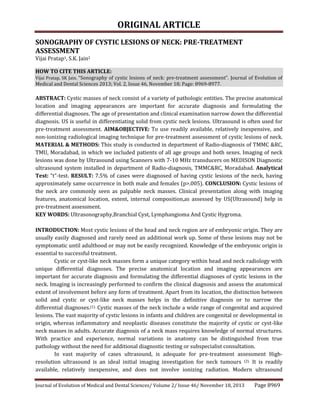
- 1. ORIGINAL ARTICLE Journal of Evolution of Medical and Dental Sciences/ Volume 2/ Issue 46/ November 18, 2013 Page 8969 SONOGRAPHY OF CYSTIC LESIONS OF NECK: PRE-TREATMENT ASSESSMENT Vijai Pratap1, S.K. Jain2 HOW TO CITE THIS ARTICLE: Vijai Pratap, SK Jain. “Sonography of cystic lesions of neck: pre-treatment assessment”. Journal of Evolution of Medical and Dental Sciences 2013; Vol. 2, Issue 46, November 18; Page: 8969-8977. ABSTRACT: Cystic masses of neck consist of a variety of pathologic entities. The precise anatomical location and imaging appearances are important for accurate diagnosis and formulating the differential diagnoses. The age of presentation and clinical examination narrow down the differential diagnosis. US is useful in differentiating solid from cystic neck lesions. Ultrasound is often used for pre-treatment assessment. AIM&OBJECTIVE: To use readily available, relatively inexpensive, and non-ionizing radiological imaging technique for pre-treatment assessment of cystic lesions of neck. MATERIAL & METHODS: This study is conducted in department of Radio-diagnosis of TMMC &RC, TMU, Moradabad, in which we included patients of all age groups and both sexes. Imaging of neck lesions was done by Ultrasound using Scanners with 7-10 MHz transducers on MEDISON Diagnostic ultrasound system installed in department of Radio-diagnosis, TMMC&RC, Moradabad. Analytical Test: “t”-test. RESULT: 7.5% of cases were diagnosed of having cystic lesions of the neck, having approximately same occurrence in both male and females (p>.005). CONCLUSION: Cystic lesions of the neck are commonly seen as palpable neck masses. Clinical presentation along with imaging features, anatomical location, extent, internal composition,as assessed by US(Ultrasound) help in pre-treatment assessment. KEY WORDS: Ultrasonography,Branchial Cyst, Lymphangioma And Cystic Hygroma. INTRODUCTION: Most cystic lesions of the head and neck region are of embryonic origin. They are usually easily diagnosed and rarely need an additional work up. Some of these lesions may not be symptomatic until adulthood or may not be easily recognized. Knowledge of the embryonic origin is essential to successful treatment. Cystic or cyst-like neck masses form a unique category within head and neck radiology with unique differential diagnoses. The precise anatomical location and imaging appearances are important for accurate diagnosis and formulating the differential diagnoses of cystic lesions in the neck. Imaging is increasingly performed to confirm the clinical diagnosis and assess the anatomical extent of involvement before any form of treatment. Apart from its location, the distinction between solid and cystic or cyst-like neck masses helps in the definitive diagnosis or to narrow the differential diagnoses.(1). Cystic masses of the neck include a wide range of congenital and acquired lesions. The vast majority of cystic lesions in infants and children are congenital or developmental in origin, whereas inflammatory and neoplastic diseases constitute the majority of cystic or cyst-like neck masses in adults. Accurate diagnosis of a neck mass requires knowledge of normal structures. With practice and experience, normal variations in anatomy can be distinguished from true pathology without the need for additional diagnostic testing or subspecialist consultation. In vast majority of cases ultrasound, is adequate for pre-treatment assessment High- resolution ultrasound is an ideal initial imaging investigation for neck tumours (2). It is readily available, relatively inexpensive, and does not involve ionizing radiation. Modern ultrasound
- 2. ORIGINAL ARTICLE Journal of Evolution of Medical and Dental Sciences/ Volume 2/ Issue 46/ November 18, 2013 Page 8970 machines equipped with high-resolution transducers provide excellent resolution. The use of Ultrasound for the initial diagnosis of neck masses in adults and children continues to increase. Ultrasound is useful in differentiating solid from cystic neck lesions in both adults and children, in recording the size of nodes (at least in the upper neck), and in discriminating high-flow from low- flow vascular malformations (3-6). In children who present with neck masses, congenital etiologies should be added to differential diagnostic considerations (7).Most cystic neck masses have non-specific ultrasound features, and these features can be complicated by infection or haemorrhage. The basic level of equipment required for head and neck ultrasound is a modern system with a high-frequency transducer ( > 7.5 MHz). High-frequency transducers allow excellent near-field resolution.. Lower frequency transducers (5 MHz) required for assessing deep-seated lesions, such as those in the deep lobe of the parotid gland. Based on anatomical location and ultrasound imaging cystic lesions of neck are identified as under as under: 1. Submental region. (a) Epidermoid/dermoid cyst: The cyst is usually well-defined and anechoic with posterior acoustic enhancement in a midline position of the neck (b) Ranula: A ranula appears as a unilocular,well-defined, cystic lesion in the submental region related to the sublingual gland. 2. Midline region. (a) Thyroglossal duct cyst: frankly cystic with homogeneous anechoic internal appearance and posterior acoustic enhancement; hypoechoic echo pattern with internal debris. (b) Acute suppurative thyroiditis: Both intra- and extra-thyroid abscesses are seen as ill-defined, hypoechoic, heterogeneous areas with internal debris. (c) Thymic cyst: Appears as a well-defined,anechoic cystic lesion below the level of the thyroid gland and related to the carotid sheath. 3. Submandibular region. (a) Second branchial cleft cyst: Most uninfected second branchial cleft cysts demonstrate the typical appearances of a cyst in that they are well-defined and anechoic with no internal debris and show posterior acoustic enhancement however, some cysts may exhibit a pseudo solid appearance with uniform internal echoes. (b) Abscess:An abscess appears as an ill-defined, irregular collection with thick walls and internal debris. It may be unilocular or multilocular. 4. Parotid region. (a) Extraglandular mass- First branchial cleft cyst: First branchial cleft cyst may appear as truly cystic with anechoic content, homogeneous hyperechoic with a pseudo solid appearance or heterogeneous hypoechoic with internal debris in infected cysts. (b) Intraglandular mass(Warthin’s tumor)-Warthin’s tumor is typically a well circumscribed, hypoechoic mass in the parotid tail with internal heterogeneity (solid and cystic components) and posterior acoustic enhancement. 5. Along cervical chain.
- 3. ORIGINAL ARTICLE Journal of Evolution of Medical and Dental Sciences/ Volume 2/ Issue 46/ November 18, 2013 Page 8971 (a)Cystic metastatic lymph node: Cystic necrosis may manifest itself as a truly cystic area or as a central, ill-defined area of relative hypoechogenicity accompanied by eccentric solid component within an enlarged lymph node. (b)Distended internal jugular vein: Ultrasound supplemented by Doppler ultrasound examination accurately identifies the dilated internal jugular vein 6. Posterior triangle. (a) Lymphangioma:Cystic hygroma appears as a compressible multiloculate cystic lesion with intervening thin septa. Vascularity may be seen within the septa. (b)Tuberculous lymphadenitis: On imaging, it may appear almost entirely cystic or necrotic and mimic a cystic metastatic lymph node MATERIAL & METHODS: This study was conducted in department of Radio-diagnosis of TMMC & RC, TMU, Moradabad. Patients of all age groups, socioeconomic strata, rural /urban & male/female were included in the study. In this study, we found 7.5% cases of cystic lesions in the neck. After we find the cystic lesions of neck we nurtured our study our by positioning the neck slightly extended, placing a pillow behind the shoulders and lower neck which allows the patient to adopt a comfortable position. We revaluated the following regions consecutively: submental, submandibular, parotid, upper cervical, midcervical, lower cervical, supraclavicular fossa, posterior triangle, and midline of the neck.Further differentiation of different type of cystic lesions was done by Ultrasonography using Scanners with 7-10 MHz transducers on MEDISON Diagnostic ultrasound system. Institutional Ethical and research committee approval was taken prior to start the study. Female subjects were examined in presence of female nursing staff and one female attendant. In case of minor consent from guardians was taken. No grant from any source was taken and there was no conflict of interest. RESULTS: Age group (In years) Male Female Total 0-10 6 3 9 11-20 3 9 12 21-30 12 24 36 31-40 9 9 18 41-50 15 6 21 51-60 9 3 12 61-70 9 - 9 71-80 3 - 3 Total 66 54 120 Table 1: Age and Sex distribution of patients with Neck Masses
- 4. ORIGINAL ARTICLE Journal of Evolution of Medical and Dental Sciences/ Volume 2/ Issue 46/ November 18, 2013 Page 8972 Nature of the lesion No. of cases Percentage of total cases Inflammatory Abscess Adenopathy 3 18 17.5% 2.5% 15% Developmental Branchial Cyst Lymphangioma 6 3 7.5% 5% 2.5% Thyroid Masses Benign Malignant 24 12 30% 20% 10% Mesenchymal Lipoma Sarcoma 6 6 10% 5% 5% Neural Schwannoma Neurofibroma 3 3 5% 2.5% 2.5% Vascular Hemangioma Carotid body tumor 3 3 5% 2.5% 2.5% Bone Osteoma Metastasis 3 3 5% 2.5% 2.5% Lymph node Masses (non inflammatory) Lymphoma Metastasis 3 9 10% 2.5% 7.5% Salivary Gland Masses Benign Malignant 6 6 10% 5% 5% Total 120 100% Table 2: Distribution of neck Masses according to the nature of the lesion On the basis of above table it seems that cystic lesions were present in 7.5% cases ,out of which 5% were Branchial cyst and 2.5% were Lymphangiomas. US Appearance Lymphangioma Branchial Cyst ECHOGENECITY Hypoechoic Anechoic Fluid-Fluid level - + - + + + POSTERIOR ENHANCEMENT + + ATTENUATION (10-25 HU) + + RIM ENHANCEMENT - +
- 5. ORIGINAL ARTICLE Journal of Evolution of Medical and Dental Sciences/ Volume 2/ Issue 46/ November 18, 2013 Page 8973 LOCULATION + - CALCIFICATION - - Table 3: Imaging features of cystic masses Branchial Cyst: six patients of different age group(2-20 yrs) and different clinical presentations were found to have branchial cysts. Ratio of male to female was approximately 1:1.Three (50%) of the lesions were truly anechoic .About 22% were predominantly homogeneously hypoechoic but showed the presence of internal,low-amplitude, freely floating debris. Remaining lesions were hyperechoic and showed a pseudo solid appearance. On real-time images, after the application of transducer pressure on the cyst, the entire contents shifted in all cases, suggesting theirtrue cystic nature. (Fig-1) Lymphangioma: Three cases of Lymphangiomas presented with different age group and different ultrasonographic appearences. Lymphangiomas presented as an echogenic, multilocular cystic mass containing septa of variable thickness and solid components. On ultrasound, three different types of sonographic features were seen (a) cystic with thin septae-5 cases (b) cystic with thick septae-3 cases (c) cystic with thick septae and solid areas-3 cases DISCUSSION: The term branchial cyst was first used by Ascherson in 1832. He suggested that these cysts were results of impaired obliteration of branchial clefts(8). Phylogenetically, the branchial apparatus is related to gill slits. In fish and amphibians, these structures are responsible for the development of the gills, hence the name branchial (branchia is Greek for gills). At the fourth week of embryonic life, the development of 4 branchial (or pharyngeal) clefts results in 5 ridges known as the branchial (or pharyngeal) arches, which contribute to the formation of various structures of the head, the neck, and the thorax. The second arch grows caudally and, ultimately, covers the third and fourth arches. The buried clefts become ectoderm-lined cavities, which normally involute around 7thweek of development. If a portion of the cleft fails to involute completely, the entrapped remnant forms an epithelium-lined cyst with or without a sinus tract to the overlying skin.(9,10,11,12 ).By the end of the 4th week of embryonic life, the branchial arches (derived from neural crest cells) and the mesenchyme (derived from the lateral mesoderm) are easily recognizable. Five pairs of ectodermal clefts (grooves) and five endodermal branchial pouches separate the six arches, with a closing membrane located at the interface between the pouches and the clefts (13-14). Many branchial cleft cysts are asymptomatic. They may become tender, enlarged, or inflamed, or they may develop abscesses, especially during periods of upper respiratory tract infection, due to the lymphoid tissue located beneath the epithelium. Spontaneous rupture of an abscessed branchial cleft cyst may result in a purulent draining sinus to the skin or the pharynx. Depending on the size and the anatomical extension of the mass, local symptoms, such as dysphagia, dysphonia, dyspnea, and stridor, may occur. No sexual predilection is recognized for branchial cleft cysts. The branchial cysts are classified into four types
- 6. ORIGINAL ARTICLE Journal of Evolution of Medical and Dental Sciences/ Volume 2/ Issue 46/ November 18, 2013 Page 8974 ▪ First branchial cleft cysts are divided into type I and type II. Type I cysts are located near the external auditory canal. Most commonly, they are inferior and posterior to the tragus (base of the ear), but they may also be in the parotid gland or at the angle of the mandible. They may be difficult to distinguish from a solid parotid mass on clinical examination. Type II cysts are associated with the submandibular gland or found in the anterior triangle of the neck. ▪ The second branchial cleft accounts for 95% of branchial anomalies (15,16,17). Most frequently, these cysts are identified along the anterior border of the upper third of the sternocleidomastoid muscle, adjacent to the muscle. However, these cysts may present anywhere along the course of a second branchial fistula, which proceeds from the skin of the lateral neck, between the internal and external carotid arteries, and into the palatine tonsil (18). Therefore, second branchial cleft cyst is in the differential of a parapharyngeal mass. ▪ The third and the fourth branchial cleft cyst are rare. At least 75% of all second branchial cleft abnormalities are cysts (19), which typically present when an individual is between 10 and 40-years-old. Second branchial cleft fistulas and sinuses are less common and usually present during the first decade of life (20,21). No gender predilection has been reported (22). Cervicofacial lymphangioma is uncommon, representing fewer than 6% of benign tumors of childhood. Incidence has been reported to be less than 2.8 per 1000 population. No sex preponderance or side predilection has been reported. The origin of lymphangiomas is controversial. Theories include lymphangiomas as true neoplasms, hamartomas, or congenital dysplasias of the lymphatics. To determine the role of angiogenesis in the pathogenesis of lymphangioma.(23).examined patients' specimens for expression of angiogenic inducer vascular endothelial growth factor (VEGF) and angiogenic inhibitor pigment epithelium-derived factor (PEDF) using immunohistochemical analysis. Staining patterns of VEGF and PEDF were evaluated. Histological evidence of increased angiogenesis, including microvascular density, stromal fibrosis, and inflammation, were graded in each group and correlated with recurrence. The different types of cervicofacial lymphangioma are classified as follows: Lymphangioma circumscriptum is a simplex superficial red macular or vesicular lesion of mucous membranes or skin. Lymphangioma capillary type is a simplex lesion of dilated capillary like channels. Lymphangioma cavernosa is a simplex lesion of dilated lymphatic channels with deep extension and without cyst formation. Lymphangioma cystica (ie, cystic hygroma) is composed of large lymphatic cysts that expand into adjacent soft tissue planes and are well defined, circumscribed, or lobulated. Lymphangioma complex is composed of multiloculated poorly defined cysts extending to more than one anatomic area, tissue plane, or organ system. Differential diagnosis includes teratomas, hemangiomas, cervical meningoceles, thyroglossal duct cysts, esophageal diverticula, dermoid cysts, brachial cleft cysts, epignathus, and congenital
- 7. ORIGINAL ARTICLE Journal of Evolution of Medical and Dental Sciences/ Volume 2/ Issue 46/ November 18, 2013 Page 8975 goiters. In some cases, it is possible to establish a diagnosis of lymphangioma with 2D sonography(24,25). CONCLUSION: Ultrasound is typically the initial imaging choice for neck masses, and usually enables the distinction between cystic and solid lesions. More detailed characterisation is often possible with ultrasound if precise anatomic localisation can be achieved. In the vast majority of cases ultrasound, is adequate for pre-treatment assessment. The multiple loculations and extension across fascial planes is very helpful distinguishing lymphangioma from other cystic neck masses. Nonspecific morphologic features, especially when complicated by infection, hemorrhage or tumour, may necessitate CT (branchial cleft lesions), MR (lymphangioma) and specialist referral including a tissue diagnosis. ACKNOWLEDGEMENT: Authors wish to acknowledge Prof.(Dr. Pankaj Mishra),Department of community medicine, SGRRIMHS, Dehradun for statistical help. REFERENCES: 1. Lev S, Lev MH. Imaging of cystic lesions. Radiol Clin North Am 2000;38:1013-27. 2. Ahuja AT. Lumps and bumps in the head and neck. In: Ahuja AT, Evans RM, editors. Practical head and neck ultrasound. London: Greenwich Medical Media Limited;2000. p. 87e104 3. Ahuja AT, Richards P, Wong KT, Yuen EH, King AD. Accuracy of high-resolution sonography compared with magnetic resonance imaging in the diagnosis of head and neck venous vascular malformations. ClinRadiol2003; 58(11):869-875. 4. Wong KT, Lee YY, King AD, Ahuja AT. Imaging of cystic or cyst-like neck masses. ClinRadiol2008; 63(6):613-622. 5. Yang WT, Ahuja A, Metreweli C. Sonographic features of head and neck hemangiomas and vascular malformations: review of 23 patients. J Ultrasound Med1997; 16(1):39-44. 6. Hohlweg-Majert B, Metzger MC, Voss PJ, Holzle F, Wolff KD,. Schulze D. Preoperative cervical lymph node size evaluation in patients with malignant head/neck tumors: comparison between ultrasound and computer tomography. J Cancer Res Clin Oncol 2009; 135(6):753-759. 7. Tanaka T, Morimoto Y, Takano H, et al. Three-dimensional identification of hemangiomas and feeding arteries in the head and neck region using combined phase-contrast MR angiography and fast asymmetric spin-echo sequences. Oral Surg Oral Med OralPathol Oral RadiolEndod2005; 100(5):609-613. 8. Golledge J & Ellis H., The aetiology of lateral cervical (branchial) cysts: past and present theories, J Laryngol Otol, 1994, 108(8):653–659. 9. Doi O, Hutson JM, Myers NA, McKelvie PA. Branchial remnants: a review of 58 cases. J Pediatr Surg. Sep 1988; 23(9):789-92. 10. Little JW, Rickles NH. The histogenesis of the branchial cyst.Am J Pathol. Mar 1967;50(3):533- 47. 11. Rickles NH, Little JW. The histogenesis of the branchial cyst. II. A study of the lining epithelium. Am J Pathol. May 1967;50(5):765-77. 12. Telander RL, Deane SA. Thyroglossal and branchial cleft cysts and sinuses. Surg Clin North Am. Aug 1977;57(4):779-91.
- 8. ORIGINAL ARTICLE Journal of Evolution of Medical and Dental Sciences/ Volume 2/ Issue 46/ November 18, 2013 Page 8976 13. Moore K., The developing human, 3rd edition, Philadelphia, Saunders, 1988. 14. Langman J., Medical embryology, 3rd edition, Williams & Wilkins, Baltimore, 1975. 15. Vogl T., Hypopharynx, larynx, thyroid, and parathyroid. In: Stark D., Bradely W. (eds), Magnetic resonance imaging, 2nd edition, Mosby–Yearbook, St. Louis, 1992, 1184–1243. 16. KoellerK. K., Alamo L., Adair C. F., SmirniotopoulosJ. G., Congenital cystic masses of theneck: radiologic–pathologic correlation, Radiographics, 1999, 19(1):121–146 ; quiz 152–153. 17. Ang A. H., Pang K. P., Tan L. K., Complete branchial fistula. Case report and review of literature, Ann OtolRhinolLaryngol, 2001, 110(11):1077–1079. 18. Zadvinkis D. P., Benson M. T., Som P. M., Smoker W. R. K., Embryology and congenital cysticlesions. In: SOM P. M., CURTIN H. D. (eds), Head and neckimaging, Mosby, St. Louis, 1996, 754. 19. Telander R. L., Filston H. C., Review of head and neck lesions in infancy and childhood, SurgClin North Am, 1992, 72(6):1429–1447. 20. Som P. M., Sacher M., Lanzieri C. F., SolodniP., Cohen B. A., Redde D. L., Bergeron R. T., Biller H. F., Parenchymal cysts of the lower neck, Radiology, 1985, 157(2):399–406. 21. Michael A. S., Mafee M. F., Valvassorri G. E., Tan W. S., Dynamic computed tomography of the head andneck: differential diagnostic value, Radiology, 1985, 154(2):413–419. 22. Faerber E. N., Swartz J. D., Imaging of neck masses in infants and children, Crit Rev Diagn Imaging, 1991, 31(3–4):283–314. 23. Sidle DM, Maddalozzo J, Meier JD, et al. Altered pigment epithelium-derived factor and vascular endothelial growth factor levels in lymphangioma pathogenesis and clinical recurrence. Arch Otolaryngol Head Neck Surg. Nov 2005;131(11):990-5. [Medline] 24. Axt-Fliedner R, Hendrik HG, Ertan K, Remberger K, Schmidt W. Course and outcome of a pregnancy with a giant fetal cervical teratoma diagnosed prenatally. Ultrasound Obstet Gynecol 2001; 18:543–546. 25. Donegan JO. Congenital neck masses. In: Cimmings CW, Fredickson JM, Harker LA (eds). Otolaryngology Head and Neck Surgery. St Louis, MO: CV Mosby Co; 1986:1604–1612. Figure 1- Common neck lesions Fig. 1: US Image of circumscribed cyst Fig. 2: True cystic appearance
- 9. ORIGINAL ARTICLE Journal of Evolution of Medical and Dental Sciences/ Volume 2/ Issue 46/ November 18, 2013 Page 8977 AUTHORS: 1. Vijai Pratap 2. S.K. Jain PARTICULARS OF CONTRIBUTORS: 1. Associate Professor, Department of Radiodiagnosis, TMMC & RC, Moradabad. 2. Professor, Department of Anatomy, TMMC & RC, Moradabad. NAME ADDRESS EMAIL ID OF THE CORRESPONDING AUTHOR: Dr. S.K. Jain, Professor, Department of Anatomy, TMMC & RC, Moradabad. Email – drskjain2005@rediffmail.com Date of Submission: 26/10/2013. Date of Peer Review: 28/10/2013. Date of Acceptance: 12/11/2013. Date of Publishing: 13/11/2013 Fig. 3: Showing heterogeneous appearance Fig. 4: Showing phlebolith Fig. 5: cystic lesion in the submental region