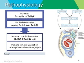
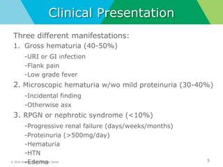
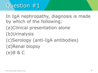
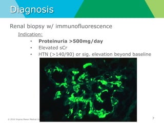
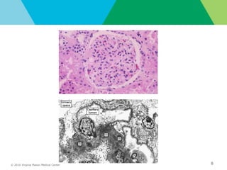
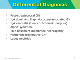
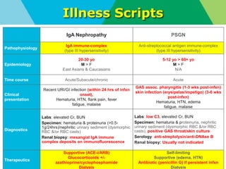
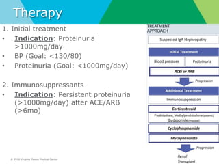
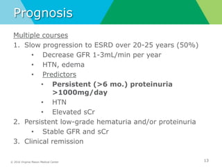
Noon Conference
Matthew Ong
01/15/2019
Ā© 2016 Virginia Mason Medical Center 2
Objectives
IgA Nephropathy
(AKA Berger’s disease)
• Definition
• Pathophysiology
• Clinical presentation
• Diagnosis
• Differential diagnosis
• Illness script
• Treatment
• Prognosis
Ā© 2016 Virginia Mason Medical Center
Definition
• Nephritic syndrome (vs. nephrotic)
• Most common cause 1˚ glomerulonephritis worldwide
• Peak age – 20-30 yo
• 2:1 male-to-female
• East Asians & Caucasians
• Painless hematuria in the setting of concurrent/recent URI
3
Ā© 2016 Virginia Mason Medical Center
Pathophysiology
4
Ā© 2016 Virginia Mason Medical Center
Clinical Presentation
Three different manifestations:
1. Gross hematuria (40-50%)
-URI or GI infection
-Flank pain
-Low grade fever
2. Microscopic hematuria w/wo mild proteinuria (30-40%)
-Incidental finding
-Otherwise asx
3. RPGN or nephrotic syndrome (<10%)
-Progressive renal failure (days/weeks/months)
-Proteinuria (>500mg/day)
-Hematuria
-HTN
-Edema 5
Ā© 2016 Virginia Mason Medical Center
Question #1
In IgA nephropathy, diagnosis is made
by which of the following:
(a)Clinical presentation alone
(b)Urinalysis
(c)Serology (anti-IgA antibodies)
(d)Renal biopsy
(e)B & C
6
Ā© 2016 Virginia Mason Medical Center
Diagnosis
Renal biopsy w/ immunofluorescence
Indication:
• Proteinuria >500mg/day
• Elevated sCr
• HTN (>140/90) or sig. elevation beyond baseline
7
Ā© 2016 Virginia Mason Medical Center 8
right lung nodules and infiltrates
Ā© 2016 Virginia Mason Medical Center
Differential Diagnosis
• Post-streptococcal GN
• IgA dominant Staphylococcus-associated GN
• IgA vasculitis (Henoch-Schonlein purpura)
• Alport syndrome
• Thin basement membrane nephropathy
• Membranoproliferative GN
• Lupus nephritis
9
Ā© 2016 Virginia Mason Medical Center
Illness Scripts
10
IgA Nephropathy PSGN
Pathophysiology
IgA immune-complex
(type III hypersensitivity)
Anti-streptococcal antigen immune-complex
(type III hypersensitivity)
Epidemiology
20-30 yo
M > F
East Asians & Caucasians
5-12 yo > 60+ yo
M > F
N/A
Time course Acute/Subacute/chronic Acute
Clinical
presentation
Recent URI/GI infection (within 24 hrs of infxn
onset),
Hematuria, HTN, flank pain, fever
fatigue, malaise
GAS assoc. pharyngitis (1-3 wks post-infxn)
skin infection (erysipelas/impetigo) (3-6 wks
post-infxn)
Hematuria, HTN, edema
fatigue, malaise
Diagnostics
Labs: elevated Cr, BUN
Specimen: hematuria & proteinuria (>0.5-
1g/24hrs)/nephritic urinary sediment (dysmorphic
RBC &/or RBC casts)
Renal biopsy: mesangial IgA immune
complex deposits on immunofluorescence
Labs: low C3, elevated Cr, BUN
Specimen: hematuria & proteinuria, nephritic
urinary sediment (dysmorphic RBC &/or RBC
casts); positive GAS throat/skin culture
Serology: anti-streptolysin/anti-DNAse B
Renal biopsy: Usually not indicated
Therapeutics
Supportive (ACE-i/ARB)
Glucocorticoids +/-
azathioprine/cyclophosphamide
Dialysis
Self-limiting
Supportive (edema, HTN)
Antibiotic (penicillin G) if persistent infxn
Dialysis
Ā© 2016 Virginia Mason Medical Center
Therapy
11
1. Initial treatment
• Indication: Proteinuria
>1000mg/day
• BP (Goal: <130/80)
• Proteinuria (Goal: <1000mg/day)
2. Immunosuppressants
• Indication: Persistent proteinuria
(>1000mg/day) after ACE/ARB
(>6mo)
Ā© 2016 Virginia Mason Medical Center
Question #2
In IgA nephropathy, which of the
following does not indicate an
increased risk of renal failure?
(a) Proteinuria
(b) High blood pressure
(c) Reduced GFR
(d) Elevated sCr
(d) Macroscopic hematuria
12
Ā© 2016 Virginia Mason Medical Center
Prognosis
Multiple courses
1. Slow progression to ESRD over 20-25 years (50%)
• Decrease GFR 1-3mL/min per year
• HTN, edema
• Predictors
• Persistent (>6 mo.) proteinuria
>1000mg/day
• HTN
• Elevated sCr
2. Persistent low-grade hematuria and/or proteinuria
• Stable GFR and sCr
3. Clinical remission
13
Ā© 2016 Virginia Mason Medical Center
References
14
• Cattran, DC, & Appel, GB. Treatment and prognosis of IgA
nephropathy. Lam AQ, ed. UpToDate. Waltham, MA: UpToDate Inc.
https://www.uptodate.com (Accessed on January 11, 2019.)
• Cheung, CK, & Barratt, J. Clinical presentation and diagnosis of
IgA nephropathy. Lam AQ, ed. UpToDate. Waltham, MA: UpToDate
Inc. https://www.uptodate.com (Accessed on January 11, 2019.)
• Cheung, CK, & Barratt, J. Pathogenesis of IgA nephropathy. Lam
AQ, ed. UpToDate. Waltham, MA: UpToDate Inc.
https://www.uptodate.com (Accessed on January 11, 2019.)
• Niaudet, P. Poststreptococcal glomerulonephritis. Kim MS, ed.
UpToDate. Waltham, MA: UpToDate Inc.
https://www.uptodate.com (Accessed on January 11, 2019.)
• Suzuki H, Kiryluk K, Novak J, et al. The pathophysiology of IgA
nephropathy. J Am Soc Nephrol. 2011; 22(10), 1795-1803.
https://www.ncbi.nlm.nih.gov/pubmed/21949093. Accessed
Jaunary 11, 2019.

Conference 1 15-19

  • 1.
  • 2.
    Ā© 2016 VirginiaMason Medical Center 2 Objectives IgA Nephropathy (AKA Berger’s disease) • Definition • Pathophysiology • Clinical presentation • Diagnosis • Differential diagnosis • Illness script • Treatment • Prognosis
  • 3.
    Ā© 2016 VirginiaMason Medical Center Definition • Nephritic syndrome (vs. nephrotic) • Most common cause 1˚ glomerulonephritis worldwide • Peak age – 20-30 yo • 2:1 male-to-female • East Asians & Caucasians • Painless hematuria in the setting of concurrent/recent URI 3
  • 4.
    Ā© 2016 VirginiaMason Medical Center Pathophysiology 4
  • 5.
    Ā© 2016 VirginiaMason Medical Center Clinical Presentation Three different manifestations: 1. Gross hematuria (40-50%) -URI or GI infection -Flank pain -Low grade fever 2. Microscopic hematuria w/wo mild proteinuria (30-40%) -Incidental finding -Otherwise asx 3. RPGN or nephrotic syndrome (<10%) -Progressive renal failure (days/weeks/months) -Proteinuria (>500mg/day) -Hematuria -HTN -Edema 5
  • 6.
    Ā© 2016 VirginiaMason Medical Center Question #1 In IgA nephropathy, diagnosis is made by which of the following: (a)Clinical presentation alone (b)Urinalysis (c)Serology (anti-IgA antibodies) (d)Renal biopsy (e)B & C 6
  • 7.
    Ā© 2016 VirginiaMason Medical Center Diagnosis Renal biopsy w/ immunofluorescence Indication: • Proteinuria >500mg/day • Elevated sCr • HTN (>140/90) or sig. elevation beyond baseline 7
  • 8.
    Ā© 2016 VirginiaMason Medical Center 8 right lung nodules and infiltrates
  • 9.
    Ā© 2016 VirginiaMason Medical Center Differential Diagnosis • Post-streptococcal GN • IgA dominant Staphylococcus-associated GN • IgA vasculitis (Henoch-Schonlein purpura) • Alport syndrome • Thin basement membrane nephropathy • Membranoproliferative GN • Lupus nephritis 9
  • 10.
    Ā© 2016 VirginiaMason Medical Center Illness Scripts 10 IgA Nephropathy PSGN Pathophysiology IgA immune-complex (type III hypersensitivity) Anti-streptococcal antigen immune-complex (type III hypersensitivity) Epidemiology 20-30 yo M > F East Asians & Caucasians 5-12 yo > 60+ yo M > F N/A Time course Acute/Subacute/chronic Acute Clinical presentation Recent URI/GI infection (within 24 hrs of infxn onset), Hematuria, HTN, flank pain, fever fatigue, malaise GAS assoc. pharyngitis (1-3 wks post-infxn) skin infection (erysipelas/impetigo) (3-6 wks post-infxn) Hematuria, HTN, edema fatigue, malaise Diagnostics Labs: elevated Cr, BUN Specimen: hematuria & proteinuria (>0.5- 1g/24hrs)/nephritic urinary sediment (dysmorphic RBC &/or RBC casts) Renal biopsy: mesangial IgA immune complex deposits on immunofluorescence Labs: low C3, elevated Cr, BUN Specimen: hematuria & proteinuria, nephritic urinary sediment (dysmorphic RBC &/or RBC casts); positive GAS throat/skin culture Serology: anti-streptolysin/anti-DNAse B Renal biopsy: Usually not indicated Therapeutics Supportive (ACE-i/ARB) Glucocorticoids +/- azathioprine/cyclophosphamide Dialysis Self-limiting Supportive (edema, HTN) Antibiotic (penicillin G) if persistent infxn Dialysis
  • 11.
    Ā© 2016 VirginiaMason Medical Center Therapy 11 1. Initial treatment • Indication: Proteinuria >1000mg/day • BP (Goal: <130/80) • Proteinuria (Goal: <1000mg/day) 2. Immunosuppressants • Indication: Persistent proteinuria (>1000mg/day) after ACE/ARB (>6mo)
  • 12.
    Ā© 2016 VirginiaMason Medical Center Question #2 In IgA nephropathy, which of the following does not indicate an increased risk of renal failure? (a) Proteinuria (b) High blood pressure (c) Reduced GFR (d) Elevated sCr (d) Macroscopic hematuria 12
  • 13.
    Ā© 2016 VirginiaMason Medical Center Prognosis Multiple courses 1. Slow progression to ESRD over 20-25 years (50%) • Decrease GFR 1-3mL/min per year • HTN, edema • Predictors • Persistent (>6 mo.) proteinuria >1000mg/day • HTN • Elevated sCr 2. Persistent low-grade hematuria and/or proteinuria • Stable GFR and sCr 3. Clinical remission 13
  • 14.
    Ā© 2016 VirginiaMason Medical Center References 14 • Cattran, DC, & Appel, GB. Treatment and prognosis of IgA nephropathy. Lam AQ, ed. UpToDate. Waltham, MA: UpToDate Inc. https://www.uptodate.com (Accessed on January 11, 2019.) • Cheung, CK, & Barratt, J. Clinical presentation and diagnosis of IgA nephropathy. Lam AQ, ed. UpToDate. Waltham, MA: UpToDate Inc. https://www.uptodate.com (Accessed on January 11, 2019.) • Cheung, CK, & Barratt, J. Pathogenesis of IgA nephropathy. Lam AQ, ed. UpToDate. Waltham, MA: UpToDate Inc. https://www.uptodate.com (Accessed on January 11, 2019.) • Niaudet, P. Poststreptococcal glomerulonephritis. Kim MS, ed. UpToDate. Waltham, MA: UpToDate Inc. https://www.uptodate.com (Accessed on January 11, 2019.) • Suzuki H, Kiryluk K, Novak J, et al. The pathophysiology of IgA nephropathy. J Am Soc Nephrol. 2011; 22(10), 1795-1803. https://www.ncbi.nlm.nih.gov/pubmed/21949093. Accessed Jaunary 11, 2019.

Editor's Notes

  • #2Ā Title your presentation ā€œNoon Conferenceā€ Prevents inadvertently giving away the case.
  • #4Ā I won’t go into detail about the difference between nephritic vs. nephrotic as we all learned it in school, but As you all know, nephritic syndrome is a type of renal disease secondary to inflammatory process damaging the glomerulus (AKA glomerulonephritis) (vs. nephrotic (non-inflammatory damage to glomeruli) Key features of nephritic – hematuria, HTN, oliguria, azotemia So just a bit on the demographic, IgA nephropathy mostly seen among 20-30 yo males of east Asian/Caucasian descent, which our pt. fits. Buzzword: painless hematuria in setting of recent URI/GI infection Most common form of primary glomerulonephritis in developed countries Occur at any age, but peak at 20-30 (just as our pt.) 2:1 male-to-female predominance (just as our pt.)
  • #5Ā So in regards to the pathophysio, there’s 4 main factors that contribute to the manifestation of this disease. The first is the IgA themselves, which stem from ………which produces pathogenic IgA (known as IgA1, which are notable for being poorly galactosylated) which have a high affinity for self-aggregation and deposition to the mesangium The second mechanism is the production of anti-IgA1 antibodies (which are mostly IgA themselves, and less frequently IgG), and these bind to the pathogenic IgA forming polymeric immune complexes and exacerbates the affinity for mesangial deposition. Autoimmune disease d/t dysregulation of IgA immune complexes. Autoantigens: poorly galactosylated IgA (AKA IgA1) – pathogenic IgA Any mucosal infection or food antigen can drive the production of pathogenic IgA. MCC CMV, H. influenzae, S. aureus, toxoplasmosis Pt.’s w/ IgA nephropathy have increase leukocytes overexpressing specific miRNA that increases proportion of poorly galactosylated IgA1 -> increased tendency to self-aggregate (form polymeric immune complexes) and increased affinity for ECM components (fibronectin & type 4 collagen) -> promotes mesangial deposition -> stimulates profibrotic/proinflammatory molecules (interleukins and cytokines) & PDGF -> mesangial cell activation & proliferation. -Increased Compared with healthy subjects without IgA nephropathy, there is an increase in the proportion of poorly galactosylated IgA1 O-glycoforms in the serum Similar findings have been reported in children with either IgA nephropathy or IgA vasculitis (Henoch-Schƶnlein purpura) nephritis [14] IgA1 molecules lacking terminal galactose units have increased in vitro affinity for the extracellular matrix components fibronectin and type IV collagen [20], which will promote mesangial deposition. Poorly galactosylated IgA1 O-glycoforms have an increased tendency to both self-aggregate (perhaps due to the different physiological environment of plasma compared with mucosal secretions) and form antigen-antibody complexes with IgA and IgG antibodies directed against an N-acetylgalactosamine residue in the IgA1 hinge region [15-18]. Both mechanisms favor the generation of macromolecular aggregates of polymeric IgA1 and IgA-immune complexes, which are more likely to deposit in the mesangium
  • #6Ā So in regards to clinical presentation, most pts. Develop gross hematuria (visible blood), and can also but not always have flank pain and low grade fever Now in our pt, he did have gross hematuria w/o flank pain or fever, but did have suprapubic pain (which is more of a confluent finding with his concurrent, self-resolved kidney stone) Less commonly, other pts can present w/ microscopic………….and this is commonly found incidentally (like on a routine annual) because they’re otherwise asymptomatic And rarely, some pts can have severe, rapidly progressive GN or nephrotic syndrome with significant proteinuria greater than >500mg/day, renal failure, hematuria, HTN, and edema. Although this is less commonly seen (<10%), we do still see it, and my pt. falls into this last category. 1. Flank pain – indicate stretching of renal capsules (since kidney swells d/t inflammation) MOST PT. FEW EPISODES OF GROSS HEMATURIA -> SELF RESOLVE AND RECUR FOR A FEW YRS AT MOST FYI: IF GROSS HEMATURIA AT AGE 40 YRS OR OLDER -> RARELY D/T IGA NEPHROPATHY QUIZ: PT. 60 YO GROSS HEMATURIA (WHAT IS THE MCC? IGA NEPHROPATHY, COMPLICATED UTI, OR KIDNEY STONES, OR CANCER?) -> YOU LOOK UP WHAT THE MCC FOR GROSS HEMATURIAT FOR ELDERLY THAN USE IT FOR ANSWER FOR QUIZ OR DDX DURING PRESENTATION: PT. HAS UTI OR UROLITHIASIS OR NEPHROPATHY? (BASED ON ABOVE SX FLANK PAIN & FEVER & HEMATURIA) 2. Incidental finding (like annual screening or for eval of CKD) 3. RPGN – AS IN OUR PT., RAPID ESRD NEED DIALYSIS
  • #8Ā FYI MATT: PROTEINURIA DEFINITION: >150MG/DAY And so for diagnosis, the key indications for being suspicious of IgA nephropathy is the clinical history.. As understood, UA for hematuria, renal function for Cr, GFR, but KEY IS PROTEINURIA As you can see here in the picture is the fluorescent staining of the IgA immune complexes limited to the mesangium PROTEINURIA is key indicator of prognosis – if >500-1000mg/day ->high risk (up to 50% develop ESRD within 20-25 years Indications for renal biopsy: proteinuria >500mg/day elevated sCr (AKA renal insufficiency) HTN
  • #9Ā As you can see on the very top image is the IF w/ apple-green bifirengence indicaing the IgA immune complex deposits IF – IgA immune-complex deposit at mesangium LM: Increased mesangial matrix and cellularity EM: Mesangial expansion noted as ā€œDā€
  • #10Ā So on the differential you’ve got PSGN, which presents with similar symptoms in the setting of URI, but there’s some key differences that I’ll go over on the next slide. And you’ve got a few more here as well…. IgA dominant staphylococcus – Hematuria/AKI/proteinuria (difficult to distinguish from IgA) – usually adult & DM w/ recent S. aureus infection
  • #11Ā PSGN: acute since onset within 10 days and self resolve within 10 days IgA nephropathy is IgA immune complex mediated reaction vs. anti-streptococcal antigen immune complex
  • #12Ā And so for therapy, as you can see here in the flow diagram…..the indication for initial treatment is having proteinuria For our pt., he was started on carvedilol & amlodipine (I’m assuming it’s because his proteinuria was already below 1000mg/day (CHECK MATT) And so lastly is therapy. For individuals presenting with more severe forms of IgA nephropathy and are progressing to ESRD, there are 2 main approaches The first is general intervention, which are non-specific ways of slowing the progression of nephropathy to ESRD by means of management of BP & proteinuria, which usually entails ACE/ARBs And for those with persistent proteinuria >1000mg/day despite optimal ACE/ARB therapy for at least 6 months, we typically add immunosuprresive therapy notably steroids to augment the slowing of the disease. Typically add cyclophosphamide/mycophenolate if steroids not enough General intervention non-specific methods to slow progression of nephropathy to ESRD BP control Proteinuria Tx: ACE/ARB – how reduce proteinuria? Combo lower BP/intraglomerular pressure (via efferent arteriolar dilation – RAAS) NEVER COMBINE ACE & ARB -> HIGH CR AND RATE OF NEED DIALYSIS Even if still proteinuria >1g/day after ACE/ARB -> still use it with steroids
  • #14Ā So the prognosis for pts. w/ IgA nephropathy varies, from a slow progression to ESRD over the course of 20-25 yrs. The key predictor of this trajectory is having persistent proteinuria greater than >1000mg/24hrs for >6 months Other pts. Are more fortunate in having only persistently low-grade hematuria and/or proteinuria w/ stable renal function (Cr/GFR), while for others it’s a self-limiting condition Monitored annually or up to q5yrs, no consensus.