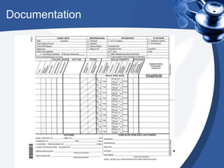
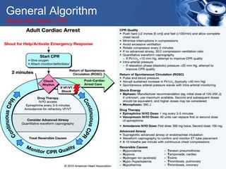
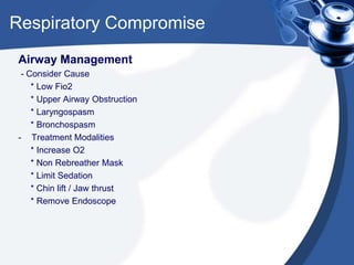
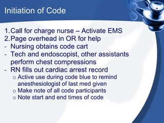
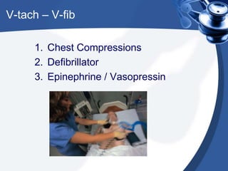
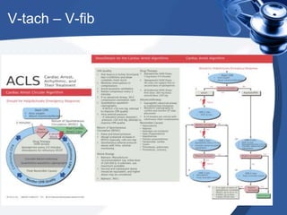
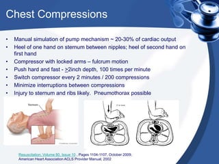
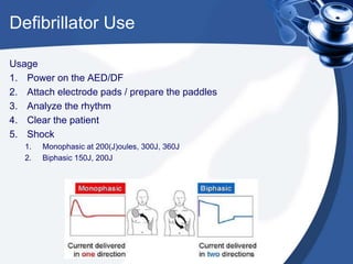
Code Blue in the OR
An Inservice for SFENDO
Staff
Goal
To create an awareness of the
vital points associated with the
preparation for, recognition of
and response to a Code Blue in
the endoscopy suite
What is a “Code Blue?”
“Code Blue” refers to a state of
cardiopulmonary arrest. The heart no
longer pumps blood, leading to anaerobic
metabolism, organ failure, and death.
Code Blue in OR
University of Pittsburg study Jan ‘89 – Dec ’01
• 218,274 anesthetics, 23 cases of cardiac arrest
• 14 patients died in the OR – total mortality = 0.64 in 10k
• Immediate overall survival rate after arrest was 39% (9/23).
• 50% of the patients (12/23) were emergency cases with 41% survival rate
(5/12).
• 25% of the arrests were trauma patients (6/23).
• Most arrest patients (87%, 20/23) were American Society of
Anesthesiologists Physical Status (ASA PS) IV and V
• One case was attributable to an anesthesia-related cardiac arrest and
recovered after successful resuscitation.
Chinese Medical Journal, 2011, Vol. 124 No. 2 : 227-232
Ethical concerns
DNR Policy – Site / organization specific
• The DNR status must be discussed by the surgeon and the patient /
family or legally designated decision maker, at the time the surgical
consent is obtained
• The order can be continued, partially suspended, or suspended during
the perioperative period.
• The physician must document the choice, as well as the time for
reinstatement if it is lifted.
• A written order detailing this must appear.
• All team members must be informed of the DNR status
• Team members uncomfortable with the choice may seek a
replacement for the procedure
• Aside from the DNR enforcement, OR care should be based on the
standard of practice
Preparation - key
1. Know your resources
2. Know your role
3. Know your patient
4. Understand the algorithm
5. Understand the potential
outcomes
Materials
Be familiar with the function and
location of the following:
1. Code Cart
2. Airway Equipment
3. Defibrillator
4. Documentation
Code Cart
Airway Cart
Defibrillator
Documentation
Roles
1. Anesthesiologist
2. RN
3. Technician
4. Endoscopist
The Patient
1. Diagnosis
2. Procedure
3. Other disease states
4. Test results (labs, EKG)
5. ASA classification
6. DNR status
General Algorithm
Starts with Basic CPR
Case Presentation
56 year old female for EGD / Colonoscopy.
• Past medical history significant for :
HTN, Obstructive Sleep Apnea, Fibromyalgia,
Restless leg syndrome, renal insufficiency,
Diabetes type 2, & anemia of chronic disease.
• Social history significant for :
20 pack year smoking history, quit 5 months ago.
Vital signs: BP of 163/78 HR of 64 SPO2 of 95% Ht: 5’1” Wt: 89 kg.
NPO: 12 hours.
Physical exam is remarkable for the following: patient’s habitus, poor
dentition, and mild end – expiratory wheeze, cleared with coughing.
What else would you like to know?
Other Info
1. Allergies: PCN, Sulfa, Vicodin,
tape
2. Medications
3. Lab values: creatinine, potassium,
glucose
4. ASA Classification
5. DNR status
Case Presentation
Pt. is transported to the OR after Albuterol therapy.
Standard monitors are placed, and the care is
assumed by the anesthesiologist. The
endoscopist proceeds in routine fashion after time
out.
At roughly 3 minutes into the case, you hear the
tone of the pulse oximeter decreasing
What should the team do?
Respiratory Compromise
Airway Management
- Consider Cause
* Low Fio2
* Upper Airway Obstruction
* Laryngospasm
* Bronchospasm
- Treatment Modalities
* Increase O2
* Non Rebreather Mask
* Limit Sedation
* Chin lift / Jaw thrust
* Remove Endoscope
Respiratory Compromise
Airway Devices
Bag ventilation
o Involves chin lift, jaw thrust and proper mask
seal pressure
o Ventilation must limit over pressure, which may
inflate stomach
LMA Placement
o Blind approach
o Limited Stimulation
o Does NOT treat laryngospasm
Intubation
o Definitive airway
o Requires laryngoscopy
o Significant stimulation
o Cricoid pressure
Case continuation
• Chin lift + jaw thrust, with O2 to 8L/min from 4L/min improves O2sat
from 81% to 94%
• The endoscopist did not need to remove the scope. The final GE
junction biopsy is performed & the scope is now removed
• The patient is turned for the colonoscopy.
• The colonoscope is advanced into the sigmoid. With stimulation,
there is significant pt movement. Sedation is augmented
• Eventually, the following are seen on EKG:
Conduction Issues
Dysrhythmias to consider
Sinus Bradycardia – Often related to Vagal stimulation
o Significant if perfusion declines – May require chest compressions
o Atropine 0.2 -1 mg. Glycopyrollate reasonable substitute
Sinus Tachycardia – Often relates to painful stimulation
o Significant with decreasing perfusion – may lead to ischemia
o Initial treatments = vagal maneuvers, beta blockade, adenosine
Dysrhythmias to Consider
Bigeminy
Ventricular tachycardia
Torsades
Case Continuation
The anesthesiologist is manipulating the airway, and listening to
the chest.
The endoscopist continues advancing the colonoscope. The bp
cuff cycles and alarms with “(---/---)” on the monitor
The EKG tracing reveals the rhythm below. Recycling the cuff the
anesthesiologist looks into the field. While the tachycardic rhythm
still appears on the monitor, the anesthesiologist confirms no
carotid pulse.
Initiation of Code
1.Call for charge nurse – Activate EMS
2.Page overhead in OR for help
- Nursing obtains code cart
- Tech and endoscopist, other assistants
perform chest compressions
- RN fills out cardiac arrest record
o Active use during code blue to remind
anesthesiologist of last med given
o Make note of all code participants
o Note start and end times of code
V-tach – V-fib
1. Chest Compressions
2. Defibrillator
3. Epinephrine / Vasopressin
V-tach – V-fib
Chest Compressions
• Manual simulation of pump mechanism ~ 20-30% of cardiac output
• Heel of one hand on sternum between nipples; heel of second hand on
first hand
• Compressor with locked arms – fulcrum motion
• Push hard and fast - >2inch depth, 100 times per minute
• Switch compressor every 2 minutes / 200 compressions
• Minimize interruptions between compressions
• Injury to sternum and ribs likely. Pneumothorax possible
Resuscitation; Volume 80, Issue 10 , Pages 1104-1107, October 2009;
American Heart Association ACLS Provider Manual, 2002
Defibrillator Use
Usage
1. Power on the AED/DF
2. Attach electrode pads / prepare the paddles
3. Analyze the rhythm
4. Clear the patient
5. Shock
1. Monophasic at 200(J)oules, 300J, 360J
2. Biphasic 150J, 200J
Vtach - Vfib
Medications:
o Epinephrine, 1mg every 3-5 minutes
o Vasopressin, IV or IO, 40U, either 1st or
second drug round in place of
epinephrine
o Amiodarone, if VF is refractory after
Shocks and Epi / Vasopressin – first
dose 300mg, second 150mg
Case Presentation 2
•Pt is a 41 year old female, for colonoscopy based
on a family history of colon cancer
•PMHx – HTN, mild obesity (BMI 31), fibromyalgia
•Meds include OCP, “water pill” & a “pain med”
•Social – international business, 2 children, smoker
•Case proceeds.
•Just prior to cecum, the bp cuff reads “(---/---)” as
the pulse oximeter tone decreases
•EKG appears as below:
Pulseless Electrical Activity
Definition: The electrical activity to generate cardiac contraction is
present, but no contraction occurs
Etiologies: H’s and T’s
o Hyper/hypokalemia
o Hydrogen ion /
acidosis
o Hypothermia
o Hypoglycemia
o Hypoxia
o Hypovolemia
oToxins / tablets
oTamponade (cardiac)
oTension pneumothorax
oThrombosis (cardiac or
pulmonary)
oTrauma
Which may apply to this patient?
PEA
1. Chest compressions
2. Search for cause
3. Medications
4. Interventions
PEA
Case Presentation 2
o With second dose of epinephrine, palpable
pulse returns
o Pt is transported to hospital, to ICU
intubated
o Further studies reveal lower extremity DVTs
o Pts family member reports that the pt had
just returned from a business trip to
Singapore 2 days earlier.
o “Pain med” = Amitriptyline – possible
overdose
End Points
1. The return to normalcy of blood pressure, heart rate, and
urine output
2. Multiple rounds of resuscitation without return to physiologic
parameters
3. Heroic measures (Cardiopulmonary bypass, etc)
Outcomes
1. Survival - goal unique to CPR is the reversal of
clinical death, an outcome achieved in only a
minority of patients
2. When to quit
1. Threat to safety of others
2. Rigor mortis
3. More than 30 minutes of cumulative efforts
4. All reversible causes have been ruled out
3. Debriefing
4. Cleanup
Debriefing
1. The Code team leader should be responsible for the debriefing
a. Included are all appropriate staff to include those staff that witnessed the event, those
initiated treatment and any other participants.
b. Discuss any issues for opportunities for improvement identified and develop potential
strategies for improvement.
2. Defibrillator data should be printed. Ensure that the printed download is placed with the code blue
form.
3. A debriefing critique may be considered by the team leader. This may be recorded for future
review or data collection
4. Administrators may later perform an analysis of code blues, identifies trends, and reports findings
during the regularly scheduled Medical Executive Committee meeting and the Nurse Executive
Committee.
Cleanup / Restock
1. Filing paperwork –
a. Send Code Blue record of drug use to pharmacy
b. Copy of Code Blue record to Education dept- Code Blue committee
c. Place copy of Code Blue record to patient chart
d. Fill out Code Blue evaluation form as mentioned above in debriefing
e. Complete an RDE
2. Charges
a. Additional drugs used by department on the drug slip – to pharmacy
b. Pharmacy replenishes meds / checks expiration of existing meds
3. Restock –
a. Call Stores for replacement Code Cart
b. Clean defibrillator with disinfectant wipes, transfer to new cart
c. Check that the new Code Cart has been locked with a tie number
d. Replace used surgical supplies
e. Reconnect defibrillator to electrical outlet

Code blue drill and didactic for endoscopy center providers

  • 1.
    Code Blue inthe OR An Inservice for SFENDO Staff
  • 2.
    Goal To create anawareness of the vital points associated with the preparation for, recognition of and response to a Code Blue in the endoscopy suite
  • 3.
    What is a“Code Blue?” “Code Blue” refers to a state of cardiopulmonary arrest. The heart no longer pumps blood, leading to anaerobic metabolism, organ failure, and death.
  • 4.
    Code Blue inOR University of Pittsburg study Jan ‘89 – Dec ’01 • 218,274 anesthetics, 23 cases of cardiac arrest • 14 patients died in the OR – total mortality = 0.64 in 10k • Immediate overall survival rate after arrest was 39% (9/23). • 50% of the patients (12/23) were emergency cases with 41% survival rate (5/12). • 25% of the arrests were trauma patients (6/23). • Most arrest patients (87%, 20/23) were American Society of Anesthesiologists Physical Status (ASA PS) IV and V • One case was attributable to an anesthesia-related cardiac arrest and recovered after successful resuscitation. Chinese Medical Journal, 2011, Vol. 124 No. 2 : 227-232
  • 5.
    Ethical concerns DNR Policy– Site / organization specific • The DNR status must be discussed by the surgeon and the patient / family or legally designated decision maker, at the time the surgical consent is obtained • The order can be continued, partially suspended, or suspended during the perioperative period. • The physician must document the choice, as well as the time for reinstatement if it is lifted. • A written order detailing this must appear. • All team members must be informed of the DNR status • Team members uncomfortable with the choice may seek a replacement for the procedure • Aside from the DNR enforcement, OR care should be based on the standard of practice
  • 6.
    Preparation - key 1.Know your resources 2. Know your role 3. Know your patient 4. Understand the algorithm 5. Understand the potential outcomes
  • 7.
    Materials Be familiar withthe function and location of the following: 1. Code Cart 2. Airway Equipment 3. Defibrillator 4. Documentation
  • 8.
  • 9.
  • 10.
  • 11.
  • 12.
    Roles 1. Anesthesiologist 2. RN 3.Technician 4. Endoscopist
  • 13.
    The Patient 1. Diagnosis 2.Procedure 3. Other disease states 4. Test results (labs, EKG) 5. ASA classification 6. DNR status
  • 14.
  • 15.
    Case Presentation 56 yearold female for EGD / Colonoscopy. • Past medical history significant for : HTN, Obstructive Sleep Apnea, Fibromyalgia, Restless leg syndrome, renal insufficiency, Diabetes type 2, & anemia of chronic disease. • Social history significant for : 20 pack year smoking history, quit 5 months ago. Vital signs: BP of 163/78 HR of 64 SPO2 of 95% Ht: 5’1” Wt: 89 kg. NPO: 12 hours. Physical exam is remarkable for the following: patient’s habitus, poor dentition, and mild end – expiratory wheeze, cleared with coughing. What else would you like to know?
  • 16.
    Other Info 1. Allergies:PCN, Sulfa, Vicodin, tape 2. Medications 3. Lab values: creatinine, potassium, glucose 4. ASA Classification 5. DNR status
  • 17.
    Case Presentation Pt. istransported to the OR after Albuterol therapy. Standard monitors are placed, and the care is assumed by the anesthesiologist. The endoscopist proceeds in routine fashion after time out. At roughly 3 minutes into the case, you hear the tone of the pulse oximeter decreasing What should the team do?
  • 18.
    Respiratory Compromise Airway Management -Consider Cause * Low Fio2 * Upper Airway Obstruction * Laryngospasm * Bronchospasm - Treatment Modalities * Increase O2 * Non Rebreather Mask * Limit Sedation * Chin lift / Jaw thrust * Remove Endoscope
  • 19.
    Respiratory Compromise Airway Devices Bagventilation o Involves chin lift, jaw thrust and proper mask seal pressure o Ventilation must limit over pressure, which may inflate stomach LMA Placement o Blind approach o Limited Stimulation o Does NOT treat laryngospasm Intubation o Definitive airway o Requires laryngoscopy o Significant stimulation o Cricoid pressure
  • 20.
    Case continuation • Chinlift + jaw thrust, with O2 to 8L/min from 4L/min improves O2sat from 81% to 94% • The endoscopist did not need to remove the scope. The final GE junction biopsy is performed & the scope is now removed • The patient is turned for the colonoscopy. • The colonoscope is advanced into the sigmoid. With stimulation, there is significant pt movement. Sedation is augmented • Eventually, the following are seen on EKG:
  • 21.
  • 22.
    Dysrhythmias to consider SinusBradycardia – Often related to Vagal stimulation o Significant if perfusion declines – May require chest compressions o Atropine 0.2 -1 mg. Glycopyrollate reasonable substitute Sinus Tachycardia – Often relates to painful stimulation o Significant with decreasing perfusion – may lead to ischemia o Initial treatments = vagal maneuvers, beta blockade, adenosine
  • 23.
  • 24.
    Case Continuation The anesthesiologistis manipulating the airway, and listening to the chest. The endoscopist continues advancing the colonoscope. The bp cuff cycles and alarms with “(---/---)” on the monitor The EKG tracing reveals the rhythm below. Recycling the cuff the anesthesiologist looks into the field. While the tachycardic rhythm still appears on the monitor, the anesthesiologist confirms no carotid pulse.
  • 25.
    Initiation of Code 1.Callfor charge nurse – Activate EMS 2.Page overhead in OR for help - Nursing obtains code cart - Tech and endoscopist, other assistants perform chest compressions - RN fills out cardiac arrest record o Active use during code blue to remind anesthesiologist of last med given o Make note of all code participants o Note start and end times of code
  • 26.
    V-tach – V-fib 1.Chest Compressions 2. Defibrillator 3. Epinephrine / Vasopressin
  • 27.
  • 28.
    Chest Compressions • Manualsimulation of pump mechanism ~ 20-30% of cardiac output • Heel of one hand on sternum between nipples; heel of second hand on first hand • Compressor with locked arms – fulcrum motion • Push hard and fast - >2inch depth, 100 times per minute • Switch compressor every 2 minutes / 200 compressions • Minimize interruptions between compressions • Injury to sternum and ribs likely. Pneumothorax possible Resuscitation; Volume 80, Issue 10 , Pages 1104-1107, October 2009; American Heart Association ACLS Provider Manual, 2002
  • 29.
    Defibrillator Use Usage 1. Poweron the AED/DF 2. Attach electrode pads / prepare the paddles 3. Analyze the rhythm 4. Clear the patient 5. Shock 1. Monophasic at 200(J)oules, 300J, 360J 2. Biphasic 150J, 200J
  • 30.
    Vtach - Vfib Medications: oEpinephrine, 1mg every 3-5 minutes o Vasopressin, IV or IO, 40U, either 1st or second drug round in place of epinephrine o Amiodarone, if VF is refractory after Shocks and Epi / Vasopressin – first dose 300mg, second 150mg
  • 31.
    Case Presentation 2 •Ptis a 41 year old female, for colonoscopy based on a family history of colon cancer •PMHx – HTN, mild obesity (BMI 31), fibromyalgia •Meds include OCP, “water pill” & a “pain med” •Social – international business, 2 children, smoker •Case proceeds. •Just prior to cecum, the bp cuff reads “(---/---)” as the pulse oximeter tone decreases •EKG appears as below:
  • 32.
    Pulseless Electrical Activity Definition:The electrical activity to generate cardiac contraction is present, but no contraction occurs Etiologies: H’s and T’s o Hyper/hypokalemia o Hydrogen ion / acidosis o Hypothermia o Hypoglycemia o Hypoxia o Hypovolemia oToxins / tablets oTamponade (cardiac) oTension pneumothorax oThrombosis (cardiac or pulmonary) oTrauma Which may apply to this patient?
  • 33.
    PEA 1. Chest compressions 2.Search for cause 3. Medications 4. Interventions
  • 34.
  • 35.
    Case Presentation 2 oWith second dose of epinephrine, palpable pulse returns o Pt is transported to hospital, to ICU intubated o Further studies reveal lower extremity DVTs o Pts family member reports that the pt had just returned from a business trip to Singapore 2 days earlier. o “Pain med” = Amitriptyline – possible overdose
  • 36.
    End Points 1. Thereturn to normalcy of blood pressure, heart rate, and urine output 2. Multiple rounds of resuscitation without return to physiologic parameters 3. Heroic measures (Cardiopulmonary bypass, etc)
  • 37.
    Outcomes 1. Survival -goal unique to CPR is the reversal of clinical death, an outcome achieved in only a minority of patients 2. When to quit 1. Threat to safety of others 2. Rigor mortis 3. More than 30 minutes of cumulative efforts 4. All reversible causes have been ruled out 3. Debriefing 4. Cleanup
  • 38.
    Debriefing 1. The Codeteam leader should be responsible for the debriefing a. Included are all appropriate staff to include those staff that witnessed the event, those initiated treatment and any other participants. b. Discuss any issues for opportunities for improvement identified and develop potential strategies for improvement. 2. Defibrillator data should be printed. Ensure that the printed download is placed with the code blue form. 3. A debriefing critique may be considered by the team leader. This may be recorded for future review or data collection 4. Administrators may later perform an analysis of code blues, identifies trends, and reports findings during the regularly scheduled Medical Executive Committee meeting and the Nurse Executive Committee.
  • 39.
    Cleanup / Restock 1.Filing paperwork – a. Send Code Blue record of drug use to pharmacy b. Copy of Code Blue record to Education dept- Code Blue committee c. Place copy of Code Blue record to patient chart d. Fill out Code Blue evaluation form as mentioned above in debriefing e. Complete an RDE 2. Charges a. Additional drugs used by department on the drug slip – to pharmacy b. Pharmacy replenishes meds / checks expiration of existing meds 3. Restock – a. Call Stores for replacement Code Cart b. Clean defibrillator with disinfectant wipes, transfer to new cart c. Check that the new Code Cart has been locked with a tie number d. Replace used surgical supplies e. Reconnect defibrillator to electrical outlet

Editor's Notes

  • #3 A code blue may be called in the OR even without a true cardiac arrest.
  • #5 Results published 2011
  • #6 This may ultimately be part of the time out
  • #26 Once the sterile field is managed / isolated or broken, other assistants may assist with chest compressions
  • #39 While there is no formal structure for a debriefing in the John Muir system, it is a possibility for the future.