
More Related Content
Similar to classificationofbonesanatomy-150725135414-lva1-app6891.pptx
Similar to classificationofbonesanatomy-150725135414-lva1-app6891.pptx (20)
More from shehlazaman1
More from shehlazaman1 (18)
Recently uploaded
Recently uploaded (20)
classificationofbonesanatomy-150725135414-lva1-app6891.pptx
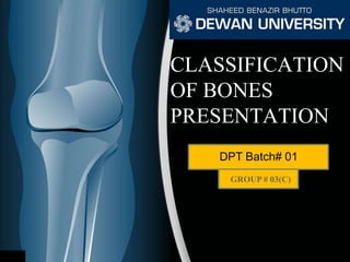
- 1. CLASSIFICATION OF BONES PRESENTATION DPT Batch# 01 GROUP # 03(C)
- 2. PRESENTED BY: 1. Saadiya Naeemi.
- 3. OUTLINE •Introduction. •Types Of Bones: A.Types of bone on the basis of shape: 1. Long bones, 2. Short bones, 3. Flat bones, 4. Irregular bones, 5. Pneumatic bones, 6. Sesamoid bones.
- 4. OUTLINE B. Types on the basis of development: 1. Membranous bones, 2. Cartilaginous bones, 3. Membro-cartilaginous bones. C. Types on the basis of region: 1. Bones of Axial skeleton, 2. Bones of Appendicular skeleton. D. Types on the basis of structure: 1. According to Macroscopic approach: a. Compact bone, b. Spongy bone.
- 5. 2. According to Microscopic approach: a. Fibrous bone, b. Lamellar bone. c. Woven bone. d. Dentine and Cement.
- 6. •Bones: Bone is the one-third connective tissue, forming the main supporting framework of the body. The in-organic Calcium salts make it hard and rigid. •Osteology: The scientific study of bones is known as Osteology. INTRODUCTION
- 7. •Functions Of Bones: a. Support, Protection & Movement: 1. Gives shape to the body. 2. Supports body weight. 3. Protects sensitive parts of the body. b. Blood Cell Formation: The red bone marrow found in the connective tissue of certain bones is the site of blood cell production.
- 8. c. In-organic Salt Storage: Functions as a storage depot for many of the body needs. For example: (Calcium, Potassium, Sodium etc).
- 9. •Cartilage: Cartilage is a connective tissue composed of cells & fibers embedded in Nucleopolysachharide. There are three main types of cartilages which are mentioned below: 1.Hyaline Cartilage: Covers the atricular Surface of synovial membrane. 2.Fibro-Cartilage: Present in the Epiglottis etc. 3. Elastic Cartilage: Present in the ear Pinna etc.
- 10. •Functional Classification of Joints: The point in human skeleton where two bones join together is called a joint. There are three types of joints which are mentioned below: 1. Synarthroses: Immoveable Joints. Example: Suture Fig. Example of Synarthroses.
- 11. 2. Amphiarthrosis: Slightly/Partially moveable joints. Example: Intervertebral disc. . Fig. Example of Amphiarthrosis. 3. Diarthrosis: Freely moveable joints. Example: Synovial membrane Fig. Example of Diarthrosis.
- 12. Types Of Bones: A. Types On The Basis Of Shape: There are 6 basic types which are mentioned below: 1. Long Bones. 2. Short Bones. 3. Flat Bones. 4. Irregular Bones. 5. Pneumatic Bones. 6. Sesamoid Bones.
- 13. 1. Long Bones: These bones typically have an elongated shaft and two expanded ends one on either side of the shaft. The shaft is known as diaphysis and the ends are called epiphyses. Examples: Humerus, femur etc. Fig. Femur, Long Bone.
- 14. 2. Short Bones: These bones are short in posture and can be of any shape. Examples: The carpal and tarsal bones. Fig. Carpal Bones, Short Bones. Fig. Tarsal Bones, Short Bones.
- 15. 3. Flat Bones: These bones are flat in appearance. Examples: Scapula, Ribs, Sternum etc. Fig. Scapula, Flat Bone. Fig. Sternum & ribs, Flat bones.
- 16. 4. Irregular Bones: These bones are completely irregular in shape. Examples: vertebrae, hip bone and bones in the base of skull. Fig. Hip bone, Irregular bone. Fig. 1st & 2nd Cervical of vertebra, Irregular bone.
- 17. 5. Pneumatic Bones: Pneumatic bones can also be categorized under the irregular bones.The characteristic difference is the presenceof large air spaces in these bones which make them light in weight and thus they form the major portion of skull Examples: Sphenoid, Ethmoid, Maxila etc. Fig. Sphenoid, Ethmoid, Maxila , Pnematic Bones.
- 18. 6.Sesamoid Bones: These are in the form of nodules embedded in tendons and joint capsules. Examples: Patella, Pisiform, Fabella etc. Fig. Pisiform, Sesamoid Bone. Fig. Patella, Sesamoid Bone.
- 19. B. On The Basis Of Development: There are three basic types which are mentioned below: 1. Membranous bones, 2. Cartilaginous bones, 3. Membro-cartilaginous bones.
- 20. 1. Membranous bones: These bones ossify in membrane from mesenchymal condensations. Examples: Bones of the vault of skull and Facial bones. Fig. Bones of Skull & Face, Membranous bones.
- 21. 2. Cartilaginous Bones: They ossify in cartilage and thus derived from performed cartilaginous models. Examples: Thoracic cage etc. Fig. Thoracic cage, Cartilaginous bones.
- 22. 3. Membrano-cartilaginous Bones: They ossify partly in membrane and partly in cartilage. Examples: Clavicle, Mandible, Temporal etc Fig. Mandible, Membrano-cartilaginous bone. Fig. Clavicle, Membrano- Cartilaginous bone. Fig. Temporal, Membrano-Cartilaginous bone.
- 23. C. On The Basis Of Region: It is divided into two types, they are: 1. Axial Skeleton. 2. Appendicular Skeleton.
- 24. 1. Bones Of Axial Skeleton: These bones forms the axial skeleton of the human body. Examples: Bones of skull, Thoracic cage & Vertebral Column Fig. Bones of Axial Skeleton.
- 25. 2.Bones Of Appendicular Skeleton: These bones forms the Appendicular skeleton of the human Body. Examples: bones of the limbs and girdles of limbs. Fig. Upper limb, Clavicle & Scapula Girdle bones of Appendicular Skeleton. Fig. Lower limb, Pelvic Girdle bones of Appendicular Skeleton.
- 26. D. On The Basis Of Structure: They are sub divided into two parts, which are: 1. Macroscopic Approach. 2. Microscopic Approach.
- 27. 1. Macroscopic Approach: a. Compact Bone: Compact bone is dense in texture but is extremely porus. Example: In the cortex of long bones. Fig. Macroscopic structure of the cortex of femur(Long Bone).
- 28. b. Cancellous OR Spongy Bone: The part of bone where there is more empty space and less bone tissue. Example: The inner part of Long Bones. Fig. The Macroscopic Structure of the inner part of Femur(Long Bone).
- 29. a. Fibrous Bone: These have more fibers in them. Also known as immature bones. Example: Found only in fetus, sockets of alveolar bones and sutures of the skull. 2. Microscopic Approach: Fig. Microscopic structure of Fetus of Human.
- 30. Fig. Microscopic structure of Lamellar arranged in piles in a cancellous bone. b. Lamellar Bone: Most of the mature human bones, whether compact or Cancellous, are composed of thin plates of bony tissue called lamellae. Example: Formed on the periosteal surface of diaphysis.
- 31. c. Woven Bone: Occurs initially in fetal bones. In adults woven bone is created after fractures. Example: Seen in fetal bone, fracture repair and in cancer of bone. Fig. Microscopic structure of Woven bone.
- 32. d. Cementum and Dentine: Cementum is a specialized calcified substance covering the root of a tooth. It hardens to act as an adhesive glue. Dentine is one of the hard tissues of the teeth which constitutes most of its bulk. Example: Occur in teeth. Fig. Dentine of a tooth. Fig. The Cementum of a human tooth.
- 33. Do you know? • Hyaline cartilage is the most abundant cartilage. • By age 25 the skeleton is completely hardened. •206 bones make up the adult skeleton (20% of body mass) • 80 bones of the axial skeleton • 126 bones of the appendicular skeleton • The largest bone in the human skeleton is Femur. • Babies are born with about 300 bones. •Almost a third of bones of babies eventually fuse together to form the 206-bone skeleton of an adult.
- 34. • Hyaline cartilage is the most abundant cartilage. • By age 25 the skeleton is completely hardened. • 206 bones make up the adult skeleton (20% of body mass) • 80 bones of the axial skeleton • 126 bones of the appendicular skeleton • The largest bone in the human skeleton is Femur. • Babies are born with about 300 bones. •Almost a third of bones of babies eventually fuse together to form the 206-bone skeleton of an adult. Do you know?
- 35. • Hyaline cartilage is the most abundant cartilage. • By age 25 the skeleton is completely hardened. • 206 bones make up the adult skeleton (20% of body mass) • 80 bones of the axial skeleton • 126 bones of the appendicular skeleton • The largest bone in the human skeleton is Femur. • Babies are born with about 300 bones. • Almost a third of bones of babies eventually fuse together to form the 206-bone skeleton of an adult. Do you know?
- 36. Do you know? • Hyaline cartilage is the most abundant cartilage. • By age 25 the skeleton is completely hardened. • 206 bones make up the adult skeleton (20% of body mass) • 80 bones of the axial skeleton • 126 bones of the appendicular skeleton • The largest bone in the human skeleton is Femur. • Babies are born with about 300 bones. • Almost a third of bones of babies eventually fuse together to form the 206-bone skeleton of an adult.