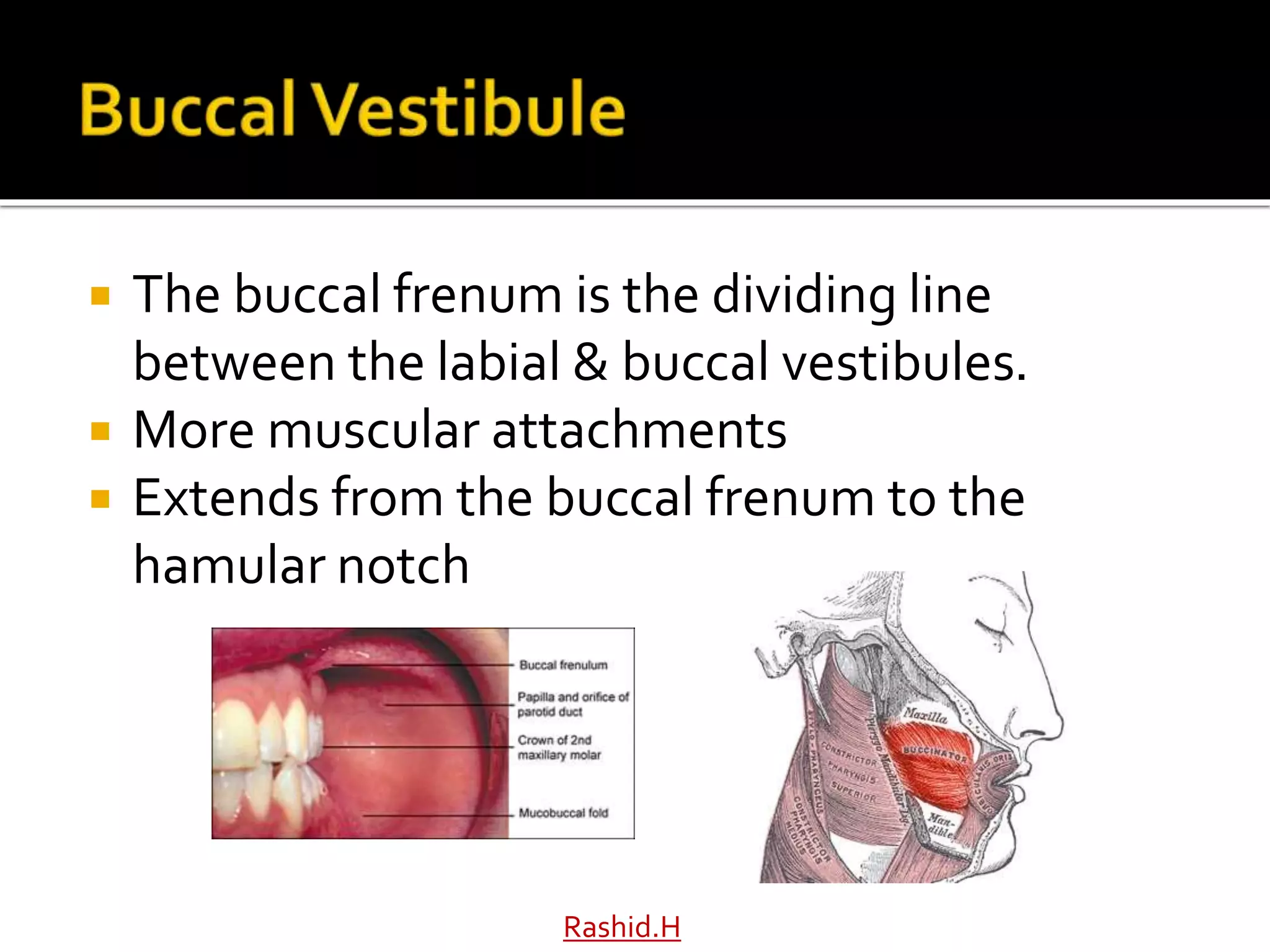
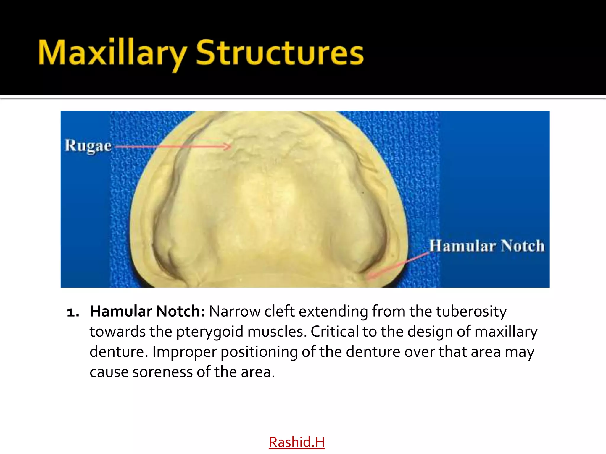
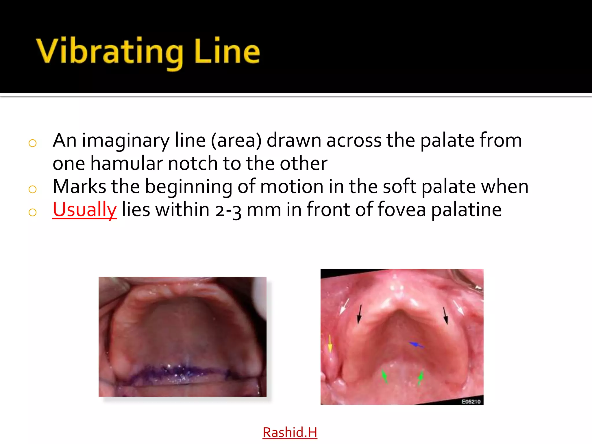
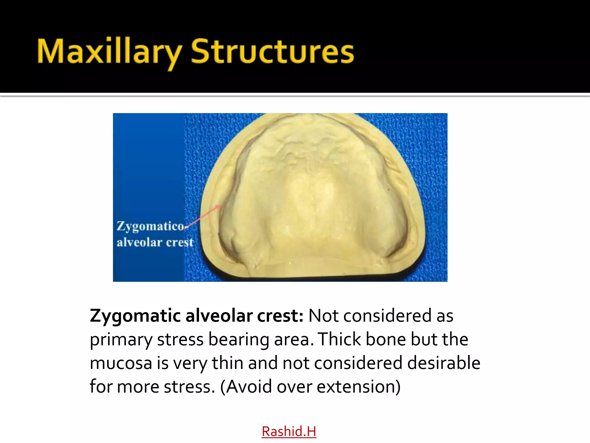
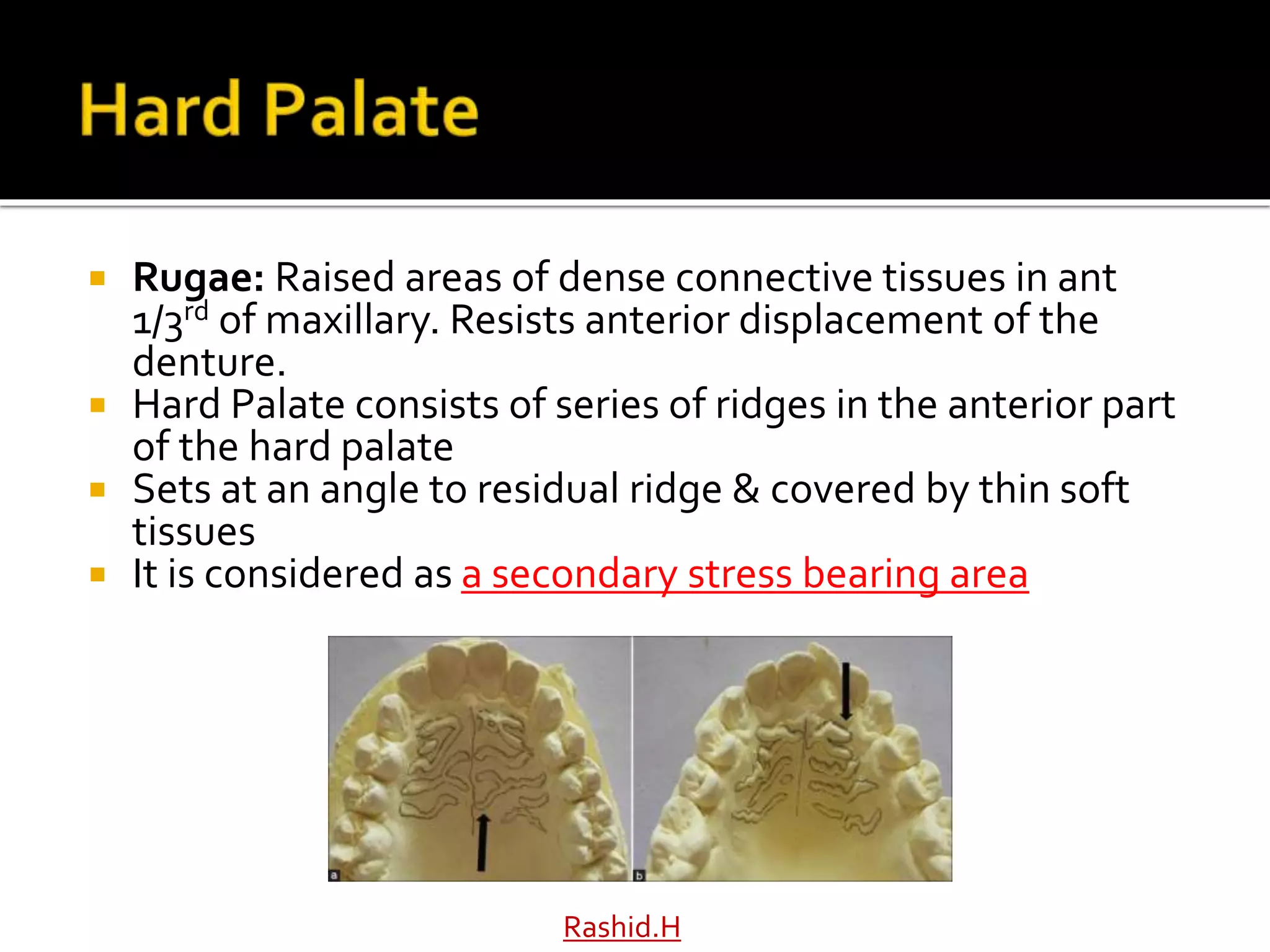
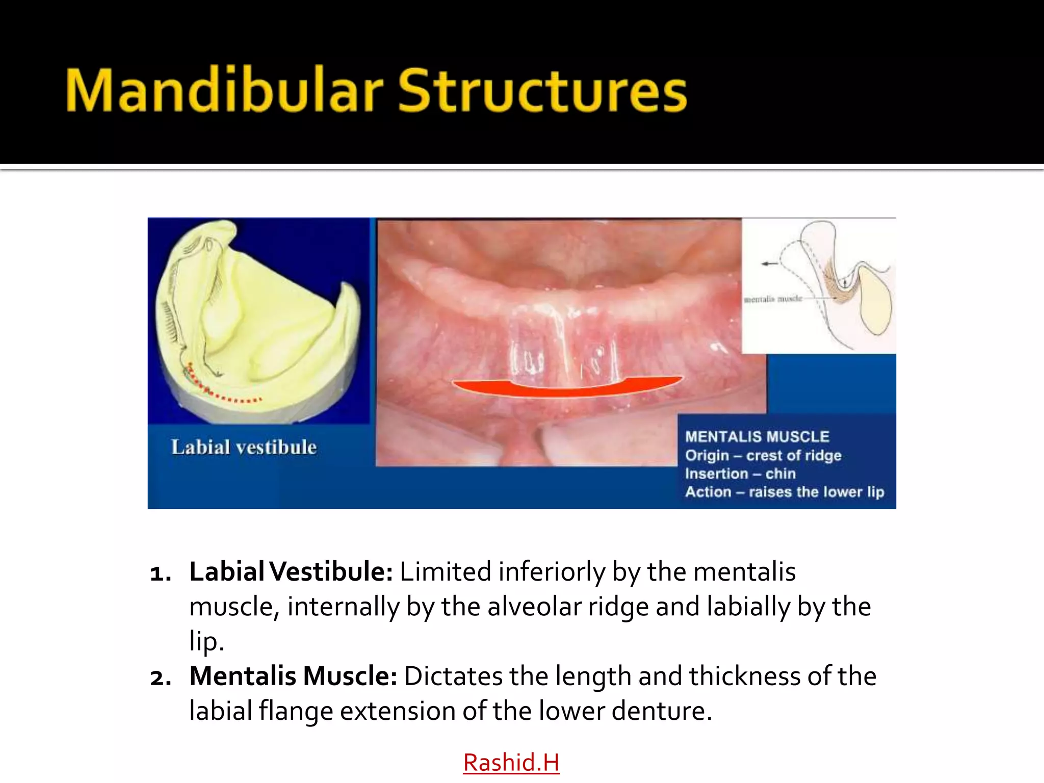
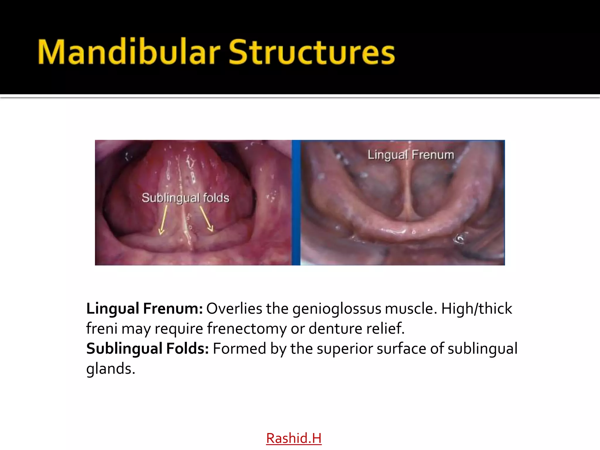
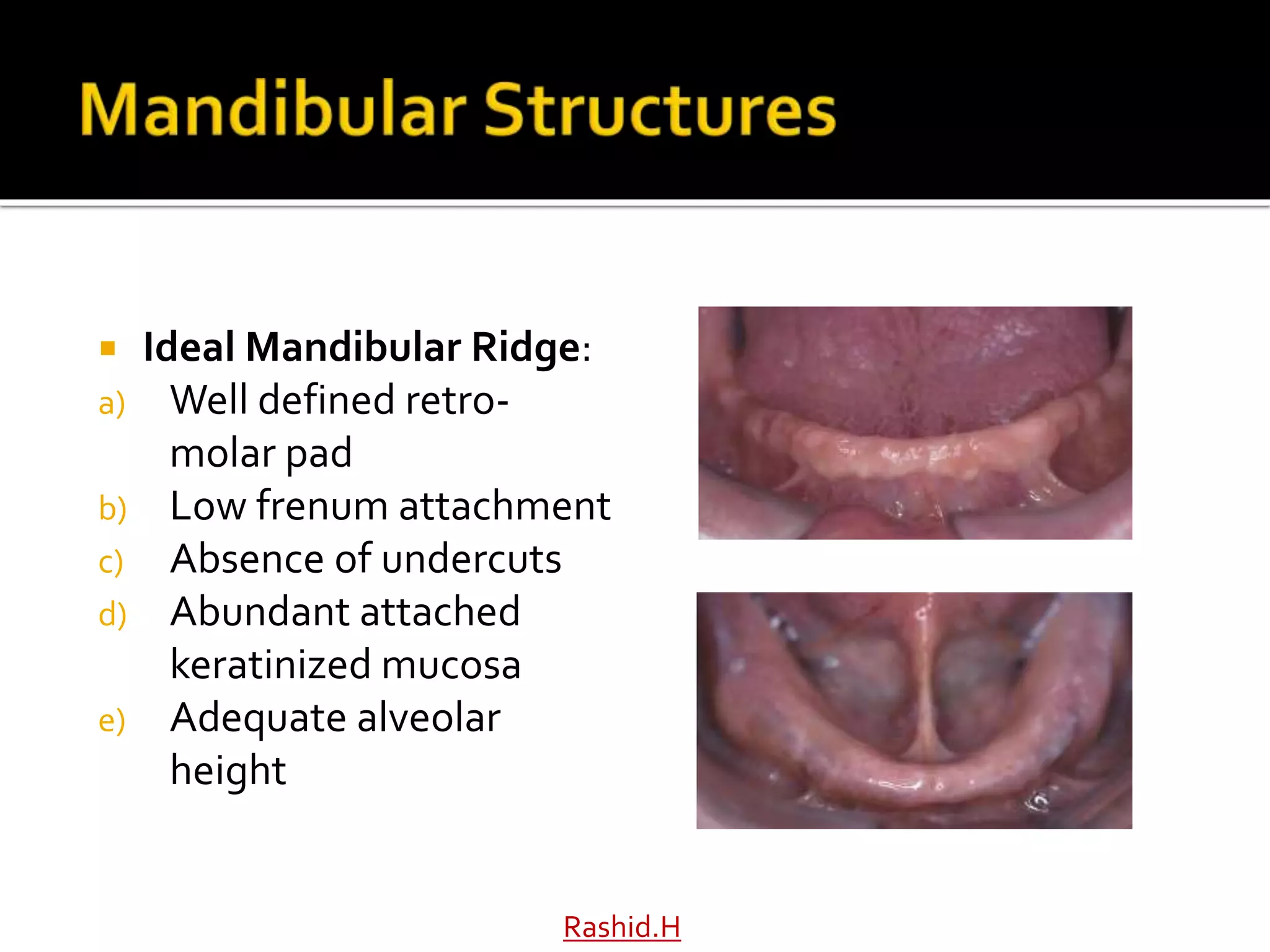
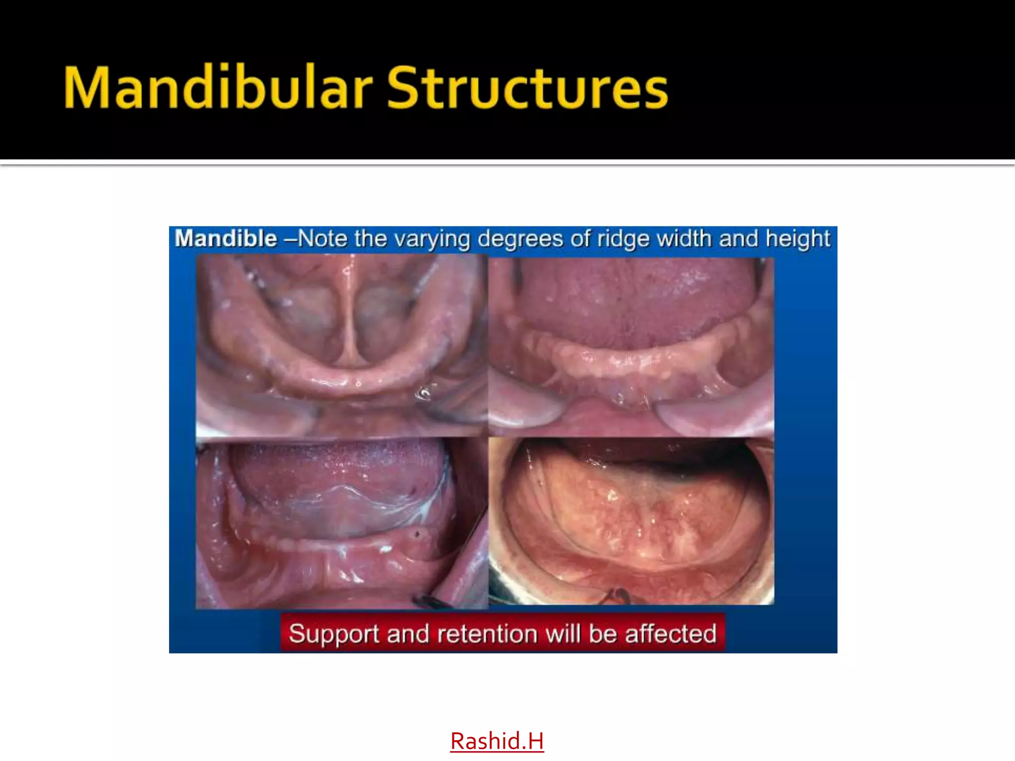
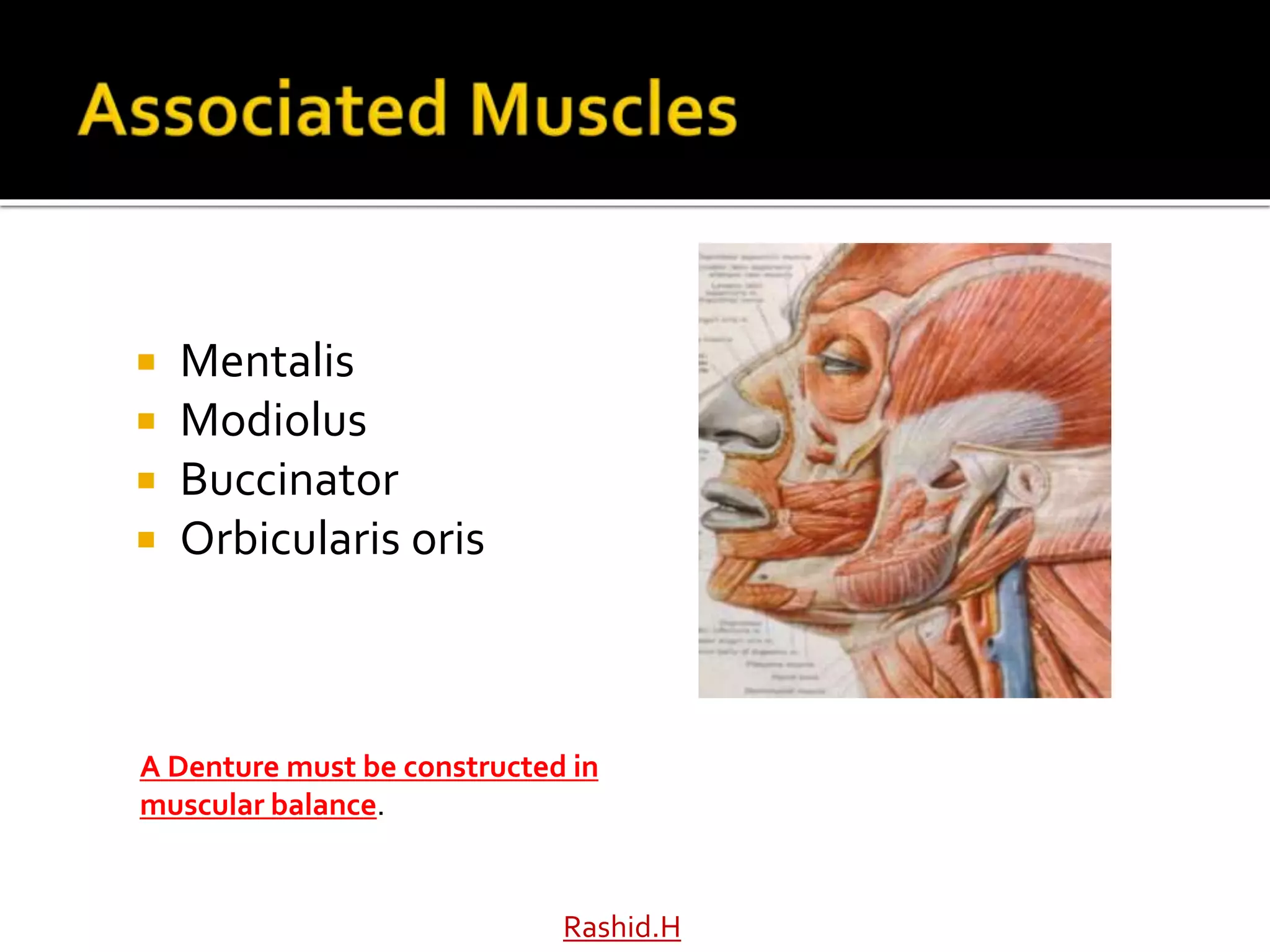
Independent Call Girls Hyderabad 💋 9352988975 💋 Genuine WhatsApp Number for R...
Anatomy for Complete and Partial Dentures (1).pptx
1. Dr Haroon Rashid Baloch
Department of Prosthodontics,
Ziauddin College of Dentistry.
2. A thorough knowledge of the anatomy of the
denture bearing surfaces is paramount to
designing and fabricating functional
dentures.
The functional anatomy of the denture
foundation areas of the maxilla and mandible
is presented in detail.
In particular, the relationship of these
anatomic structures that impact retention,
stability and support.
Rashid.H
3. Retention
Stability
Support
“When the key anatomical landmarks and
their role with retention, stability and support
are understood, dentures can be fabricated
as integral part of oral cavity and not just
artificial mechanical substitutes”
Rashid.H
5. Osseous structures support the overlying
denture
Affects the impression making
Maxillary denture is supported by two main
bones:
a) Maxilla
b) Palatine bone
Mandibular Denture is supported mainly by:
a) Mandible
Rashid.H
6. The cushion between the denture and the
supporting bone
Mucous membrane consists of
a) Mucosa
b) Sub mucosa
The submucosa is formed by connective
tissue that may be:
a) Dense
b) Loose
c) May vary in thickness
Rashid.H
7. Makes the bulk of overlying mucous
membrane
Firmly attached to periosteum
Thin submucosa Non resilient soft tissue
mucous membrane easily traumatized
Rashid.H
8. Masticatory covers the hard palate and the
crest of the alveolar ridge
Lining Mucosa covering the cheek, inner
aspect of the lips, soft palate, ventral surface
of the tongue.
Specialized Mucosa dorsum of the tongue
Rashid.H
10. a) Labial frenum
b) Labial vestibule
c) Buccal frenum
d) Buccal vestibule
e) Hamular notch
f) Posterior palatal seal area
Rashid.H
11. 1. Frenum: Freni are folds of mucous membranes and do not
contain significant muscle fibres.They need to be excised
(frenectomy) or maybe relieved.They may compromise denture
stability and retention.
2. Vestibule: Space between the cheeks and the teeth.When filled
with a denture, greatly enhances retention and stability.
Rashid.H
12. 1. Canine Eminence: This prominent bone provides denture support.
Area may also prevent denture rotation thus provide stability.
2. Incisive Papilla: A pad of fibrous tissue overlying the orifice of
naso-palatine canal. Excessive pressure over this area may cause
disruption of blood flow and compression of the nerve. Relief may
be provided if necessary.
Rashid.H
13. Divided into right & left by labial frenum *
The outer surface of the labial vestibule is
attached with the orbicularis oris.
The fibres of orbicularis oris run in a
horizontal direction.
The muscle has an indirect effect on the
denture base
14. The buccal frenum is the dividing line
between the labial & buccal vestibules.
More muscular attachments
Extends from the buccal frenum to the
hamular notch
Rashid.H
15. Rashid.H
1. Hamular Notch: Narrow cleft extending from the tuberosity
towards the pterygoid muscles. Critical to the design of maxillary
denture. Improper positioning of the denture over that area may
cause soreness of the area.
17. 1. Tuberosity: Important primary denture support area in
maxilla. Provides resistance to horizontal denture
movements.
2. Posterior Palatal Seal: Is at the junction of hard and soft
palate near the vibrating line. Marks the posterior extension
of the denture.
Rashid.H
18. Rashid.H
o An imaginary line (area) drawn across the palate from
one hamular notch to the other
o Marks the beginning of motion in the soft palate when
o Usually lies within 2-3 mm in front of fovea palatine
19. Rashid.H
The direction of the vibrating line usually varies
with the shape of palate
Higher the vault the more abrupt and forward the
vibrating line
Flatter palatal vault, the vibrating line is usually
farther posterior and has a gradual curvature
20. Usually two in number, present just behind the
junction of hard and soft-palate
Are indentations near the midline of the palate
Formed by the coalescence of several minor
mucous gland ducts
21. Rashid.H
Coronoid Process: Patient is told to open, protrude and perform lateral
movements to get the impression of coronoid process.The width of the
disto-buccal flange will then be contoured by the coronoid process. (Avoid
over extension)
Minor Salivary Glands: Present at the posterior 1/3rd of the palate.The
impression may appear irregular because of salivary gland secretions that
may adhere to the impression material.
22. Rashid.H
Zygomatic alveolar crest: Not considered as
primary stress bearing area.Thick bone but the
mucosa is very thin and not considered desirable
for more stress. (Avoid over extension)
23. Rashid.H
1. Hard Palate: Considered to be primary stress bearing area of maxillary
denture. Provides retention, stability and support.
2. Midline Palatal Suture:Thin line extending from incisive papilla towards the
posterior end of the palate. Mucosa is tightly attached and very thin. Relief
may be provided to prevent soreness.
3. Major Palatine Foramen: Carries anterior palatine nerve and vessels. Relief
is NOT provided because of presence of abundant overlying tissues.
24. Covered by keratinized stratified squamous
epithelium .
In the region of medial palatal suture , the
submucosa is extremely thin ; relief should
be provided to avoid trauma or rocking of
the denture
Rashid.H
25. Antero-laterally, the submucosa contains
adipose tissue.
Postero-laterally, it contains glandular tissue.
The horizontal portion of the hard palate
provides the primary stress-bearing area.
Rashid.H
26. Rugae: Raised areas of dense connective tissues in ant
1/3rd of maxillary. Resists anterior displacement of the
denture.
Hard Palate consists of series of ridges in the anterior part
of the hard palate
Sets at an angle to residual ridge & covered by thin soft
tissues
It is considered as a secondary stress bearing area
Rashid.H
27. Covered by keratinized stratified squamous
epithelium.
Considered as a secondary stress-bearing area
because it is subject to resorption contrary to
horizontal portion of hard palate.
Rashid.H
28. Ideal Maxillary Ridge
Should have:
a) Abundant attached
keratinized tissue
b) Broad and well developed
ridge.
c) Moderate palatal vault
d) Absence of undercuts
e) Well defined hamular
notches
Rashid.H
29.
30. Maxillary denture influenced by following
muscles:
a) Anterior Labial Flange influenced by
orbicularis oris muscle as far as the pre-
molar region
b) Buccal Flange influenced by the
buccinator muscle
Rashid.H
35. 1. LabialVestibule: Limited inferiorly by the mentalis
muscle, internally by the alveolar ridge and labially by the
lip.
2. Mentalis Muscle: Dictates the length and thickness of the
labial flange extension of the lower denture.
Rashid.H
36. Rashid.H
1. Alveolar Ridge: Secondary support area. High rate of
resorption when excessive pressure is applied.
2. Buccal Frenum: Histologically and functionally same
as that of labial frenum.
37. Rashid.H
Buccal Shelf: Bordered externally by external oblique ridge and
internally by the slope of the alveolar ridge. PRIMARY STRESS
BEARING AREA IN MANDIBLE.
38. The mucous membrane covering the buccal shelf area is loosely attached
Less keratinized
Contains thick sub mucosal layer.
Considered as a primary stress-bearing area because it is covered by a
layer of cortical bone
Lies at right angles to vertical occlusal forces
40. The buccal vestibule extends from the
buccal frenum to the back corner of the
retromolar pad
Influenced by the presence of buccinator
The external oblique ridge does not
govern the extension of the buccal flange
The denture border can be extended 1-2
mm beyond this ridge
Rashid.H
41. Rashid.H
External Oblique Ridge: A ridge of dense bone arising from the
mental foramen, coursing superiorly and distally to become
continuous with the anterior border of the ramus. It is the site of
attachment of buccinator muscle and anatomic guide for lateral
termination of the buccal flange of mandibular denture.
42. Rashid.H
Mental Foramen: The anterior exit of the mandibular canal and the
inferior alveolar nerve. In cases of severe resorption, the foramen
occupies more superior position and the denture should be relieved
from the area to prevent nerve compression.
43. Located on lateral surface of mandible
Most common location between 1st & 2nd
bicuspid
If the loss of residual ridge is extensive,
foramen occupies superior position and
denture base should be relieved over the area
to avoid numbness or paresthesia of lower lip.
Rashid.H
44. Rashid.H
Retromolar Pad: Contains glandular tissue, loose connective tissue, the
lower margin of pterygomandibular raphe, fibres of buccinator and
superior constrictor.The bone beneath does not resorb due to denture
use.
One of the Primary Support Area of the Denture.
45. Rashid.H
Masseter Groove: The action of masseter muscle reflects
the buccinator muscle in a superior and medial position.
The disto-buccal flange of the denture should be
contoured to allow freedom of this action otherwise the
denture will be dislodged.
46. Rashid.H
Mylohyoid Muscle:The muscle forms the floor of the
mouth. Arises from the mylohyoid ridge of the
mandible. Determines the lingual flange extension of
the mandibular denture.
47. Irregular rough bony crest extending from 3rd
molar to lower border of mandible in region
of chin.
If mylohyoid ridge is prominent and sharp it
surgical intervention is indicated.
Mucosa in this region is poorly keratinized
and prone to trauma denture may require
relief in this area.
Rashid.H
49. Rashid.H
•Present on the anterior surface of the mandible and serve as
attachment site for genioglossus muscle and geniohyoid muscle.
50. When the loss of residual ridge is extensive, the
spines are sometimes superior in position than
the crest of ridge; so relief or surgical procedure
is implicated
Rashid.H
52. Mandibular denture limited distally by:
a) Ramus of the mandible
b) Buccinator
c) Superior constrictor
The denture base should extend one half to
two thirds over the retromolar pad
Rashid.H
53. Terminal border of the denture
base
Compressible soft tissue and
provides:
a) Comfort
b) Peripheral seal
Must be captured in impression
Rashid.H
54. One half to two thirds over retromolar pad
because:
a) Thin mucosa
b) Non keratinized
c) Sub-mucosa has loose alveolar and
glandular tissue
d) Contains fibers of buccinator, superior
constrictor and tendon of temporalis.
Rashid.H
55. The mylohyoid muscle has:
a) an indirect effect on anterior lingual
border up to second premolar
b) Direct effect on posterior lingual border
in molar region
c) The muscle elevates the floor of the
mouth in the first stage of deglutition
d) May also elevate hyoid bone
Rashid.H
56. Space between the residual ridge & tongue
Divided intro three regions (anterior, middle &
posterior)
The anterior region extends from the lingual
frenum up to the mylohyoid muscle
Rashid.H
57. Following structures may be found in the
lingual sulcus:
a) Sublingual gland
b) Mylohyoid muscle
c) Geniohyoid muscle
Rashid.H
58. Rashid.H
Lingual Frenum: Overlies the genioglossus muscle. High/thick
freni may require frenectomy or denture relief.
Sublingual Folds: Formed by the superior surface of sublingual
glands.
59. Ideal Mandibular Ridge:
a) Well defined retro-
molar pad
b) Low frenum attachment
c) Absence of undercuts
d) Abundant attached
keratinized mucosa
e) Adequate alveolar
height
Rashid.H
61. Consists of intrinsic and
extrinsic muscles
A denture flange must be
contoured so that the
tongue moves freely
Approximately 35% of the
tongues are abnormal is
size & shape.
Rashid.H
62. Mentalis
Modiolus
Buccinator
Orbicularis oris
Rashid.H
A Denture must be constructed in
muscular balance.